CurveBeam HiRise – Live Demo of the Next Generation of WBCT Imaging

At CurveBeam, we pride ourselves on  being at the forefront of weight bearing CT (WBCT) technology. The next product in CurveBeam’s family of innovative systems has arrived.

Introducing the HiRise, the next level of weight bearing CT imaging. HiRise is investigational only and is not available for sale.

Vinti Singh, CurveBeam’s own Director of Marketing, demonstrated the HiRise for visitors at RSNA 2019.

What makes HiRise so revolutionary?

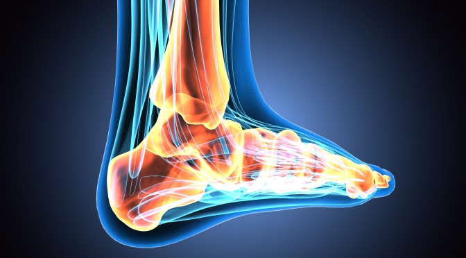
The CurveBeam HiRise is the industry’s first solution for capturing bilateral, weight bearing CT imagery of the hip and pelvis.

The HiRise will provide high-resolution, high-contrast imaging of the hip and pelvis that allows for cutting-edge orthopedic care.

Leveraging a rising gantry that surrounds the patient being scanned, the HiRise allows for long-leg views of the entire lower extremity.

The HiRise is also self-shielded, plugs into a standard wall outlet, and offers the largest patient platform in its class at 35 centimeters. The gantry rises high enough to accommodate patients well above 6 feet tall.

Best of all, like CurveBeam’s other solutions, the HiRise is simple to use, offering a fast track to revolutionary orthopedic care.

Providing Complete Care with CurveBeam

The HiRise is yet another addition to CurveBeam’s industry-leading solutions for weight bearing extremity imaging, joining the pedCAT, LineUP and InReach in helping you achieve the weight bearing difference.

With 3D, weight bearing imagery and software that can help you analyze and act upon the insights it provides, you can improve diagnostic information, achieve better patient outcomes and streamline your practice’s workflow and operations.

To learn more about CurveBeam and the HiRise, visit curvebeam.com/.

IFFAS Award of Excellence Goes to Study Using Weight Bearing CT to Examine Residual Effects of Misalignment in TAR Patients

An international research team of orthopedic surgeons was awarded the prestigious International Federation of Foot & Ankle Societies (IFFAS) Award for Excellence for a study that utilized weight bearing CT imaging  to analyze the effect of residual misalignment on cyst formation in total ankle replacement (TAR) patients.

The award was presented to the researchers at the American Orthopedic Foot and Ankle Society (AOFAS) Annual Meeting in Chicago on September 14 .

Courtesy of AOFAS. Researchers suggested the establishing a "safe zone" of ankle alignment to reduce the risk of peri-prosthetic cysts after total ankle replacement surgery.
Courtesy of AOFAS. Researchers suggested the establishing a “safe zone” of ankle alignment to reduce the risk of peri-prosthetic cysts after total ankle replacement surgery.

The authors hypothesized that longer follow-up and increased residual misalignment would correlate with increased peri-prosthetic cyst (PPC) volume and worse AOFAS scores. They found that residual varus cases had a more medial distribution of PPC and residual valgus cases had a more lateral distribution of PPC. During a presentation the next day, the authors explained that a using weight bearing CT to calculate a “Safe Zone” for  3D alignment of the foot prior to a TAR procedure could be instrumental in preventing  osteolysis, a condition that can eventually lead to implant failure.

The study was carried out at institutions using CurveBeam weight bearing  imaging solutions,  which provided the researchers with automatic 3D  measurement tools such as TALAS. The authors explained this research contributes to the comprehension of how peri-prosthetic cysts develop in the ankle.

The award recipients were Francois Lintz, MD, FEBOT; Jef Mast, MD; Mazim Medhi, MD; Alessio Bernasconi, MD; Cesar de Cesar Netto, MD, PhD; and Kristian Buedts, MD.

WBCT & MRI Study Sheds Light on Flat Foot Degeneration

Some degree of subtalar joint subluxation, as well as sinus tarsi impingement, was found in approximately 70 percent of patients with flexible adult acquired flat foot (AAFD) in a recent imaging study using MRI and weight bearing CT (WBCT) imaging.

Investigators used the imaging to assess which soft tissue structures demonstrating MRI signs of degeneration would correlate and influence more positive findings of bone collapse.

The study results were on display in the poster section of the AAOS Annual Meeting  and summarized by the AAOS Daily News.

Investiagators obtained MRI and WBCT imaging for 55 patients. Patients were evaluated for markers of arch collapse including increased valgus alignment of the hindfoot and forefoot abduction. Subfibular impingement was found in only 9 percent of patients.

“The significant and isolated influence of pronounced degeneration of the ligaments as positive indicators of more severe collapse of the bony architecture in AAFD patients, represented by subtalar joint subluxation and subfibular impingement, must be considered essential findings,” Dr. Cesar de Cesar Netto, a coauthor of the study, told AAOS News Daily. “Since we don’t really know which structures degenerate first in AAFD, our results point toward an increased importance of the ligamentous structures in the development and progression of the pathology, with the degeneration of the PTT potentially representing a late finding and only the tip of the ice berg.

The CurveBeam pedCAT is the only weight bearing CT imaging system with a field of view large enough to capture a bilateral foot and ankle scan. Flat foot degeneration is often a bilateral condition.

CurveBeam Plays an Important Role in the Best Research Awards at the EFAS Congress

Dr. Francois Lintz posted about being nominated for best research award at EFAS 2018 and congratulated Dr. Cesar de Cesar Netto for winning the award with his paper on Weight Bearing CT.
Dr. Francois Lintz posted about being nominated for best research award at EFAS 2018 and congratulated Dr. Cesar de Cesar Netto for winning the award with his paper on weight bearing ct.


The 12th annual European Foot and Ankle Society (EFAS) Congress was recently held in Geneva. As a part of this prestigious event, Dr. Cesar de Cesar Netto and Dr. Francois Lintz presented papers that were both nominated for research awards. CurveBeam is proud to have played a part in both of these important projects.

Dr. Cesar de Cesar Netto Wins Awards for Research and Best Oral Presentation

 Dr. Cesar de Cesar Netto, Clinical Fellow of Foot and Ankle Surgery at MedStar Union Memorial Hospital, was nominated and won a distinguished research award for his paper titled, “Weightbearing CT and MRI findings of Stage II Flatfoot Deformity: Can We Predict Patients at High-Risk for Foot Collapse?” Earlier in the year, he also won the best oral presentation at the FLAMeCiPP meeting, organized by the Latin American Foot and Ankle Federation.

Dr. de Cesar Netto’s research involved the prediction of soft tissue failure using weight bearing cone beam computed tomography (WBCT), an imaging modality that allows excellent evaluation of the relative three-dimensional positioning of the tarsal bones in dynamic deformities such as Acquired Adult Flatfoot Deformity (AAFD). The study indicated that WBCT can predict ligamentous injuries and dynamic bone deformity in AAFD patients.

Dr. Francois Lintz was Nominated for his Research

 Dr. Francois Lintz an orthopedic Surgeon specializing in Foot & Ankle Surgery in Toulouse, France, was also nominated for a research award for his paper, “3D Biometrics: A Prospective Comparative Evaluation of the Foot Ankle Offset Using Weight Bearing CT Semi Automatic Software.” The objective of Dr. Lintz’s study was to prospectively assess the clinical relevance and reproducibility of the Foot and Ankle Offset (FAO) value for hindfoot alignment, which requires WBCT for measurement, and compare it with previous findings. The study concluded that hindfoot alignment can be measured reliably using WBCT and demonstrated that FAO has good repeatability, and correlates well with clinical examination, 2D findings, and previous literature.

The EFAS Congress  

 The EFAS Congress provides a platform for professionals of all disciplines dealing with foot and ankle surgery and research to share their knowledge and advance the specialty. This year’s program, themed “State of the Art Foot and Ankle Surgery,” included a line-up of internationally renowned surgeons and scientists in the foot and ankle community. For this year’s gathering, the EFAS also invited representatives from two established national foot and ankle societies—the SFAS (Swiss Foot and Ankle Society) and the KFAS (Korean Foot and Ankle Society)—to share their expertise and innovative approaches in foot and ankle pathologies and treatments. The result was a program that provided a multidisciplinary, comprehensive overview of the latest scientific and clinical findings in foot and ankle surgery, state of the art surgical techniques, biomechanics, efficient conservative treatments, and the most recent imaging modalities in the field.

To watch a recording of the Weight Bearing CT International Study Group’s open scientific meeting at EFAS, click here.