pedCAT CubeVue: Automatic X-Ray Views

CurveBeam pedCAT Weight Bearing CT

It takes less than a minute to scan a patient in the pedCAT, but that’s enough time for the pedCAT system to collect enough data to create a 3D reconstruction of the foot and ankle, as well as .3 mm slices in all three planes.

COMING SOON: A new feature will let you take that data one step further – pedCAT’s CubeVue software will automatically generate the data into common X-Ray views, including the Saltzman hindfoot view, the dorsoplantar view, and left and right lateral views.

The automatic X-Ray view feature will save the physicians valuable time when analyzing their patient data.

To learn more about this and other CubeVue features,  contact your CurveBeam representative!

Distinguishing Categories of Medial Column Ligamentous Fracture

Weight bearing CT scans offer new, three-dimensional perspectives that reveal a better understanding of the biomechanics of flat foot. That was a finding on an academic poster on display at The American Orthotic and Prosthetic Association’s National Assembly in Las Vegas (Sept. 4 – 9).

“IRD, PTTD, Posterior Tib, Flat Foot, Pronation…these are all terms that various professions use to describe some sort of medial column ligamentous breakdown,” according to the poster. “The purpose of this study is to better distinguish some types of pathomechanics of the foot. By categorizing and redefining them, it makes the description of pathology more useful for Orthotic Treatment. This was possible through the use of 3D rendering of the pedCAT machine and CurveBeam software.”

The study was conducted by Ian Engelman, M.S. CPO, and Harold Chamberlin, DPM. They looked at 3D renderings of the bony structures in flat feet, and came up with some new descriptions, as well as their possible ligamentous causes.

For example:

Lisfranc Breakdown
Lisfranc Breakdown – Tarsometatarsal Ligamentous Failure
Lateral Foot Dislocation - Deltoid Ligament Fracture
Lateral Foot Dislocation – Deltoid Ligament Fracture


The study concluded that while the exterior contours of the foot suggest certain bony pathologies, the 3D renderings provided by the pedCAT would allow for orthotists to better distinguish the pathomechanics of the foot and ankle.

An unexpected result showed a moderately strong correlation (-0.68) between the vertical position of the navicular and medial displacement in a patient population with ranging degrees of medial column breakdown.

Moderately strong correlation of -0.68


CurveBeam Reception at AOFAS: You’re Invited!

If you’re attending the AOFAS/IFFAS Annual Meeting 2014 in Chicago from September 19 – 23, be sure to plan to attend the CurveBeam reception on September 21 at 7:30 p.m.

Sip cocktails and dine on hors d’oeuvres while mingling with colleagues who have integrated the pedCAT into their practices. Meet like-minded forward thinking peers who are considering a pedCAT acquisition at their facilities.

This informal event will feature brief talks by Prof. Dr. med. Martinus Richter, who has published studies on the pedCAT’s efficacy, as well as Dr. Erik Nilssen, MD and Dr. Martin O’Malley, MD.

When: Sept. 21, 7: 30 p.m.

Where: Hyatt Regency Wrigley’s Room (Bronze Level, West Tower)

If you can’t attend the seminar, you can visit us at booth #307 all conference long.

pedCAT: Fracture & Fusion Assessments

X-rays of the foot and ankle may not always provide conclusive assessments for post-operative fusions such as of the tarso-metatarsal joint or hind foot joints. Similarly, the physician is often left guessing if a fracture has properly healed.

“The pedCAT takes all the variability out, all the guesswork out of it,” Dr. Martin O’Malley, MD, an Associate Attending Orthopedic Surgeon at the Hospital for Special Surgery in New York, New York, said.

Dr. O’Malley said pedCAT scans allow him to clearly determine if a fusion has healed more than 50 percent, and he decides when to ambulate his patient accordingly.

“I let people walk on it earlier than before, and I keep them off longer than before,” Dr. O’Malley said.

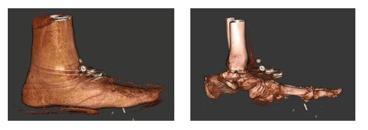
The pedCAT provides a three-dimensional view of fractures that changes the way O’Malley sees this common diagnosis.

“These posterior pieces are often bigger than we thought were based on plain X-ray and they often travel all the way around the medial side as well, which we never thought they did,” Dr. O’Malley said. “You know, we thought it was an infrequent fracture, but now we see it routinely. Now most of my ankle fracture work, I’d say more than half the time, is through a posterior approach. For the first 15 to 18 years of my practice I would do medial/ lateral incisions. Now I’m going to the back of the ankle. And a lot of it is driven by the pedCAT.”


pedCAT Stress Fracture Cross Section


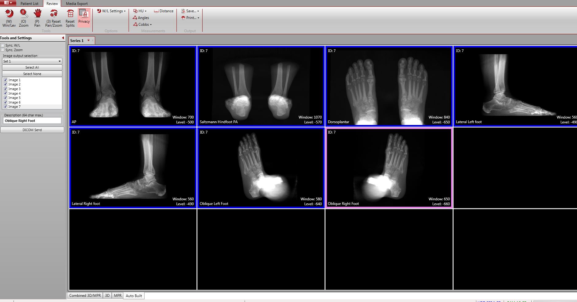
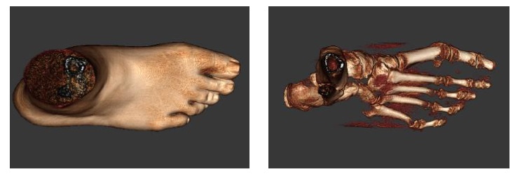
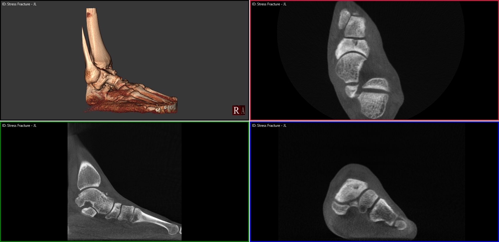
Above is an example of a navicular stress fracture in a collegiate runner that had not healed at all after six weeks of casting. Were it not for the conclusive pedCAT scan, this patient would have been allowed to ambulate.

Watch Dr. O’Malley talk more about the pedCAT here .

To offer your patients state-of-the-art fracture & fusion assessment, consider a pedCAT for your practice.


Made in the USA

At CurveBeam, we are proud that the pedCAT is manufactured entirely in the United States. Our factory headquarters are located in Warrington, Penn., just north of Philadelphia. The factory floor is housed in the same building as our administrative, sales, and research & development offices. We are committed to creating opportunities in our community. Most of the pedCAT’s components are sourced from local manufacturers as well.

CurveBeam’s manufacturing process is certified by ISO, a developer of voluntary industry standards. Each pedCAT goes through a rigorous testing process to ensure it produces high quality images.

An engineer surveys inventory at CurveBeam headquarters.

Engineers on the factory floor follow the principles of lean manufacturing, developed by Toyota. The process is easily scalable, meaning CurveBeam can react quickly to large orders or orders that require a fast turnaround time.

We would like to extend an open invitation to all foot and ankle specialists to visit us. We’d be happy to give you a tour and show you the pedCAT “behind the scenes.”

Tweet: Check out this example of high tech medical device manufacturing on American soil: http://ctt.ec/HPlnc+

Welcome to the CurveBeam Blog!

Welcome to the CurveBeam Blog!

2foot-with-platform1.jpg

It’s an exciting time at CurveBeam. Almost two years ago  we received FDA-approval for our first product, the pedCAT. The in-office 3D weight bearing imaging system for the foot and ankle has been integrated into orthopedic and podiatric practices around the world.  Foot and ankle specialists, considered leaders in their field, have told us that the pedCAT will become the standard of care for lower extremity imaging in the near future. In fact, in some of their practices, it already is.

We’re dedicated to continuing the improvement of foot & ankle imaging and are excited to share with you the technology advancements, right here on this blog in the upcoming months. So check back often!