CurveBeam Announces First pedCAT Installation at an Accredited College of Podiatric Medicine

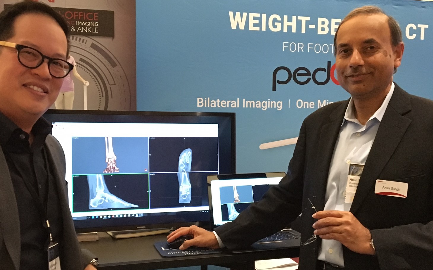
CurveBeam is proud to announce the installation of a pedCAT cone beam CT imaging system at Kent State University College of Podiatric Medicine.

Kent State is the first member of the American Association of Colleges of Podiatric Medicine to acquire weight bearing CT imaging technology.

“Kent State University is proud to be the first facility in the Northeast Ohio region to provide this ‘state- of-the-art” imaging modality,” said Dr. Allan Boike, Dean & Professor of Foot & Ankle Surgery at KSUCPM. “The CurveBeam pedCAT will allow the college to improve the foot and ankle health of the community while providing the highest quality education for our students and research opportunities for our faculty and residents.”

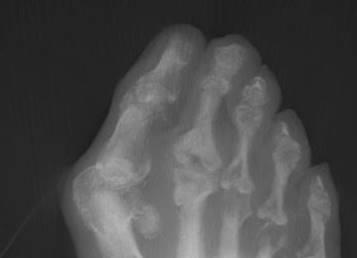
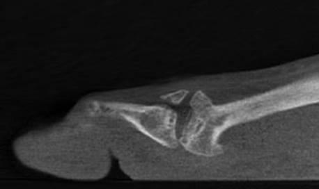
The pedCAT is the only cone beam CT imaging system that allows for bilateral, weight bearing CT images of the foot & ankle. Due to its compact size and low radiation exposure, the pedCAT is an an ideal solution for CT imaging at the podiatric point-of-care. Recent articles in orthopedic journals demonstrate weight bearing CT imaging is a valuable research tool that is shedding new light on even our basic understanding of foot & ankle biomechanics and deformities.

“Our initial euphoria in acquiring the pedCAT weight-bearing cone beam CT has only been eclipsed by actually getting to use it first-hand,” said Dr. Lawrence Osher, Director of the Radiology Department. “Simply put, this is an utterly amazing tool in the podiatric diagnostic and research armamentarium.  The ability to do 3D and multi-planar reconstructions on a weight-bearing foot and ankle, coupled with the prospect of marrying structure and function, opens up a seemingly endless array of research opportunities.  Bounded only by our creativity, we at KSUCPM look forward to adding significantly to the pool of knowledge in the medical literature.”

The pedCAT was funded through a research grant from the Ohio College of Podiatric Medicine (OCPM) Foundation, which was established to promote podiatric medical education and research.

“Dean Boike is to be commended for his foresight in procuring this advanced apparatus which further enables the College to maintain and expand its competitive edge in podiatric medical education,” said Dr. David Nicolanti, Executive Director of the OCPM Foundation. “In addition, this weight bearing CT imaging system provides a basis for enhancing collaboration between the College and foot and ankle specialists, from all medical stratums throughout the state of Ohio and the encompassing region.”

The College has a number of weight bearing CT research initiatives planned.

The pedCAT is located at the Cleveland Foot and Ankle Clinic’s midtown office at 7000 Euclid Ave in Cleveland. Weight bearing CT services will be offered to the clinic’s patients.

Need Bunion Surgery? A Weight Bearing CT Scan Could Help You Decide

When a patient appears to have a bunion, a physician typically orders traditional foot X-Rays as part of the clinical evaluation. The X-Rays capture three views of the foot- dorsoplantar, medial oblique, and lateral. But is this enough information to understand complex, three-dimensional object such as the foot? 

A growing number of foot & ankle specialists are advocating that there may be better way to evaluate this common deformity.

“Weight bearing computed tomography scans are beginning to take our understanding to the next level,” Dr. Paul Dayton, DPM, of Des Moines, IA, said in a roundtable discussion on bunions published in the Foot & Ankle Specialist academic journal. “Once you see the connection between coronal rotation and what we have traditionally evaluated on AP radiographs, it opens up a whole new understanding.”

On X-Rays, physicians have to “mentally interpolate” the valgus component of the bunion because they do not adequately capture the frontal plane, said Dr. Robert Weinstein, DPM, FACFAS, of Atlanta, explained in a CurveBeam case study.  

“Since the condition is a tri-plane deformity, we need to understand all of the components and their angular values contributing to the deformity,” Weinstein said. “Better deformity analysis leads to better pre-operative planning, surgical execution, and post-operative results.”

The new data on the coronal position is “exciting” because it explains so many of foot and ankle specialists’ previous questions, Dayton said.  

Weight bearing CT imaging also allows for evaluating deformities that involve external rotation. Rotation of one or more metatarsals, including displacement of the sesamoids, cannot be clearly seen in standard foot X-ray imaging. The lack of the weight-bearing aspect in traditional medical CT imaging means the physician cannot evaluate displacement and rotation under load.

Hallux Valgus Blog Post Image 1
One component of the deformity that is seldom quantified is external rotation, or frontal plane deviation. The presence of rotation of the hallux implies a more complex deformity. A very careful assessment of the first metatarsophalangeal joint architecture, including sesamoid position, their condition, and erosion or flattening of the crista on the inferior first metatarsal head is essential.

It has long been known that in hallux valgus or bunions the relationship between the metatarsal head and the sesamoids is altered,” Andy Goldberg, MD, MBBS, FRCS (TR and Orth), of Stanmore, United Kingdom, told Lower Extremity Review magazine. he said. “The sesamoid bones should sit underneath the metatarsal head, while in hallux valgus the big toe drifts off the sesamoids and the tip of the big toe points outwards. But our research has shown that in many cases the cartilage is worn, which in essence is localized arthritis, and we believe that this could affect the outcome of surgery.”

rotated first met and sesamoids

CubeVue, the pedCAT weight bearing CT imaging system’s custom visualization software, allows physicians to create oblique and frontal-plane images of varying angles and thicknesses from the weight bearing CT data.

Weight bearing CT images could have an impact on the technique a physician selects to repair a bunion, Dr. Dayton said.

“The knowledge that the sesamoids can be in normal position medial and lateral to the crista yet look dislocated on the AP X-ray because of pronation completely changes our mindset about the need for capsular balancing,” Dayton said. “We can see that in those cases supination corrects the deformity.”

pedCAT: Early Diagnosis of Osteomyelitis in the Diabetic Patient

The pedCAT weight bearing CT imaging system could identify bone infection at an early stage, and possibly prevent amputations, researchers at the California School of Podiatric Medicine at Samuel Merritt University determined in a report.

The researchers outlined two cases where “the use of CBCT device enabled us to diagnose and treat osteomyelitis in a timely manner, preventing its spread to adjacent bone and soft tissue, and minimizing the amount of required surgical resection.”

Plain radiograph is the primary imaging modality for the osteomyelitis diagnosis, the report states, but X-Rays may not reveal osteolytic changes for up to 20 days from the onset of infection or until the bone density is reduced by 30 – 50 percent.

Osteomyelitis is one of the most feared complications of diabetic foot ulceration, which often leads to lower extremity amputation and disability. Early diagnosis of osteomyelitis increases the likelihood of successful treatment and preserving ambulatory function. Unfortunately, most of the currently available imaging modalities are of limited use in assessing early stages of bone infection due to their low specificity and sensitivity for early osteolytic changes.

Magnetic resonance imaging (MRI) is more sensitive and specific than X-Ray, and yields greater accuracy in detecting soft tissue abscesses or early osteomyelitis in patients with high clinical suspicion and negative radiographs. In controversial or uncertain cases where MRI is not available, other imaging techniques such as indium-labeled leukocyte imaging combined with radionucleotide bone scan can be used as an alternative. (“Preventive and Therapeutic Strategies for Diabetic Foot Ulcers” – Foot & Ankle International® 2016, Vol. 37(3) 334– 343 – Chris C. Cychosz, BS, Phinit Phisitkul, MD, Daniel A. Belatti, BS, and Dane K. Wukich, MD).

Nuclear Imaging and MRI perform well in detecting early onset of osteomyelitis; but they are expensive to own and operate, are time-consuming in their acquisition of images.

What is desirable for early detection is a method that is accurate, inexpensive, and readily available. Cone beam CT, and the pedCAT in particular, fills this gap nicely.  The device is small enough to fit into most practices, offers high-resolution 3D imaging capabilities, and has a reduced radiation dose compared to traditional CT, according to the report.

Lead author Alexander M. Reyzelman, DPM,  and his associates reported on two diabetic patients who presented with infected neuropathic foot ulcers and were evaluated for potential osteomyelitis using plain film radiographs and the pedCAT CBCT scanner. In both cases, the “pedCAT was instrumental in identifying bone infection. The diagnosis of osteomyelitis was later confirmed by positive findings on bone biopsy. The use of CBCT device enabled us to diagnose and treat osteomyelitis in a timely manner, preventing its spread to adjacent bone and soft tissue, and minimizing the amount of required surgical resection.”

Case 1

A 49 year old diabetic female presented with an infected neuropathic ulcer at the lateral aspect of her fourth digit. The ulcer demonstrated malodor, cellulitis that extended to fourth metatarsophalangeal joint and positive probe-to-bone test. The plain film radiographs and CBCT were utilized in order to rule out osteomyelitis and assess the extent of soft tissue infection. The weight-bearing X-rays of the affected foot revealed subtle lucency at the lateral aspect of the proximal phalanx of the fourth digit, which was contiguous with the ulcer location. However, this finding alone was not sufficient to yield a conclusive diagnosis.  The images obtained using PedCAT clearly demonstrated the break in the cortex and the area of osteolysis involving the proximal phalanx of the fourth digit. The head of the fourth metatarsal and adjacent digits appeared intact. These findings, in conjunction with the clinical appearance of the affected digit, led to a preliminary diagnosis of osteomyelitis. The patient was treated with an arthroplasty of the fourth proximal interphalangeal joint, and has fully recovered. The bone specimens obtained intraoperatively were sent for biopsy, which confirmed our preliminary diagnosis of osteomyelitis.

Case 2

A 53 year old diabetic male presented with an infected neuropathic ulcer at his fifth metatarsal head, which exhibited malodor, edema and erythema extending through tthe plantar lateral aspect of  fifth metatarsal shaft and probed to joint capsule. The X-rays demonstrated no signs of bone involvement, while CBCT revealed distinct areas of cortical lysis and bony fragmentation of the fifth metatarsal head. The proximal two thirds of the shaft of the fifth metatarsal appeared unaffected, with intact cortex, uniform bony density and lack of osseous fragmentation. The patient was treated with partial resection of the fifth metatarsal. The bone biopsy has confirmed our preliminary diagnosis of osteomyelitis.

In the concluding discussion, Dr Reyzelman noted: “Though in our case studies we have not taken advantage of the option allowing to scan the patient in both, a weight-bearing and a non-weight bearing positions, this option could be highly useful for evaluation of complex fractures and dislocations of the foot and ankle.”

Orientation of the Subtalar Joint: Measurement and Reliability Using Weight Bearing CT Scans

Is there a reliable method to predict the type and perhaps the extent of osteoarthritis one might find in the ankle? Based on a recent article, which the examined the varus and valgus orientation of the talus and the configuration of the subtalar joint under weight bearing conditions, the possibility is there.

“A majority of the patients with ankle osteoarthritis present with an asymmetric wear pattern (eg, varus or valgus type),” according to a study published in 2009 by Valderrabano V, Horisberger M, Russell I, Dougall H, Hintermann B. titled, “Etiology of ankle osteoarthritis.”

Evaluation of these wear patterns, however, remained a challenge until recently, when Nicola Krähenbühl, MD, Michael Tschuck, Lilianna Bolliger, MSc, Beat Hintermann, MD, and Markus Knupp, MD published “Orientation of the Subtalar Joint: Measurement and Reliability Using Weightbearing CT Scans.” (Foot & Ankle International® 2016, Vol. 37(1) 109–114.)

Osteoarthritis of the ankle joint is relatively common and found in 1 percent of the world’s population, and a majority of those patients present with an asymmetric wear pattern (eg, varus or valgus type), according to the authors. Furthermore, up to 60 percent of the patients suffering from an osteoarthritic ankle joint develop talar tilt with progression of the osteoarthritic process.

Current research suggests this condition is caused by deformities of the lower leg and knee joint, ligamentous laxity, tendon dysfunction and neurologic disorders. Recently, it has been proposed that the adjacent joints and, particularly, the subtalar joint may have a major influence on this process.  “However, it is rather difficult to evaluate the orientation

of the subtalar joint using conventional radiographs; CT scans would be more appropriate,” the authors posit.

To distinguish between varus/valgus configuration of the subtalar joint, Van Bergeyk et al introduced the subtalar vertical angle (SVA) in 2002 using non weight bearing CT scans. “Today, weight bearing CT scans can be performed, leading to a better understanding of the functional anatomy of the hindfoot,” the article states.

Weight bearing CT technology became available in 2012. Weight bearing imaging only had been available in 2-dimensional X-Ray imaging prior to this, but weight bearing combined with computed tomography was needed to properly measure the SVA without superimposition of non-relevant anatomy that might throw off the measurement, including analyzing the shape of the subtalar joint. The subtalar joint is especially difficult to clinically and radiographically assess in 2D, due to the superimpositions, and attempts to artificially stress the joint and then scan using a conventional (non weight bearing) CT produced inconsistent results.

“Using weight bearing CT scans, we assessed the reproducibility of the SVA and analyzed the orientation of the subtalar joint in patients with asymmetric ankle osteoarthritis. We hypothesized that the SVA would provide reliable and reproducible measurements in varus ankles presenting with a varus subtalar joint and valgus ankles with a valgus orientation of the subtalar joint, respectively,” the authors said.

Using the new technology to view the joints, including utilization of the SVA measurement, the authors concluded the SVA measurements were reliable and consistent. “In our cohort, varus osteoarthritis of the ankle joint occurred with varus orientation of the subtalar joint whereas in patients with valgus osteoarthritis, valgus orientation of the subtalar joint was found,” the study said.

The authors found the results for the healthy cohort were significantly different, suggesting the orientation of the subtalar joint may play an important role in the development of ankle joint osteoarthritis.

Weight bearing CT not only allowed the authors to clinically and radiographically assess the ankle joints under the patient’s normal weight bearing conditions, but it also enabled them to make consistent and reproducible measurements.

Panel Recap: Dr. Michael Chin, DPM, Speaks about pedCAT at FABI

Dr. Michael Chin, DPM, and Arun Singh, President & CEO of CurveBeam

His decade old X-Ray system was failing, and feeling like a warrior without his weapon, Dr. Michael Chin, DPM, knew it needed to be replaced fast.

“We could have gotten a DR system, but at the end of the day, I wanted something that was unique, and something that would change the way I practice,” Dr. Chin said.

Dr. Chin participated in a panel discussion about new technology at the Foot and Ankle Business Innovations meeting in Chicago on Jan. 30. Dr. Chin practices at The Running Institute in downtown Chicago.

Dr. Michael Chin, DPM, at FABI

Dr. Chin uses the pedCAT for all of his X-Ray and CT imaging. His X-Ray revenues cover the device’s monthly capital lease payment, and the approximately 20 CTs he and his associate order every month provide his practice with an additional revenue stream.

Dr. Chin said he is able to order a CT scan and his staff can get payer authorization in the same day. This saves his patients from having to come in for a follow-up visit.

“We can fill that slot that we would have used for a follow up with another patient,” Dr. Chin said.

Weight Bearing CT Imaging for Cuboid Subluxations

Dr. Michael Chin, DPM, presented how weight bearing CT imaging has changed how he evaluates cuboid subluxations at the American Academy of Podiatric Sports Medicine meeting held near the West Point Military Academy campus in early September, 2015.

Dr. Chin began using the pedCAT in his office in February of this year.

Not much research is out there on how to use plain radiographs to measure cuboid subluxations, Dr. Chin said in his lecture, titled, “Cuboid Syndrome…The Other Side of Heel Pain.”

Dr. Chin has tested using a bilateral oblique projection to understand the cuboid/ metatarsal relationship, and has been able to observe a slide between the head of the fourth metatarsal and the head of the cuboid.

An MRI could be ordered to see the condition of the peroneal tendon, but the study would be limited because the scan would not be weight bearing, he said.. A traditional CT scan would provide great  visualization of the bone, but would provide no information on anatomic alignment.

The pedCAT weight bearing CT imaging system is excellent for evaluating stress fractures, sesamoids, periosteal changes, or anything medullar, Dr. Chin said. Another benefit is he can measure the exact degree of subluxation between the cuboid and the fourth metatarsal head.

Dr. Chin displayed pedCAT images depicting  pre and post-reduction views of a cuboid subluxation.

chin-lecture
pedCAT scan of a pre-surgical patient with cuboid subluxation. Dr. Chin was able to reduce the subluxation to 2.18 mm.

Dr. Chin practices at The Running Institute in Chicago.

AOFAS Annual Meeting – CurveBeam Symposium Recap

Weight bearing CT is a vital tool for determining the cause of inexplicable pain, and also for avoiding painful surgical complications.

That was the takeaway message from a talk by Dr. Phinit Phisitkul, a clinical associate professor of orthopaedics at the University of Iowa. He shared some of his most interesting cases at a CurveBeam sponsored symposium held during the AOFAS Annual Meeting in Long Beach during the evening session.

We’ve selected three of his cases to share on this blog:

18-year-old male with Noonan Syndrome & severe flat foot: The patient presented with an unusual amount of pain that was difficult to diagnose on plain X-Ray. A weight bearing CT scan revealed he had a severe deformity – a congenital vertical talus. He also had severe impingement.

Vertical Talus - Weight Bearing CT
Vertical Talus – Weight Bearing CT
Impingement - weight bearing CT
Impingement – weight bearing CT

58 year-old male with ankle arthritis: The patient presented with a lot of pain in the ankle joint. A weight bearing CT scan showed a subluxation of the ankle joint and dramatic impingement of the calcaneal fibula. Interestingly, the subtalar joint was in pristine condition. Dr. Phisitkul determined the patient was a good candidate for ankle replacement and hindfoot realignment, and that his subtalar joint could be spared.

Calcaneal-fibular impingement and arthritis - Weight Bearing CT
Calcaneal-fibular impingement and arthritis – Weight Bearing CT

41-year-old female with Hallux Valgus: A weight bearing CT scan revealed a bone spur on the patient’s first metatarsal head. If the doctor had done a normal release, the spur may have ended up pinching the sesamoid. Instead, he performed a lateral release and excised the bone spur.

1st MT Bone Spur - Weight Bearing CT
1st MT Bone Spur – Weight Bearing CT


Weight Bearing CT in Everyday Practice

“In my opinion, weight bearing CT will be the standard of care in the next couple of years,” Dr. Alex Tievsky, DPM, said in a lecture at the Graham International Implant Institute 8th Annual Symposium in Miami, on April 17. “This is really the future of not just foot and ankle, but the future of medicine.”

“2D is beginning to be phased out,” Tievsky said. “Now we’re beginning to see the problem from all angles and all planes, so this is super helpful from that respect.”

Dr. Tievsky presented a number of cases where he benefited from access to weight bearing 3D technology in his office.

Clinical Case #1

A 50-year-old female presented with bilateral flat feet for 20 years. She had heard about the HyProCure procedure, which corrects hindfoot misalignment through a minimally invasive procedure. She was eager to have the procedure done, no matter the cost. Dr. Tievsky took a pedCAT scan, and found she had a severe talar coalition.

talar calc coalition coronal left

“How many times do you catch a coalition on an X-Ray? It’s hard,” Dr. Tievsky said. “Sometimes you can see a halo sign, but it’s often missed. On the first visit, I was able to tell her, we either have to resect this coalition or we have to do a fusion. It’s impossible to get this level of information on an X-Ray.”

Clinical Case #2

A 16-year old girl came in with first metatarsal head pain. She had already been to two other podiatrists in the last six months, and they had prescribed steroid injections.

The pedCAT revealed a fracture on her fibular sesamoid that is extremely easy to miss on X-Ray.

fractured sesamoid

“Within her first 10 minutes in the office, we had a diagnosis,” Dr. Tievsky said. “We treated her appropriately. We immobilized her for eight weeks and gave her a bone stimulator. And she was pain free, three podiatrists later. She was happy, her mom was happy, and she never came back.”

Clinical Case #3

A patient presented with a lateral plantar fasciitis, a talo-tarsal dislocation, back pain for five years, knee pain, and hip pain.

A pedCAT revealed she had a tumor in her bone and it was eroding it. “There was no way we would have caught it on X-Ray,” Dr. Tievsky said. “We sent her out for oncology. It could have a malignant tumor, and we could have saved her life.”

Cyst Coronal

The scan is quick to take and you have a full work-up in about three minutes, Dr. Tievsky said as a closing statement. “This is a super important tool,” he said. “I’m kind of biased. I love this now. I can’t practice without it.”

Weight Bearing CT Scans for the Evaluation of Implant Arthroplasty Candidates

Weight bearing CT scans can be critical to a proper diagnosis, even for routine procedures.

In the following case, for example, a patient’s X-Rays indicated that he would be a good candidate for a metatarsal head hemi-implant arthroplasty. However, when the patient sought a second opinion, a weight bearing CT (pedCAT) scan revealed the true condition of the metatarsal head, and the surgical plan was considerably altered as a result.

A 60 year-old male presented complaining of a many year history of 2nd metatarsophalangeal joint pain, especially joint stiffness and pain. His pain increased with attempted 2nd MTPJ dorsiflexion.  In gait, he felt pain when rolling onto the ball of the foot.  His first surgical opinion recommended a metatarsal head hemi-implant arthroplasty .

Due to the excessive bony superimposition on the patient’s lateral X-Ray, it is difficult to accurately assess the shape of the 2nd metatarsal head.  A bone fragment can be visualized over the dorsum of the first or second metatarsal heads.  The AP weight bearing images demonstrate 2nd metatarsophalangeal  joint space narrowing.

Implant Arthroplasty Candidate X-Ray X-Ray 2

The patient sought a second opinion from a podiatric surgeon who offers in-office weight bearing CT services. The podiatrist performed a pedCAT scan and found the 2nd metatarsal head had sustained an old fracture. The pedCAT scan revealed that the dorsal 50% of the 2nd metatarsal head had been avulsed dorsally and a portion of the metatarsal head presented as a dorsal loose body.  The 2nd metatarsal head didn’t have the bone stock or bone volume to support a hemi-implant. The second opinion recommended recontouring the metatarsal head and performing an interpositional arthroplasty. The patient chose to have the second surgeon perform his surgery.

fractured 2nd MT head pedCAT weight bearing CT

A CT scan is not typically ordered to evaluate feet preoperatively.

“We are all trained to believe our eyes and to believe the information present in X-Ray images.  In this case it is assumed that the 2nd metatarsal head has a normal contour, length and bone volume.  The X-Rays demonstrate joint space loss and justify the hemi-implant arthroplasty, but adequate bone volume is required for implant stability and fixation. You just assume it’s going to be OK,” said Dr. Kent Feldman, DPM. “And if you do that as a routine, you’re going to get caught over and over and over in the operating room making mistakes or making assumptions that aren’t necessarily true.”

Dr. Feldman integrated a pedCAT into his surgical practice in 2012.

Dr. Gary Briskin: The pedCAT is the Future

“It’s the future – embrace it. It’s your future – control it.”

Dr. Briskin lectures on weight bearing CT imaging for foot 7 ankle

Dr. Gary Briskin delivered this motivating message at the Foot & Ankle Business Innovations conference in Chicago on Jan. 23.

Dr. Briskin, of University Foot & Ankle Institute in Santa Monica, Cali,  discussed how the pedCAT weight bearing CT system has improved his practice since his group acquired one last year.

His main take-away points were:

– The pedCAT gives you the advantage when it comes to diagnosing pathology.

“You can solve that Lisfranc case no one else can,” Dr. Briskin said. “It opens up the midfoot, which historically has been a problem for us.”

– The pedCAT allows him to clearly determine if bones and joints are completely fused after surgery.

“I do a lot of lapidus procedures,” Dr. Briskin said. “My concern; is this patient fused adequately to start bearing weight?” The exact level of fusion is visible with a pedCAT scan 5 – 6 weeks later, and with minimal scatter.

– Radiologists like the pedCAT images

“I think I’m getting a better CT, because it’s weight bearing and I think the quality is far superior. We get all of our CTs read by a radiologist. We have everything sent digitally. And the feedback we get is they are also impressed with the quality of the images.”