The Hospital for Special Surgery Utilization of pedCAT

The Hospital for Special Surgery (HSS) located in Manhattan is ranked as the #1 hospital for orthopedics by the prestigious US News and World Report (2016-2017). And when the surgeons and clinicians at this elite medical institution need to make a proper foot and ankle diagnosis, they rely on CurveBeam’s revolutionary pedCAT system for fast and accurate 3D imaging.

Assessing the root cause of a patient’s pain is essential for developing a comprehensive treatment plan. At HSS, this evaluation process begins with an interview so that specialists can learn a patient’s unique history and obtain information regarding the primary care physician’s prognosis. Then the patient steps into the pedCAT scanner, where expert radiologists can conduct foot and ankle imaging faster than X-rays and CT scans, resulting in fewer hospital trips and significantly reduced exposure to radiation. The pedCAT system delivers the highest quality images and robust data, allowing physicians to make the most accurate diagnosis of the malady, and guiding the surgeon in the operating room for a superior treatment outcome. Watch below and visit curvebeam.com to learn more about the pedCAT.


The Top 3 Reasons to Visit CurveBeam at the AAOS Annual Meeting

The American Academy of Orthopaedic Surgeons (AAOS) is holding its annual meeting at the San Diego Convention Center from March 14-18, 2017. More than 30,000 Academy Fellows, AAOS members, international guests, allied medical professionals, and exhibitors are expected to attend. Attendees can learn about the latest industry trends and medical advancements in educational sessions, and speak with representatives from top equipment vendors. So why stop by the CurveBeam booth?

1. You’ll learn about new automatic measurement tools and experience firsthand the many ways CurveBeam can help you harness the power of artificial intelligence in your practice. Get a hands-on demonstration of the pedCAT, CurveBeam’s compact, ultra-low dose CT imaging system, which is ideal for orthopedic and podiatric clinics, and see how to harness the full power of the datasets with CurveBeam’s visualization software solutions. Watch below.


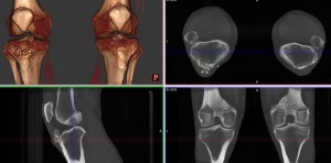
2. Get an exclusive look at the LineUP (investigational only), which is not yet available for sale in the United States. LineUP is CurveBeam’s revolutionary new system that allows bilateral, weight-bearing CT imaging of the knee and lower extremities in less than one minute. LineUP will redefine orthopedic care and you will be among the first to see it.

bilateral knee

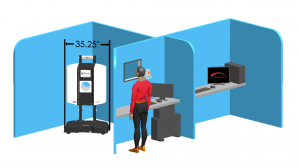
3. Witness for yourself the compact design of InReach, which provides point-of-care CT imaging for the upper extremities. InReach is a cone beam CT scanner that images the hand, wrist, forearm and elbow, as well as the lower extremities, providing the perfect reformat, every time. Best of all, a scan takes less than 20 seconds, improving both workflow and patient care. With InReach’s ability to provide 3D renderings, 0.3 mm multi-planar slices, and X-rays, point-of-care extremity CT imaging will soon be “in reach” to everyone.inreach room layout

CurveBeam’s automatic measurement tools lead to better patient outcomes, streamlined practice workflow, and increased revenue. Stop by booth 510 at the AAOS Annual Meeting and find out how our orthopedic imaging solutions can help you and, more importantly, help your patients.

Announcing a CurveBeam Webcast: “Weight-Bearing CT of the Foot & Ankle – Literature Review and Indications for Use in the Clinic”

To delve into the effect Weight-Bearing CT scans can have on modern orthopedic medicine, we are excited to host a live webinar. Dr. Alexej Barg, an orthopedic surgeon at University of Utah Health Care, will go over recent studies and analyze their findings. He will also demonstrate case studies of how this technology can best be used to revolutionize patient care in clinics.

For those who wish to join us, the webinar will be held on February 23, from 8-9pm (EST). You can register at any time, but it must be at least fifteen minutes before the webcast begins.

You can register for the webinar on FOOTInnovate here. We hope you will join us as Dr. Barg lends his insight and expertise to improving the field of orthopedics, and making all clinics a better place to practice.

Lower Limb Influence on Hindfoot Alignment

A recent study headed by L. Baverel, a distinguished French M.D., came out of the European Foot and Ankle Society in 2016. The study, published in Elsevier, examines the effect lower limb placement, specifically rotation, has on hindfoot alignment.

The implications for the study are still being sorted through, but a few standouts are worth mentioning. This study points out a major problem with 2D radiographic assessments of hindfoot alignment: there are numerous factors that influence measurements. Rotation of the lower limb is the first and foremost of these factors. “In the near future, it is very likely that 3D, weight bearing CT technology will enable the development of new ways to measure hindfoot alignment, based on 3D hindfoot-forefoot divergence angles or offsets rather than tibia-calcaneus angles.”

The constant goal of all orthopedic surgeons is to facilitate the correct alignment of bones. Even the slightest error in measurement can have devastating consequences on post-surgery recovery and future mobility. As the study notes, “Abnormal coronal alignment of the hindfoot has been proven to increase the risk of degenerative pathology or surgical failure in the foot and ankle.” The results of this study bring us that much closer to understanding foot and ankle dynamics, and are already impacting X-ray, stabilization and recovery protocol around the world.

For the purposes of study, foot alignment depends on basic two-dimensional radiographic techniques. However, apart from two recent radiographic techniques, the Ground Reaction Force Calcaneal Offset (GRFCO) and Arunakul’s Tripod Index, all others depend on lower limb rotation. This is important, as lower limb rotation necessarily affects foot alignment. The study describes rotation as one of the variables explaining lack of reproducibility in assessing hindfoot alignment: “The hypothesis for this study was that a mathematical model predicts how [rotation] modifies radiographic hindfoot alignment measurements.”

The study concluded, “The mathematical model accurately predicted the variations of the hindfoot angle, which was maximum when the foot was aligned with the X-ray source. It then decreased when the foot rotated away, following a parabolic curve.” The Meary angle was at maximum value when incident X-rays were aligned according to the second metatarsal bone, then declined along a parabolic curve as the lower limb rotated away from this axis. The study plainly illustrates this dynamic at play. “In practical terms, during the Meary procedure for shooting an AP ankle view, misalignment of the foot (caused either by incorrect positioning of the patient or an actual deformity), results in underestimating the value of the hindfoot angle.”

Until 3D weight bearing CT is an available option, it is recommended foot & ankle specialists exercise caution when planning from 2D radiographs. Radiographic procedures must account for correct alignment of the foot. That means checking that the second metatarsal is aligned with the X-ray source. This procedure appears to most consistently approximate true alignment. If alignment isn’t based off that axis, the deformity could be underestimated and the radiography should be done again or carefully questioned. For more detail, access a link to the article here.

Improved CT Foot and Ankle Analysis with True 3D

This video takes a look at EchoPixel’s latest technology, True 3D. A few studies are used as examples of what this powerful visualization platform can allow specialists to see. Using a simple stylus tool, analysts can take full advantage of the program’s 3D rendering capabilities. The program provides intuitive displays for complex tasks, such as making measurements in a 3D space to determine the depth of an implant.

Please enjoy this video as it shows the power of True 3D on a non-weight bearing CT dataset, and imagine the possibilities of pairing EchoPixel technology with bilateral, true weight bearing pedCAT datasets.  Visit echopixeltech.com to learn more.


The Opportunity for Private Office Imaging

Changes in healthcare insurance, regulations, and other factors have shifted the bulk of imaging services to hospitals, but the opportunity for private radiologists is still very promising.

“Hospital outpatient imaging has been migrating from private offices since about 2010,” says Dr. David C. Levin of Thomas Jefferson University, as reported by Aunt Minnie. “But that doesn’t mean that radiologists still can’t make private office ventures work.”

Dr. Levin goes further than just his diagnosis of the medical imaging service industry by prescribing a solution to private radiology offices interested in increase their imaging business.  His business model suggestions include a mix of customer service, marketing, and cost competitive advice that can turn around a struggling radiology office and build a successful and sustainable private practice.

Implement a Cost-Effective Payment Schedule for Patients with High Deductibles

The increasing number of high deductible healthcare plans has created a patient incentive to shop around for low price imaging services. Understanding these plans will help your office develop a fee schedule that is attractive from a cost perspective.

“Patients are going to have higher and higher deductibles, and that’s going to make them shop around for healthcare,” says Dr. Levin. “If you can offer, say, an MRI of the knee for $600, while the local hospital has it at $4,000, you can be sure patients will come to you.”

Marketing – It Really Works

Make prospective patients aware of your services by advertising an elevated quality of care and customer service.

“Hospitals generally don’t market their radiology services, but private practices certainly can,” says Dr. Levin. A well-designed advertising campaign can make your practice stand out and drive new customers to your location.

Don’t Neglect Customer Service

There is so much more that goes into the patient experience besides the treatment itself. Examine the way patients are treated, from the initial contact all the way through follow up, to identify opportunities to improve the experience. Patient frustration can arise at any step in the process. Time saving processes as well as friendly and helpful engagement go a long way to improving the overall patient experience.

Efficiency.  Efficiency.  Efficiency.

Dr. Levin stresses the importance of efficiency throughout the business.

“The more efficient your practice, the lower your costs will be,” he says. Fully utilizing assets and people in a streamlined operation brings down overhead and operating costs, and makes for a lean, cost competitive business with higher margins.

At the end of the day, Dr. Levin emphasizes private radiology practices have an amazing opportunity to improve their imaging services and overall business. Taking the time to evaluate the situation, develop an action plan, and implement constructive changes will certainly pay dividends now and for years to come.

Curvebeam at RSNA 2016

In November many of the greatest minds and innovators in radiology convened in Chicago for the 2016 RSNA. We had our talented and skilled team representing us and our innovations. While there we unveiled the prototype for the LineUP, a bilateral weight-bearing Cone Beam CT imaging system for the knees and lower extremities. We also showcased  InReach and TALAS. Watch below to recap the show!


CurveBeam’s Top 5 Moments of 2016

Founded in 2009, the news of CurveBeam and its advanced, compact 3D imaging devices spread quickly among orthopedic and podiatric specialists. Just three years later, the CurveBeam pedCAT system cleared the FDA. Without losing any momentum, CurveBeam Europe LTD’s office was opened in London in 2013. Even with all of these notable accomplishments, 2016 shines as the most impressive year for CurveBeam thus far. From our President and CEO, Arun Singh, receiving a lifetime achievement award to our expansion into the Asian market, 2016 has been a year to remember.

Take a look at the top five most significant moments for CurveBeam in 2016.

  1. President and CEO Arun Singh presented the Lifetime Achievement Award

“In recognition and appreciation of [his] visionary contributions to the advancement of Cone Beam Computed Tomography,” the American Association of Dental Maxillofacial Radiographic Technicians presented Arun Singh with the Carl Gugino Lifetime lifetime-achievement-awardAchievement Award on October 7th. The award was presented to him by Mr. Jerry Peck, who owns dental imaging centers in the San Francisco area. In his speech, Mr. Peck described when Arun installed a Tomax system in one of his offices 27 years ago. He described how Arun worked overnight to create the software code Mr. Peck needed to run the calculations  for the measurements he was looking for.

“This is the type of person, how bright, smart, ingenious, he is. I mean, overnight, all of a sudden, the program is written,” Mr. Peck said.

Mr. Peck went on to describe how Arun cofounded Imaging Sciences and developed both the CommCAT and I-CAT, which were game changers for dental professionals.

“Today, you look at what is the most successful CBCT company in the world, and you built it Arun,” Mr. Peck said.

2. First Weight Bearing Users Meeting is Held

Focusing on the applications of the weight bearing cone beam CT, the first pedCAT Weight Bearing CT Scientific User’s Meeting was held at the 2016 Foot International in Berlin, Germany. “We can’t have non-weight bearing CTs and say anything about alignment – that’s over. What do we want to measure, and how do we know what we’re measuring represents reality?” said Dr. Charles Saltzman as he emphasized the importance of WBCT at the meeting.

3. First Podiatry School is Outfitted with pedCAT System

The Kent State University College of Podiatric Medicine became the first podiatry school to implement the revolutionary pedCAT cone beam CT imaging system. “Kent State University is proud to be the first facility in the Northeast Ohio region to provide this state-of-the-art imaging modality,” said Dr. Allan Boike, Dean and Professor of Foot and Ankle Surgery at KSUCPM. “The CurveBeam pedCAT will allow the college to improve the foot and ankle health of the community while providing the highest quality education for our students and research opportunities for our faculty and residents.”
ben-franklin4. Ben Franklin and Susan B. Anthony See the pedCAT System

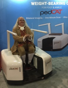
Philadelphia is a city well known for its rich history, and CurveBeam is proud to be in its next chapter, as the company is now headquartered in this remarkable town. At the APMA “The National” meeting this summer, historical interpreters acting as Ben Franklin and Susan B. Anthony visited the CurveBeam booth to learn more about the revolutionary pedCAT technology.

5.CurveBeam Expands International Presence to Asia

Not only has CurveBeam expanded its domestic presence, the use of the technology worldwide is also growing. The pedCAT system received notable attention at the 2016 National Foot & Ankle Surgery Symposium in Qingdao, China, as CurveBeam’s Chinese partners presented the weight bearing CT technology to the audience of medical professionals.

It’s been quite a ride since our original team pioneered cone beam CT imaging technology for the podiatric specialty. As we gear up to expand into new orthopedic specialties, we could not be more excited for what 2017 will bring!
Exclusive Video From The First pedCAT Weight Bearing CT Scientific Users’ Meeting in Berlin

meeting-83519_960_720 (1)The first pedCAT Weight Bearing CT Scientific User’s Meeting recently convened at the 2016 Foot International (EFAS, DAF, I-FAB) in Berlin. The meeting focused on weight bearing cone beam CT (WBCT) technology and the potential applications and challenges it presents.

Fill out this form to access the video.

Look for several interesting items as you watch:

  • Professor Martinus Richter’s work included a time workflow study on the use of X-Ray,  conventional medical CT, and the pedCAT; the results will surprise you. His study also showed how weight-bearing  CT measurements are more accurate than X-Ray measurements.
  • Dr. Arne Burssens’ study on hindfoot alignment answered critical questions: How do we identify varus and valgus, how do we accurately measure them, and how can we be sure that the measurements are clinically useful and reproducible?
  • Dr. Francois Lintz gives us a sneak peek at a new weight-bearing measurement CT tool that will be available exclusively in CubeVue, pedCAT’s software. It will enable users to measure alignment in three dimensions, and distinguish between normal and pathological cases.
  • Dr. Cesar Netto’s study focused on adult acquired flatfoot deformity, comparing  measurements on weight bearing CT vs non-weight bearing CT.
  • Dr. Michael Wachowsky’s study focused on the use of weight bearing CT  with pediatric patients, and asks: With the new weight bearing CT technology, what will be the exact definitions of the measurements?

The challenge created by this new technology is clear: How do we now define what we are measuring, since the current X-Ray methods have just been rendered obsolete?

“We can’t have non-weight bearing CTs and say anything about alignment – that’s over,” concluded Dr. Charles Saltzman. “What do we want to measure, and how do we know what we’re measuring represents reality?”

Fill out this form to access the video.

The Face of Technical Support at CurveBeam

When people think of Technical Support when they have a  problem that needs to be solved –  and the sooner, the better. While CurveBeam’s Technical Support can help with on-the-spot issues, it does much more than that. The support comes in many forms: software updates, assistance with calibration, online coaching, and more.

Andrew


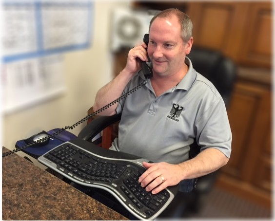
All these tasks are headed up by Andrew Kochansz, theTechnical Support Manager at CurveBeam. He joined the CurveBeam family in May of 2013, and has been a long time resident of eastern Pennsylvania.

Andrew has specialized in cone beam CT medical devices since 2006, and previously supported dental/maxillofacial cone beam CT systems. Before that he held positions involving quality assurance, software testing, and computer repairs – all which help make him uniquely qualified to provide support for computer and software dependent imaging systems like the pedCAT. When he is not directly assisting customers, Andrew is also involved in the software testing and verification processes.

When support is required, Andrew is able to remotely log in to most systems and provide assistance within minutes of receiving a call. Sometimes the solution can be as simple as turning the pedCAT off and turning it back on again (“power cycling” it).  If on site required, he coordinates with Customer Relations Director Tami Alexander to arrange for a service technician to travel to the customer site.

Andrew can be reached directly at 267-483-8097 when in his office, from 9:30am – 6:00pm Eastern Time Zone (USA); but he also is available through call forwarding for a couple of hours before and after that. If for some reason he is busy, you can always leave a message with Tami at 267-483-8089, or at the main CurveBeam number 267-483-8081.

For those who need assistance with performing Calibration and QA scans, Andrew would like to remind everyone that video tutorials on how to use CubeVue software, including performing the “manual” QA procedure, can be found on the CurveBeam website.

Andrew can be reached at 267-483-8097 or andrew.kochanasz@curvebeam.com.