Weight-Bearing CT Can Help Doctors Zoom in on the Problem

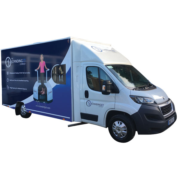
Sometimes the simplest solution to a difficult challenge is a change of perspective. That’s exactly what Dr. Selene Parekh and his colleagues at the North Carolina Orthopaedic Clinic have learned after using the CurveBeam pedCAT. In his talk titled “Standing CT: Zooming in on the Problem,” Dr. Parekh outlines how his experiences at the clinic and as a professor of orthopaedic surgery at Duke University have demonstrated the importance of weight-bearing computed tomography (WBCT) scans.

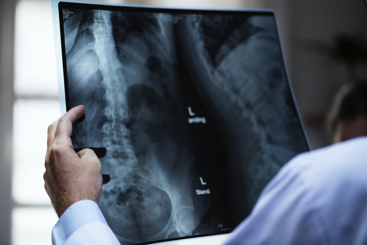
All too often, clinics rely solely on x-rays for initial diagnosis. If CT scans are used, the patient is usually lying horizontally, with weight and pressure completely removed from the area in question. As a result, the treatment plans or pre-operation strategies are developed from incomplete information. With WBCT scanners, doctors can accurately view the interaction of elements in the patient’s lower extremities, allowing them to better diagnose the issue.

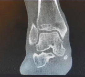
To illustrate this point, Dr. Parekh outlines several of his own real-world cases, one of which occurred within the last week. A 35-year-old woman visited the clinic complaining of medial ankle pain. While an initial x-ray revealed a minor increased talar tilt, a review of the patient’s history prompted her doctors to look again with the weight-bearing pedCAT. The test revealed the presence of osteochondral lesions. This refined diagnosis resulted in more appropriate treatment options, such as a resurfacing procedure to repair the issue at the heart of the problem.

35 xray35 wb

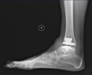
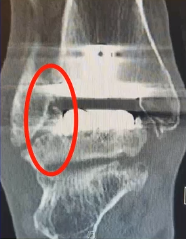
Another case involved a 61-year-old man who liked to stay active by biking and running. He presented with pain in his right foot. An x-ray revealed a malunion of the fibula and some arthritis. A total ankle replacement was subsequently performed, and the man resumed his active lifestyle. After five years, however, he returned complaining of increasing pain in the same foot. This time, a WBCT was taken, and the clinic saw the full extent of the problem. They discovered an impingement of the bone as well as a settling of the talar component into the talus, which was causing a talar fracture. Without this scan, it is likely that minor (and insufficient) treatments would have been considered. The patient chose to undergo an entire talus replacement, so he could keep cycling, a healthy activity that he enjoyed.

61 xray61 wb

These cases, as well as others outlined by Dr. Parekh, illustrate the significant difference that WBCT scanners such as the CurveBeam pedCAT can make in a physician’s ability to treat patients. Even beyond these new perspectives offered, there are other benefits to the patient, most significantly, reduced radiation exposure. While traditional CT scans have radiation levels around 2000 sieverts, the pedCAT only emits about 2 sieverts of radiation. Curve Beam is also releasing the LineUP, which will facilitate total lower body scans, from floor to knees.

By giving physicians a complete view of a patient’s extremities and the interactions of the bones, ligaments, and joints within, CurveBeam aids in providing the best treatment possible to patients. You can learn more about CurveBeam here!

Why CurveBeam Technology is Even Changing the Way We Talk

Processed with VSCOcam with f2 preset

Languages are constantly shifting and changing. As our experiences change, new terminology and ways to express the world around us are required. In a time when the medical field is experiencing technological breakthroughs that redefine the way we treat patients, it should be no surprise that the language used in some medical fields may quickly become outdated.

Nowhere is this truer than in orthopedic pathology diagnoses. In January’s issue of the Lower Extremity Review (LER), Ian Engelman’s article “Saying ‘PTTD” Is Misleading: It’s time for a new lexicon to distinguish pathologies” focuses on just one segment of the field to show the issues inherent in the current vocabulary. One of the main problems, for instance, is that the words used to describe what is occurring vary from profession to profession. This means that an orthopedic surgeon, a podiatrist and a physical therapist could all be discussing the same issue, but each use a different, and sometimes conflicting, term for the same pathology.

Engleman argues that as our ability to understand what is occurring in the foot progresses, our language should to. He refers to the organizing principle, stating that, “Better and more specific terms lead to better, more specific understanding of the origin of the pathological condition.” (Engelman, 2018). So much of our language regarding the movement of and connections within the foot regard on comparisons to mechanics in our everyday lives, such as a set of hinges or a mechanical bridge. By merely making analogies, rather than creating a specific and universal set of terms based on careful analysis, misdiagnoses or improper treatment can occur.

According to Engelman we are coming up on a paradigm shift in the world of orthopedics. This is primarily due to the integration of 3D imaging into the treatment of the lower extremities. CurveBeam’s pedCAT technology, Engelman says, is particularly groundbreaking as it allows for 3D, weight-bearing analysis, eliminating much of the guesswork about how the parts of the foot are operating in real-life conditions.

Engelman’s article highlights the limitations placed by utilizing a 2D vocabulary in a field that is beginning to integrate 3D technology. He gives suggestions for how he would linguistically alter the terms used by the industry:

  • “Normal foot” – This one may seem obvious, but before the capabilities provided by 3D scanning technology there was no way to establish a true baseline. Now there is a way to establish the ideal interactions and alignments within a patient’s foot to determine how far away from the norm they are.
  • “Talar escape” – This term is used to describe a spring ligament failure. While this aberration is easier to identify than most, the terms for it have traditionally varied across specialties
  • “Lateral Column Flat Foot” – In the past, much of the reason given for plantar ligamentous failure has been directed towards the medial column within the foot. When examined closer using modern technology, however, Engelman notes that there can be healthy medial columns while flat footedness still occurs, meaning the causality lies more with the ligament interacting with the lateral column. Engelman suggests several terms like this, where there is a linguistic need for events previously not recognized with 2D scans.

As well as making efforts to improve and expand the professional vocabulary, Engelman also urges clinics to begin to implement devices like the CurveBeam pedCAT and CubeVue software. That way there can at least be a visual representation of exactly what is occurring in a patient’s foot, even if there is not yet a way to put it into words. To learn more about CurveBeam’s innovative products, visit http://www.curvebeam.com/ and see how 3D imaging can revolutionize your clinic’s treatment practices.

When Billing CT Scans, a Little Effort Goes a Long Way


pexels-photo-461077

It should come as no surprise that providing a patient’s clinical history can help verify and interpret results from a CT scan. Including as much information as possible when submitting an imaging order seems like an obvious practice, and yet researchers from the University of Chicago Medical Center found that many physicians needed a reminder. Led by Dr. Saad Ali, the team was able to show that the effort physicians put into writing clinical history reports has benefits not only for the patient, but for the doctor’s own hospital or clinic as well. Interventions implemented by Ali’s team led to payments and reimbursements arriving on average 21 days earlier.

To obtain these results, Ali’s team took a look at their own emergency department and analyzed 1,000 requests for head CT scans. They began by simply analyzing the practices that were common in the department at the time. Not only did poor histories complicate the radiology department’s efforts to interpret results, but they also negatively impacted the hospital’s ability to get reimbursed.

According to their findings, inadequate histories can lead to confusion for the coders and billers. With a high rate of inaccurate billings, the denied claims begin to pile up significantly. Seeing this, the researchers were determined to find a way to rectify the situation. There had been plenty of studies that found poor history practices to impact image interpretation and billing, and yet bad habits still persist.

The team put together a program they called an “intervention.” This primarily involved educating the emergency staff about how to write high quality histories. PowerPoint presentations were displayed in the department with helpful tips reinforcing the concepts outlined in the lectures. After this program, not only did image requisitions for CT scans receive higher scores, but payments and reimbursements arrived a full 21 days earlier on average than they had before Ali and his team stepped in.

Any medical practice can always use some improvements. For some, this might mean improving clinical histories, for others it may just mean ensuring that necessary equipment is on hand to properly care for patients. At CurveBeam, we are constantly pushing ourselves to find new ways to provide our customers with the best in weight-bearing CT technology. Just as our customers seek to provide the best care they can to their patients, we work hard to provide them with the equipment they need to do so. To learn more about CurveBeam, visit our About page.

CurveBeam Goes to Europe: ECR 2018

The European Congress of Radiology (ECR) is the annual meeting of the European Society of Radiology (ESR). The next ECR congress will be held from February 28 – March 4, 2018, in Vienna, Austria, and CurveBeam is excited and honored to be among those participating this year.

ECR-2018

Founded in 2005, by merging the European Congress of Radiology (ECR) and the European Association of Radiology (EAR), the European Society of Radiology (ESR) is an apolitical, non-profit organization dedicated to strengthening and unifying European radiology. With more than 75,500 members worldwide, it is the largest radiological society in the world.

The Society works closely with radiological societies across Europe while establishing relationships with major international organizations from other related fields. It also monitors developments on the EU level, with a significant influence in radiology research and practice. The mission of ESR is to serve the healthcare needs of the general public by supporting scientific research, education, and training, while constantly striving to improve the quality of radiological practice.

CurveBeam, founded in 2009, is an innovator and industry leader in weight-bearing CT. A study performed in 2012 in Finland, concluded that “Cone-beam CT technology with new design and flexible gantry movements allows both supine and weight-bearing imaging of the lower extremities, with a reasonable radiation dose and excellent image quality. Weight-bearing CT of joints can provide important new clinical information in musculoskeletal radiology.”1 It was the first major study to demonstrate the viability of weight-bearing CT.

CurveBeam will be exhibiting several of their products in Booth X1/110. Medical professionals can learn about pedCAT, InReach, and the currently investigational, LineUP.

The pedCAT’s bilateral, weight-bearing 3D views of the foot and ankle give specialists the information they need to create comprehensive treatment plans. Its compact size and ultra-low dose CT imagine boasts a scan time of one minute. InReach is the most compact extremity cone beam CT scanner, and it images the hand, wrist, forearm, elbow and the lower extremities. In the early stages of development is the LineUp, a bilateral, Weight-Bearing CT device for imaging of the knee and lower extremities, designed with patient comfort in mind. LineUp is easy to operate and offers quick scans of less than 30 seconds each, producing 3D Reconstructions, Multi-Planar slices, and X-Ray views.

CurveBeam can’t wait to network with medical professionals and other imaging specialists in Vienna. It won’t be our only connection to Europe, however. CurveBeam has had the good fortune of partnering with The UK’s Standing CT, who has created a mobile solution for patients to utilize pedCAT.

CurveBeam looks forward to showcasing its exceptional products and talking with you about the imaging needs of your orthopedic or podiatric practice. We are eager to introduce you to our exceptional imaging products: pedCAT, InReach, and LineUp.

Learn more about CurveBeam here!


1Department of Diagnostic Radiology, Medical Imaging Centre of Southwest Finland, Turku University Hospital, Kiinamyllynkatu 4, Turku FI-20520, Finland. Address correspondence to E. K. J. Tuominen (esa.tuominen@iki.fi)

Takeaways from the 2018 OrthoForum Annual Meeting

The 2018 OrthoForum Annual Meeting concluded this past weekend in Orlando, and the agenda was once again packed with useful sessions for orthopedic executives.

During the popular CEO breakout sessions, executives shared some timeless principles that are worth pausing and reflecting on. Here are two of the key takeaways we picked up:

1. Approach internal and external communication with continuous improvement

There was a strong, consistent message that orthopedic executives and surgeons will need to increasingly work together as a unified group in reforming healthcare delivery. Foster communication with other orthopedists and care providers embrace opportunities to collaborate with healthcare systems, not just compete with them. Now is the time to act rather than taking a wait-and-see approach. Be involved, participate in discussions and be open to change.

Internal communication is important as well. Several administrators shared how their operations suffer from “leakage” when surgeons don’t follow practice guidelines for utilization of in-house ancillary services. Instead, they tend to stick to old habits, including outsourcing services, even if they capture less revenues and end-up with studies of inferior value. Repeatedly communicate the true value and enhanced quality of ancillary services to minimize leakage.

2. Customer Service really does matter

Speakers suggested implementing training to offer better to best in Customer Service. Healthcare has become more competitive and consumers are shopping around. How patients feel they are treated matters now more than ever. Train, monitor and measure customer/patient satisfaction and respond to all comments by acknowledging them, showing appreciation for feedback and addressing any concerns with a goal to improve perceptions. Incentivize your staff to buy-in to this goal.

Differentiating your practice with customer service delivers the most impact. It’s what patients and the general public understand more than anything else. People remember how you made them feel. Reinforce positive and engaging staff attitudes, and keep patient-treatment paramount.

CurveBeam is a proud sponsor of the OrthoForum Annual Meeting.

Curvebeam Corporate Webcast: Standing CT—Zooming in on the Problem with Dr. Selene Parekh

Untitled designMark your calendars for an upcoming FOOTInnovate lecture with Dr. Selene Parekh.  Titled, “Standing CT—Zooming in on the Problem”, the lecture will detail Dr. Parekh’s own experience with incorporating weight bearing CT imaging into his own clinical practice. Be sure to register at Foot Innovate, and join your colleagues for what’s sure to be an informative evening on Wednesday, February 28, at 8:00 PM EST.

Dr. Selene Parekh, M.D. is an orthopedic surgeon and expert on sports injuries. He currently serves as Professor of Orthopaedic Surgery at the North Carolina Orthopaedic Clinic at Duke University. Prior to his time at Duke, Dr. Parekh was a foot and ankle surgeon at the University of North Carolina (UNC), where he not only taught medical students and residents, but he also consulted in foot and ankle cases for the athletic department. He treats athletes at various levels in his current practice.

After earning an MBA in Health Care Management as well as an MD from Boston University, Dr. Parekh completed both his surgery internship and his orthopedic surgery residency at the Hospital of the University of Pennsylvania. While in residency, he was honored with multiple awards for his research in the field, and most recently was awarded the Ranawat Award by the Eastern Orthopaedic Association.

Dr. Parekh prides himself in learning about his patients to understand their specific needs and their overall functioning. He considers himself conservative in his surgical approach, and believes in maximizing non-surgery methods, only operating when necessary.

More popularly known as the “Fantasy Doctor,” Dr. Parekh has combined his passion for sports medicine with his love of fantasy football, utilizing his impressive skills to accurately predict how injuries will affect a player’s career and in turn, impact the Fantasy football league and its teams. He has even written two foundational research papers using Fantasy football data to track the epidemiology and outcomes of tendon and ligament ruptures in the NFL.

Examining the Added Value of Preoperative CT for Determining Cartilage Degeneration in Patients with Osteochondral Lesions of the Talar Dome

Osteochondral lesions of the talar dome (OLTs) involve the articular cartilage and subchondral bone. These lesions can cause deep ankle pain as well as impaired daily activities and sports activities. Surgical treatments are conducted as repair or replacement strategies to achieve biological healing of the OLT. The surgical procedure depends on the size, location, and stability of the lesion, as well as the extent of cartilage damage and the condition of the subchondral bone.

The usefulness of magnetic resonance imaging (MRI) has been reported to interpret the condition of the lesion ahead of a surgical procedure. It is not a perfect solution, however. An MRI might over- or underestimate the staging of OLT owing to bone edema and thinning of the articular cartilage. Computed tomography (CT) can provide more precise information than MRI on the subchondral bone, such as bone sclerosis, absorption, and cystic lesion.

To evaluate the unique features of the CT findings that relate to the condition of the articular cartilage in OLT, a study was conducted by the Department of Orthopaedic Surgery at Hiroshima University’s Graduate School of Biomedical Sciences in Hiroshima, Japan. The lead researcher on this study was Tomoyuki Nakasa, MD, PhD. The findings were published in the American Journal of Sports Medicine. Thirty ankles in 29 patients who had OLT with an osteochondral fragment were retrospectively reviewed to find out the extent to which CT image prediction of the histological findings on OLT will be useful to determine the most appropriate therapeutic strategy.

 The osteochondral fragment of 19 ankles could be preserved by fixation or drilling at surgery. This was the preservation group. In the remaining 11 ankles, the osteochondral fragment was removed. This was the excision group. Preoperative CT findings were compared between the two groups to determine the relationship between the CT and histological findings. Biopsies of the osteochondral fragment from 13 ankles were also performed.

The area of lesion in the preservation group was significantly larger than that in the excision group. The CT images of the lesion showed the rate of absorption of the subchondral bone plate (SBP) in the preservation group to be lower than that in the excision group. The lesion bed absorption was higher in the preservation group than in the excision group. All cases in the excision group showed bed sclerosis, compared with 42.1% in the preservation group. The specimens with disruption of the SBP exhibited cartilage degeneration and abundant chondrocyte cloning. OLT with absorption of the SBP on CT showed severe cartilage degeneration, while the remaining SBP on CT showed low-grade cartilage degeneration.

In conclusion, while the condition of the SBP affects cartilage degeneration, CT findings provide important information for the determination of surgical treatment.

 CurveBeam designs and manufactures Cone Beam CT imaging equipment for the orthopedic and podiatric specialties. Learn about CurveBeam’s Cone Beam CT systems here!

A Mobile Solution for Weight-bearing CT in the UK

Industry-Workshop-featured-image

Britain’s national healthcare system can make getting a CT scan difficult, and weight-bearing scanners are almost unheard of. However, an inexpensive mobile option which will allow more doctors to perform the scans they need is now available. Weight-bearing CT scanners are available to visit in three hospitals around the UK and growing thanks to The Standing CT Company. Their goal is to be able to provide every doctor in the country with access to these scans.

Standing CT uses the CurveBeam pedCAT, the only weight-bearing scanner that scans both ankles and feet in a single pass. The pedCAT is used by leading surgeons and hospitals around the world, providing easier access for patients and superior results for clinicians. The pedCAT has everything a radiographer is looking for. It’s a compact, ultra-low dose CT imaging system that provides a bilateral, weight-bearing, 3-Dimensional view of the foot and ankle. With a less than one-minute scan time the pedCAT generates not only a CT image but digitally reconstructed X-ray views as well.

Unless a patient is standing, it is difficult to capture the proper relationships among the bones, muscles, and ligaments. For too long hospitals did not have the proper technology to create optimally useful images of feet and ankles

The Standing CT Company was founded in 2016 to solve this problem, bringing doctors and experienced entrepreneurs together to develop a solution. They created mobile units so hospitals could provide high-quality foot and ankle scanning. Standing CT Company gained regulatory approvals in May, and received their first patient in June 2017. While the first pedCAT unit in the UK was integrated four years ago at Royal National Orthopaedic, Standing CT Company is the only company offering a mobile option. By working with surgeons to learn what and where their greatest needs lie, Standing CT Company will continue to expand their reach throughout the UK over the next 3-5 years.

The mobile scanning unit is brought directly to the doctors and patients in a van containing a mobile pedCAT visiting one hospital on specific days of the week. A radiographer and healthcare assistant work together to transport the van and assist with patients. The van can run on generator power for 10 hours. This new technology offers specialists a standard model, serving hospitals and communities that would otherwise not have the capital to purchase the technology on their own.

Hospitals throughout the UK need weight-bearing scanners, and this mobile technology allows surgeons to perform the scans they need without the hospital having to purchase the expensive technology.

Learn more about Standing CT today at www.standingct.com. To learn more about the CurveBeam pedCAT, visit https://www.curvebeam.com/products/pedcat.

Study Finds Low Levels of Radiation May Actually Help Fight Off Cancer

anatomy-3003099_1280It has long been accepted that there is no such thing as a safe dose of radiation, and to suggest that radiation could actually have positive health benefits would be laughable. The standard belief was that our bodies had natural cancer barriers and that doses of radiation would significantly diminish these barriers, leading to a higher risk of cancer. Recently, however, two articles published in the Journal of American Physicians and Surgeons included findings that show low-dose radiation to be safe and even has the potential to be beneficial to patients.

In the article “Small Radiation Doses Enhance Natural Barriers to Cancer,” Dr. Bobby R. Scott outlines the current literature available on the topic. Through his analysis of studies concerning the effects of x- and gamma rays on tissue interactions, natural immunity and cancer-facilitating inflammation, Scott found that low doses are actually far safer than previously thought. In fact, especially in studies involving tumor rates in mice, Scott’s research pointed to the possibility that low levels of radiation can actually improve the body’s natural cancer barriers.

The main theory at play here is known as the adaptive response theory. This posits that these low doses actually help the body repair double-stranded DNA breaks. Under higher levels of radiation these breaks increase in frequency, hence the previous belief that any level of radiation put patients at an increased risk of cancer.

In the same issue, a team of researchers led by Dr. Jerry Cuttler published “Thyroid Cancer Following Childhood Low Dose Radiation Exposure: Fallacies in a Pooled Analysis” with findings that lead to the same conclusion as that of Dr. Scott. While the article primarily focuses on the overdiagnosis of thyroid cancer and the negative consequences that arise from that, the safety of radiation also receives some attention. Like Dr. Scott, this team also found low levels of radiation to be safe, and that the “all radiation is bad radiation” approach was a fallacy. Of significance was the finding that levels below 500 mSv were even safe for children.

At CurveBeam, we believe it is important to stay up to date with the latest findings in our field. In order to provide ground breaking technology to our customers, it is necessary to be familiar with studies like those reviewed by Drs. Scott and Cuttler. To learn more about CurveBeam and our pedCAT technology, visit www.curvebeam.com today!

Literature Review and Case Studies from CurveBeam’s “Weight-Bearing CT of the Foot and Ankle” Webinar

On February 23, Dr. Alexej Barg, an orthopedic surgeon at University of Utah Health Care, shared an overview of the latest literature on the use of weight-bearing CT scans (WBCT) with foot and ankle patients. WBCT scans provide several benefits to doctors and patients, according to Dr. Barg. For example, there are reasonably low radiation doses. In addition, patients are in a natural standing position. Also, the relatively small scanner size allows for convenient office storage. Finally, these scans have lower capitalization costs than conventional CT scans.

The first professional paper on this topic was published in 1999. It described the significantly different radiographs with WBCT scans compared to the then-current CT scans. The paper described a simulated weight-bearing environment in which the patient was on his back with the foot held in a frame. While today’s literature focuses on true weight-bearing scans, the 1999 publication was, nonetheless, a revolutionary one.

In today’s medical publishing world, there are seven established medical literature data bases (PubMed, MEDLINE, Embase, CINAHL, Cochrane Central Registrar of Controlled Trials, ScienceDirect, SpringerLink). Also accessible are the digital contents of orthopedic and radiology journals. The biggest takeaway from each review in the webinar was the significant differences between WBCT and non-weight-bearing CT scans in patients who experienced pain or had a deformity, while, on the other hand, the control groups saw little to no differences between the two scans.

In one study, Dr. Burrsens[1] observed that, in addition to the importance of a weight-bearing stance, the measurement methods used are also critical. He performed three different types of measurements on the patients in the study to get a comprehensive view of the ankle and foot.

Highlighting the differences between WBCT and non-weight-bearing CT scans allows doctors to better understand the pain point(s) and provide an accurate treatment plan. In the case of a 57-year-old female complaining of forefront pain and overloading, doctors could recognize that the pain was caused by natural alignment of the forefoot. Instead of surgery, they recommend shoe insoles, and the patient was pain free within three months.

In another case, doctors used WBCT scans to determine that reconstruction surgery was not applicable for a 63-year-old male with charcot arthropathy, since he didn’t have the appropriate bone stock. Instead, they recommended a below-knee amputation to better address the patient’s concern.

Dr. Barg also shared some future considerations when using WBCT scans, including standardizing measurements and understanding the correlation between WBCT and conventional weight-bearing radiographs. While the industry has advanced in utilizing WBCT scans, ongoing research is necessary to take full advantage of all the WBCT scan benefits.

To discover more about the exciting research supported by the CurveBeam team and its innovative products and goals, visit the website at CurveBeam.com. For more webinars presented by Curvebeam, visit here.


[1] Burrsens et all, Foot Ankle Surg, 2016.