New Case Study Showcases Advancements in Measurement of Joint Space Width in the Knee

jsw
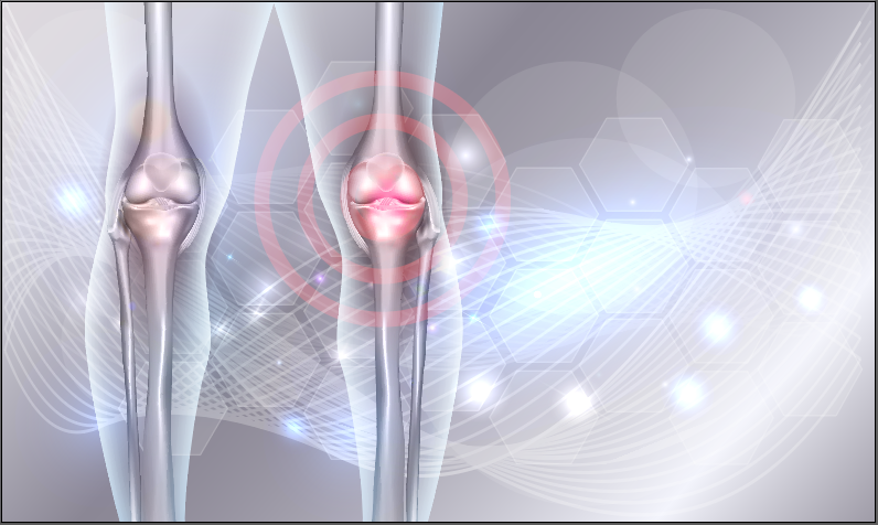
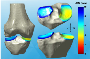
Under the current standard of care, joint space width (JSW) measured on Weight Bearing radiographs suffers from poor sensitivity to detection of knee osteoarthritis (OA). It also does a poor job of tracking symptom progression. 2D radiographic JSW is limited by dependence on X-Ray beam alignment with the medial tibial plateau as well as temporal and spatial heterogeneity of structural progression of knee osteoarthritis. Fortunately, 3D JSW measured on Weight Bearing CT images has the potential to overcome these limitations.

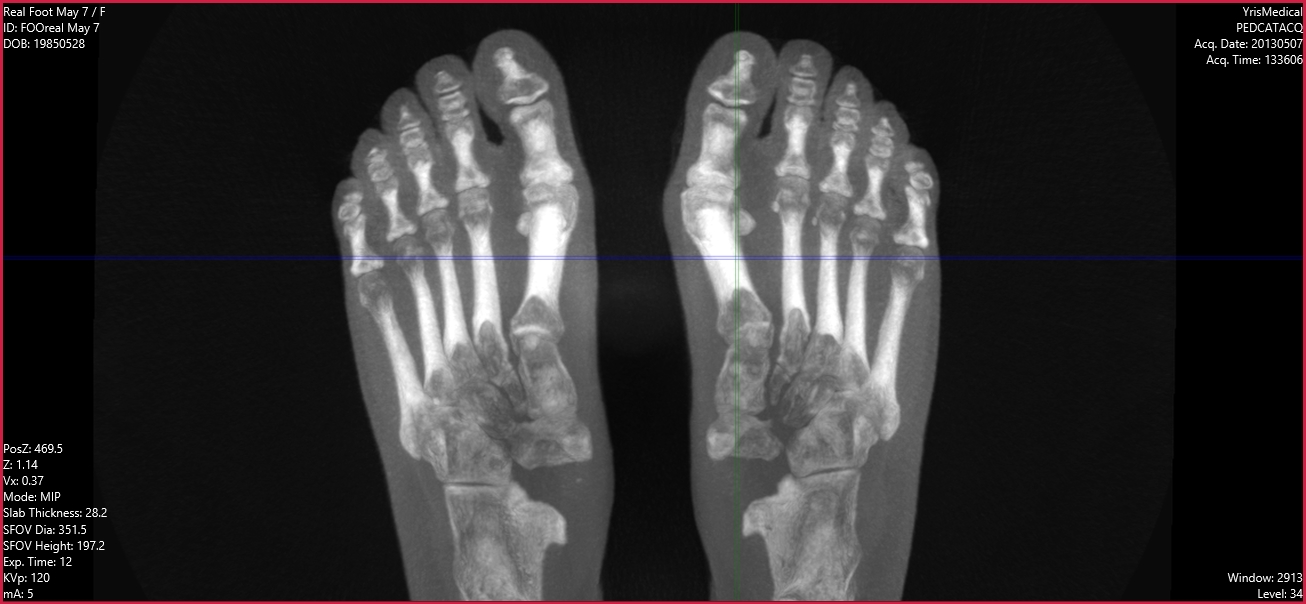
LineUp Scanner from CurveBeam Used to Create 3D Datasets

To test this assertion, 3D JSW measurements were collected on 11 participants in the Multicenter Osteoarthritis Study. Then, 3D datasets were reconstructed from Cone Beam CT projections. These images were captured using the LineUP scanner manufactured by CurveBeam. Standardized response means (SRM) were calculated to assess the ability of Weight Bearing CT to detect changes in joint space width over time.The preliminary data presents evidence that 3D JSW is sensitive to changes in joint space comparable to what was reported in other studies measuring JSW by radiographs or MRI.

Weight Bearing CT: More Sensitive and Accurate

Weight Bearing CT is showing the potential to offer better data, even when overlapping anatomy is a factor. It is proving to be more sensitive and accurate for detecting osteophytes and subchondral cysts when compared to conventional fixed-flexion radiography. This technology provides 3D biomechanically accurate views of bone morphology, alignment, and joint spaces.

“Weight Bearing CT could replace radiographs as the recommended means of assessing knee OA,” says Dr. Neil Segal, MD, Professor, and Director of Clinical Research in the Department of Rehabilitation Medicine at the University of Kansas Hospital, and the lead researcher on this study.

To learn more, download the case study by completing the form below.

Name *

Email *

New Algorithm Uses CT Data to Create 3D Map and Better Assess Osteoarthritis

knee arthritis stock

Engineers, radiologists and physicians based in the UK have developed a new technique for improving clinical imaging. It uses an algorithm to create a detailed three-dimensional map of a patient’s joint using computed tomography (CT) data. The team, led by co-author Tom Turmezei, Ph.D. of Cambridge University, recently reported on their breakthrough with a journal article titled “A new quantitative 3D approach to imaging of structural joint disease,” which appeared in the June 2018 edition of Scientific Reports.

The team knew that the ability to assess OA with 2D radiographs was not good enough to detect subtle changes that could have important implications for patients, doctors, and researchers. To meet this challenge, they worked to develop a new algorithm for the structural assessment of joints including hips, knees and ankles in 3D. Their recent technical validation study showed the technique was successful.

The algorithm maps joint space width to the nearest tenth of a millimeter. Mapping joint space width in 3D from clinical CT data has the potential to enhance understanding of osteoarthritis and how patients progress to joint failure—leading to better and earlier interventions.

Early Detection Gives Osteoarthritis Patients More Options

The 3D mapping technique has the potential to be more than twice as sensitive as radiographs in detecting small changes in joint space widths. Using the algorithm, clinicians will be able to use CT scans to map a marker of osteoarthritis across a joint. Why does it matter? Because early detection leads to earlier treatment. Doctors could identify patients with disease earlier than the current gold standard, allowing important interventions to be started before the joint fails. Patients will have the opportunity to try lifestyle changes and physiotherapy instead of immediately facing surgery.

All clinical CT imaging was performed on a 64-slice Siemens Definition AS system.

“It will be an important next step to use JSM to investigate whether differences in positioning and load-bearing have any effect on joint space width in 3D,” the authors wrote. “A straightforward solution for prospective studies would be to perform knee and ankle CT in a standing position (as used in clinical practice), and to standardise supine hip positioning by strapping feet together.”

Evaluating New Therapies

In addition, the new 3D method will likely allow researchers to determine whether new therapies in development are effective in a realistic timescale for clinical trials. This is something that has not been possible using radiographs.

CurveBeam designs and manufactures Cone Beam CT imaging equipment for the orthopedic and podiatric specialties. Bilateral, weight bearing scans of the foot and ankle give physicians the information necessary to assess the biomechanical spatial relationships and alignment of the lower extremities.

To learn about a recent grant awarded to the University of Kansas to study joint space biomarkers via weight bearing CT, click here.

ESSR in Amsterdam: Muscles & Nerves

Are you heading to Amsterdam to attend ESSR this week? The focus of this year’s meeting is “Muscles & Nerves”, and there are some great opportunities to expand your knowledge on some critical issues at this event.

ESSR Overview

The European Society of Musculoskeletal Radiology (ESSR) is an organization whose mission is to advance the knowledge, diagnosis and treatment through imaging of both the normal and abnormal musculoskeletal system. The ESSR is the primary organization in Europe in this field, and they provide a European forum for education and research of musculoskeletal radiology and cooperation among medical and non-medical professionals in these endeavors. The annual meeting is the major European forum for sharing advances in musculoskeletal radiology and offers excellence in education.

Recommended Sessions

The team at CurveBeam would like to recommend some interesting sessions as you plan your schedule at ESSR:

  • Tibial Tuberosity—Trochlear Groove Distance: Basic Positioning Errors of Current Measurement and Methods of Correction

Presented: Saturday, June 16, at 2:12 p.m. in Room 2.

  • Low Dose CT Technique and Radiation Dose: Does the ALARA Principle Still Apply?

Presented: Saturday, June 16, at 12:50 p.m. in Room 1.

  • Standardised Reporting: Guidelines for the Radiologist, Benefit for the Clinician

Presented: Saturday June 16, at 12:20 p.m. in Room 2.

CurveBeam Commitment

CurveBeam makes it a priority to stay abreast of the latest research and development in the field of imaging in order to best serve your practice and your patients.

Learn more here about the innovative imaging and diagnostic technologies available at CurveBeam.

The Weight-Bearing CT International Study Group, Brought to You by CurveBeam

This year CurveBeam will be co-sponsoring the Weight-Bearing CT International Study Group Thursday, July 12, 5:00-6:30 PM at the Hynes Convention Center, Room 310, in Boston, MA.

Dr. Martinus Richter from Krankenhaus Rummelsberg will be moderating a panel of experts from around the world. Professor Alberto Leardini from the Rizzoli Institute, Dr. Francois Lintz from Clinique de L’Union, Dr. Cesar de Cesar Netton from the Hospital for Special Surgery, and Dr. Alastair Younger from the Footbridge Centre for Integrated Orthopedic Care will join and discuss the latest thinking in weight-bearing CT.

The goal of the study group is to promote dialogue and collaboration on weight-bearing CT research, as well as create standardizations for weight-bearing CT measurements and analysis.

Weight-bearing CT scans use low doses of radiation while creating an image of the foot and ankle while the patient is standing in a natural standing position. Having patients in a natural standing position is beneficial for doctors because they can see the bones and tendons of the foot and ankle under normal conditions. Patients with pain and deformities seem to benefit most from using weight-bearing CT scans.

While many years of research have shown the benefits of these types of scans, we know there are many out there who still have questions. This International Study Group will provide you with answers. We would also love to have orthopedics and podiatrists with experience with weight-bearing CT to join us and share your own experiences with the technology.

Anyone interested in learning more about weight-bearing CT, including the Cone Beam CT imaging equipment for orthopedics and podiatrists designed by CurveBeam, is invited to join us for presentations from these experts.

Space is limited. RSVP today at wbctstudygroup.com!

Co-Sponsored by Curvebeam

Weight-Bearing CT May Improve Diagnoses in Hallux Valgus Patients

Hallux Valgus

A recent analysis of studies titled Imaging of Hallux Valgus by James Welck and Naji Al-Khudairi has shown that newer, three-dimensional imaging techniques including weight-bearing computer tomography (WBCT) may permit a more thorough understanding of the hallux valgus (HV) deformity and provide greater information to foot and ankle surgeons prior to corrective surgery.

Traditional two-dimensional AP, lateral, and oblique radiographs are the current standard for imaging HV deformity. Two-dimensional radiographic images can be used to measure several radiographic angles which are commonly used to quantify the extent of HV deformity. However, recent research shows that some of these angle measures show low levels of reliability and accuracy. Coughlin et. al. found that measures of the hallux valgus angle and the intermetatarsal angle assessed using 2-D imaging had high intraobserver and interobserver reliability. In contrast, the distal metatarsal articular angle was measured to 5° or less in only 58.9% of cases, and measurement of metatarsophalangeal joint congruency led to a wide variation in cases identified. The primary reason that two-dimensional imaging techniques may fail to be reliable is that HV is a triplanar deformity with rotational aspects that two-dimensional images cannot account for.

Fortunately, research shows three-dimensional HV imaging techniques have promise. Conventional CT imaging is not weight-bearing, but weight-bearing cone beam CT provides better insight into HV. This means that many patients imaged through traditional CT may not have been diagnosed correctly.

  • Welck and colleagues have found WBCT to better display the sesamoids, which are difficult to represent accurately using traditional AP radiographs.
  • Collan and colleagues demonstrated the importance that CT is weight-bearing by finding that there were significant differences in HVAs and IMAs between HV and control groups only when patients were in a weight-bearing stance.

Two other research groups found that WBCT provided insight into the effects of joint hypermobility in HV patients which would not typically be measurable using two-dimensional imaging techniques.

To learn more about how three-dimensional imaging techniques like WBCT can benefit your foot and ankle practice, visit http://www.curvebeam.com/products/pedcat/. CurveBeam’s PedCAT systems are at the cutting edge of WBCT innovation and can help provide your patients with the best HV care that current science has to offer.

i-FAB Awarded to Hindfoot Misalignment Research Study

i-FAB award

Hindfoot malalignment is a major cause of pathology in the foot and ankle and impacts the biomechanics of these areas of the body. Standard 2D radiograph measurements are plagued by anatomical and operator bias due to inaccuracies with 2D plane projections. Weight Bearing Computed Tomography (WBCT) and semi-automated 3D measurements of the hindfoot have shown promise for increased accuracy. They do have a critical shortcoming, however; they cannot offer information related to surface-to-surface interactions throughout various parts of the ankle and foot.

Sorin Siegler, Maui Jepsen, and Francois Lintz recently completed a study with the goal of addressing these inadequacies and finding solutions. They applied a 3D biometric tool based on distance mapping, which describes this interaction to WBCT data to characterize the effect of hindfoot valgus and varus deformities. 30 bilateral data sets were obtained, and the images were then processed to create 3D models of the foot and ankle structures. 3D CAD software calculated surface-to-surface interactions at various joints and created color-coded distance maps. The team then compared the images from the deformed feet to those of the normal feet.

With respect to hindfoot valgus, they discovered the antero-medial side of the talus is closer to the medial malleolus as this bone moves into slight external rotation and plantarflexion. The calcaneus is externally rotated relative to the talus, resulting in surface approximations on the lateral side of the posterior articular facet and an impingement of the sinus tarsi. In addition, a strong approximation on the superior side of the calcaneocuboid joint and the lateral side of the talonavicular joint occurs at the Chopart joint. At the cunei-navicular joint, there is a significant approximation of the lateral cuneiform to the navicular. At the metatarsal-cuneiform joints is a strong approximation at the 2nd and 3rd metatarsals joint surfaces.

In hindfoot varus, it was evident that the medial and lateral sides of the talus are closer to their respective malleoli, and anteromedial side of the talar dome approximates the tibial plafond, as the talus moves into inversion and slight dorsiflexion. The calcaneus is internally rotated and inverted relative to the talus, resulting in surface approximations on the medial side of the calcaneus bridging the gap between the posterior and middle articular facet and at the lateral side of the posterior articular facet of the calcaneus. The cuboid is displaced inferior to the navicular resulting in contact on the lateral facet of the calcaneocuboid joint. Also, a strong approximation at the talo-navicular joint can be observed on the medial side. At the cunei-navicular joint, the lateral cuneiform surface is displaced away from the navicular surface compared to normal.

It was concluded that WBCT can provide accurate classification of these foot and ankle deformities. Distance mapping allowed the team to characterize the specific surface-to-surface interactions at the joints and distinguish more readily between the hindfoot deformities. This information can be valuable in understanding certain pathologies associated with hindfoot varus and valgus deformities. In addition, the color-coded distance maps provided an effective method for assessing the effect of hindfoot deformities on articular joint surface interaction in the foot and ankle. The maps also enhance diagnostics, with future applications in evaluating therapy efforts to restore normal alignment.

Not only was this study highly successful in its future applications to diagnosing and treating these deformities, it also was the recipient of the distinguished i-FAB award. The mission of i-FAB, or International Foot and Ankle Biometrics Community, is to improve our understanding of foot and ankle mechanics as it applies to health, disease, and the design, development and evaluation of foot and ankle surgery, and interventions such as footwear and insoles/orthotics.

Learn about CurveBeam’s ongoing commitment to advancing foot and ankle care here.

Studies Point to Many Benefits of Weightbearing CT Scanning Technology

A-Recent-Study-Demonstrates-the-Need-for-CT-Scans-in-Complex-Ankle-Fractures 

For patients whose lives are impacted by medical technology breakthroughs, there is a significant improvement in the quality of care their doctors are then able to provide. This is especially true when it comes to cone beam computed tomography (CT) scanning. In the last decade, the innovations made in this field have allowed for true weightbearing CT scans to be taken.

In order to present a picture of the difference weightbearing CT scanning has made on foot and ankle patients, authors from the University of Utah, as well as a team of international researchers, presented a series of studies showing the benefits of the new technology. In their article, Weightbearing Computed Topography of the Foot and Ankle: Emerging Technology Topical Review, Drs. Alexej Barg and Charles Saltzman show that true weightbearing CT scans are far more effective than their simulated counterparts.

The article covered 8 studies performed between 2013 and 2017, each of which covered alignment analysis within the foot. While previous attempts to replicate weightbearing environments involved rigging up weights to affect the foot while the patient was lying down, modern technology is allowing doctors to observe the interactions within the foot in actual weightbearing scenarios with the patient standing and even walking. Regardless of whether the studies focused on talar anatomy, hindfoot alignment, valgus deformities or flat-footedness, the studies all had a similar theme. The cone beam weightbearing CT scan offered a more complete view of the relationships in the foot than what was attainable with traditional radiography.

Perhaps most importantly, the overview of the literature available pointed to several key benefits offered by this scanning technology. Not only does it allow for scans to be taken while the patient is standing, but there is increased spatial resolution and faster imaging times due to cone beam scanning. Plus, the side benefits of having lower radiation than traditional CT scans coupled with modest costs can help alleviate many patient concerns. The authors also advocate for utilizing the images provided through weightbearing CT scanning to establish standardized forefoot, midfoot and hindfoot alignment positions using anatomical landmarks, which could greatly enhance both diagnoses and care plan development for the patient.

Leading the charge in this innovative technology is CurveBeam. With a line of cone beam scanning devices like the InReach, LineUP and their standard PedCAT, CurveBeam allows clinics to offer weight bearing CT scanning to their patients to improve their quality of care. To learn more about the possibilities offered by devices like the PedCAT, or to see their products for yourself, visit http://www.curvebeam.com/products/pedcat/ today!

Weight Bearing CT Finds Important Displacement in Patients with Hallux Valgus

It’s difficult to understand how much a bunion can affect your life until you experience it. Known by the medical term hallux valgus, this deformity is painful and can be extremely disruptive. While the traditional solution has been to perform a fusion of two key bones in what is known as the Lapidus Procedure, a team of researchers believes this overlooks a secondary displacement that can cause lasting postoperative problems.

In their study, Comparison of Intercuneiform I-2 Joint Mobility Between Hallux Valgus and Normal Feet Using Weightbearing Computed Tomography and 3-Dimensional Analysis, Dr. Tadashi Kimura and fellow researchers looked at an understudied joint to find out if there is more that can be done in the treatment of hallux valgus. They looked at the feet of 11 women with the condition, as well as the feet of 11 healthy women, and found a significant difference in the displacement of the I-2 joint between the two groups.

To conduct their study, the team used CT scanning with simulated weightbearing effects on the foot to see the mobility of the joint. The images portrayed enough difference in the two groups to suggest that hallux valgus does affect the I-2 joint. Previously, a Lapidus Procedure had been used to fuse the first metatarsal with the medial cuneiform, but an additional operation may be needed to fuse the I-2 joint, a process known also as arthrodesis, in order to relieve the patient’s symptoms.

While this might not be the case for all patients with hallux valgus, it can be significant for those who experience hypermobility of this joint. To determine that this is the case, utilizing true weightbearing CT scanning technology may be critical. This study used CT scans that simulated a weightbearing environment with the intention of replicating the effects, but the authors state that the displacement may be even more severe once true weightbearing interaction is observed.

An industry leader in weight bearing, 3D scanning technology, CurveBeam’s innovations give doctors the ability to develop comprehensive treatment plans that address this entire issue. The authors of the study write that simple radiography would be unable to capture the displacement of the I-2 joint and traditional CT scanning is unable to account for the shifts caused once the full weight of a patient’s body is placed on the foot. Hallux valgus treatment can greatly improve the quality of life for a patient, and it is important to ensure that the treatment provided eliminates the problem. The PedCAT can give doctors the ability to do exactly that. To learn more about CurveBeam and the pedCAT, click here.

CurveBeam Throws A Curve at Conventional CT Scans for Orthopedic Practices

Often used for orthopedic and podiatric use when plain x-rays do not provide the visualization needed, a CBCT (Cone Beam Computed Tomography) scan is a quick, pain-free, noninvasive radiology diagnostic imaging test used to accurately generate comprehensive images of bones. These images can then be reformatted into three-dimensional images which can be easily shared through PACS. Quicker than the spiral motion of a typical CT scan, CBCT imaging exposes patients to less radiation than traditional CT scanners.

Watch the below video to learn Dr. Josef Zoldos, MD, DDS, one of the founders of the Arizona Center for Hand Surgery, explain how CurveBeam CBCT aids their doctors in detecting inflammatory conditions, tumors, and evaluating other abnormalities of the hand, wrist, and elbow, improving overall diagnoses, treatment plans, and patient engagement in their facility.

Health Canada Approves CurveBeam’s InReach Cone Beam CT

March 26, 2018 – Warrington, Penn. – CurveBeam announced the InReach cone beam CT system for orthopedics has been approved by Health Canada regulatory authority for sale in Canada.

CB_CanadaGraphic

The InReach is primarily designed for the hand, wrist &elbow; & lower extremities in non-weight bearing position. The InReach is an ultra-compact CT scanner that provides high-contrast 3D datasets of bony anatomy, which could potentially replace radiographs as a first line of diagnosis.

The InReach is ideal for the point-of-care, imaging centers, and hospital orthopedic departments because of its small footprint, its self-shielded design, and standard power requirements.

“The InReach will revolutionize the speed and accuracy of assessment of upper extremity conditions that specialists have traditionally found challenging to diagnose with plain X-Ray, such as scaphoid fractures,” said CurveBeam President & CEO, Arun Singh. “The InReach continues the company’s mission to elevate advanced diagnostic imaging capabilities to enhance orthopedic care.”

The InReach is designed with patient comfort in mind. Patients’ hand, wrist or elbow is positioned in a height-adjustable bore while in standing or sitting position. The unit can also accommodate non-weight bearing, lower limb imaging. Scan times are less than 30 seconds.

The InReach device is supplemented by CubeVue, CurveBeam’s custom visualization software.  CubeVue gives orthopedic specialists to multi-planar slice navigation tools and vivid 3D renderings of the anatomy previously not easily accessible to specialists. CubeVue’s Insta-X feature provides Digitally Reconstructed Radiographs, potentially eliminating the need for radiographic exams altogether.

The InReach is the second extremity CT imaging system CurveBeam has introduced to Canada. CurveBeam’s pedCAT system permits weight bearing CT imaging of the feet and ankles.

The InReach system was cleared by the US FDA for sale in the United States in 2017.

CurveBeam is the leader in Weight-Bearing extremity CT imaging, starting with the introduction of its pioneer product, the pedCAT, in 2012. The pedCAT is the only CT system that allows for bilateral, true weight-bearing imaging of the lower extremities. Since 2012, the pedCAT has been integrated into leading foot & ankle orthopedic and podiatric practices around the world.

CurveBeam is currently developing its next generation multi-extremity device, the LineUP, which will provide bilateral Weight-Bearing images of the knees in addition to feet, as well as hand, wrist & elbow.