Conference Will Feature Full Day Agenda on Weight Bearing CT

An upcoming conference at the Royal Society of Medicine – London will feature a day-long program on weight bearing CT Imaging. “Bipedalism: Defining Humanity and Foot & Ankle Surgery” is aimed at surgeons, podiatrists and radiologists. CurveBeam is proud to be the primary sponsor of this prestigious event.

The conference will be held on July 12. Sponsored registration is available for attendees who register prior to May 30th.

The conference is organized by Standing CT Company, which provides weight bearing CT services via mobile imaging centers to hospitals and orthopedic practices in the United Kingdom and Europe. Standing CT Company exclusively utilizes CurveBeam’s weight bearing CT systems.

Several members of Standing CT’s medical advisory board will be lecturing at the conference. Advisory board member Dr. Sajid Butt, MB BS, FCPS, FCPR, a respected radiologist at Royal National Orthopedic Hospitals in London and Stanmore, will present imaging best practices in the morning session.

Another member of the advisory board, Mr. Stephen Bendall, MBBS, FRCS, FRCS (Orth), an orthopedic foot & ankle surgeon at Princess Royal Hospital in Haywards Heath will present on Lisfranc injuries during the section “Trauma: Better Imaging, Better Outcomes.”

If you are interested in attending, call 0800 047 1010 or email kirsty.collins@standingct.com.

WBCT Research On Display at OARSI 2019

Osteoarthritis Research Society International (OARSI) 2019  World  Congress, the pre-eminent multidisciplinary global forum for all those interested in cutting edge OA research from academia and industry around the world, kicks off May 2 in Toronto, Canada.

OARSISocialGraphic2

Make sure to visit poster #512 titled, “The Relationship of Three-Dimensional Joint Space Width Measured on Standing Computed Tomography with Concurrent Pain and Physical Function in the MOST Study” (Dr. Neil Segal, M.D. et al).

If you are unable to attend this year and would like to view the poster on display at OARSI 2019  click here to download it now.

Hugentobler: pedCAT Improves Practice Workflow

Dr. Nicholas Hugentobler, DPM

It is feasible for a single-practitioner podiatry practice to add weight bearing CT (WBCT)  imaging and realize economical and operational benefits, Dr. Nicholas Hugentobler, DPM, explained in a webinar titled, “Incorporating Weight Bearing CT Imaging into the Daily Podiatric Practice” on FOOTInnovate.

Dr. Hugentobler took over Animas Foot and Ankle in Durango, Colo., in 2012 and purchased a pedCAT WBCT system three years later. Since then, Animas has grown to seven total locations and eight doctors.

Dr. Hugentobler said the pedCAT makes financial sense if a podiatrist orders on average one CT scan per clinic day, and noted “my criteria for ordering a WBCT has expanded as I have been able to see the benefit.”

Dr. Hugentobler said he regularly orders WBCT studies for post-surgical fusion verification, fracture repair verification, post-surgical alignment verification and pre-surgical planning.

Dr. Hugentobler advised to always bill only what is appropriate and to always provide adequate documentation that would stand up in an audit or a peer-to-peer review.

An unintended benefit of adding the pedCAT to his practice was increased efficiency, Dr. Hugentobler said. An X-Ray study can take as much as 8 minutes depending on the technologist, he said. Often after reviewing the radiographs, a podiatrist will have to send the patient back for additional views. A pedCAT scan takes a little more than a minute, and provides unlimited X-Ray views via its Insta-X feature.

“I have a practice in an adjacent city which has three doctors at any given time,” Dr Hugentobler said. “They order about half as many X-Rays as I do alone [with the pedCAT]. I am earning more on X-Ray than anticipated.”

Dr. Hugentobler reviewed several cases from his clinical practice. 

Sesamoid grinding on crista cropped

In the case above, the patient’s pedCAT Insta-X of the foot showed the patient had a mild bunion. Dr. Hugentobler said the X-Ray view did not explain why the patient had severe pain to the point where he could not walk. Navigating through the coronal slices on the the CT volume showed the patient’s sesamoid was directly underneath the crista and was grinding against it. “It won’t take long before that starts deteriorating in the wrong direction,” Dr. Hugentobler said.  The pedCAT allowed for prompt and responsive treatment. 

In another example,  Dr. Hugentobler showed a 34-year-old female who had gotten an open reduction and internal fixation (ORIF) after a motorcycle accident. After surgery, her foot was fixed in a varus position. She was “bounced around” from doctor to doctor after surgery to try to figure out why. Providers suggested bracing, a calcaneal slide, etc. When the patient came to Dr. Hugentobler, he ordered a WBCT, which revealed a large bone mass in the subtalar joint was causing the misalignment. Further evaluation showed arthrosis and subchondral schlerosis.

“This gives you a better understanding of what really will be beneficial,” Dr. Hugentobler said, who recommended a fusion and injections to the patient.


pedCAT saves 800+ Hours of Imaging Time a Year

A study found that performing 2D radiographs & traditional medical CT imaging for foot & ankle exams took an extra 800 hours of imaging time per year, as compared to performing only a pedCAT weight bearing CT scan for the same indications.

pedCAT image acquisition is 70% faster than radiographs and 35% faster than CTs. Patient positioning on the pedCAT does not require X-Ray emitter or sliding adjustment.

Click to expand the graphic below and see how busy orthopedic practices can save time with weight bearing CT imaging.
Time Savings Study Comparison

Demehri: WBCT Preferable Over MRI for Syndesmosis

demehriInjury to the syndesmosis often requires advanced imaging. Up until recently, the most common advanced imaging modality to evaluate this injury has been magnetic resonance imaging (MRI). Dr. Shadpour Demehri, MD, Associate Professor, Russell Morgan Department of Radiology, Johns Hopkins University, discussed the current imaging standards and the evolving role of weight bearing cone beam CT (WBCT) in a presentation he delivered to the WBCT International Study Group in July 2018.

In reviewing recent literature, Dr. Demehri explaind that MRI can reliably and accurately detect syndesmotic injury. There does not appear to be any difference between 3T and 1.5T MRI technologies, and further, there is no advantage to using contrast (MRA) when evaluating the syndesmosis. The biggest limitation of MRI studies is the lack of information regarding the significance of anatomic derangement and biomechanical instability. This limitation is where WBCT technology has emerged as a new tool for evaluation.

Dr. Demehri and his colleges conducted a study of asymptomatic patients to create an atlas of diastasis, rotation, translation, and ankle mortise measurements and range of normals. During this study, they were able to make three conclusions. First, defined syndesmotic measurements can be reliably performed on WBCT. Second, except for medial clear space, syndesmotic measurements did not vary with normal weight bearing. Finally, under physiological load, the relationship between the distal tibia and fibula remains unchanged in the presence of an intact syndesmosis.

The biggest limitation of MRI studies is the lack of information regarding the significance of anatomic derangement and biomechanical instability. This limitation is where weight bearing cone beam CT technology has emerged as a new tool for evaluation.

Considering the conclusions drawn from this initial study, Dr. Shadpour Demehri decided to further evaluate weight bearing versus non weight bearing studies in symptomatic patients. He explains that images were gathered in natural, weight bearing stance to allow for evaluation of comparative anatomy. If apparent syndesmotic injury was evident on plain x-rays or exam, those subjects were removed from the study. This allowed them to evaluate subtle injury more critically and assess the efficacy of typical patients sent for advanced imaging. The findings of this study revealed that in patients with prior ankle injuries, syndesmotic measurements using WBCT are feasible, reproducible, and had moderate to perfect level of agreement between observers. Values of syndesmotic measurements were significantly different between weight-bearing and non-weight bearing images.

As much of the radiology field continues to evolve with technology, Dr. Demehri sees great potential for the utilization of WBCT technology. He hopes to further investigate the role of automated measurements as well as automated bone segmentation and anatomical landmark localization. He believes that using these semi-automated techniques will allow measurements to be obtained using pre-defined anatomic landmarks. Ultimately, this will significantly improve workflow, intra-observer reliability, and level of agreement between radiology readers.

Watch Dr. Demehri’s full presentation here:

Webinar: Incorporating Weight Bearing CT into Daily Practice

computer-desk-electronics-238118

HugentoblerOn Thursday, February 28, 2019, at 9 pm EST, CurveBeam will be sponsoring an exclusive seminar titled Incorporating Weight Bearing CT Imaging into the Daily Podiatric Practice”, featuring Nicholas L. Hugentobler DPM, FACFAS. A graduate of Des Moines University, College of Podiatric Medicine and Surgery, Dr. Hugentobler has over 10 years of experience in the field, with offices located in Moab, UT, Durango, CO, and Farminton, NM. As a specialist in Podiatry and Foot & Ankle Surgery, Dr. Hugentobler will be discussing his unique experience incorporating weight bearing CT imaging into his own clinical practices. He will also highlight the benefits that actual weight bearing examinations can provide in enhancing biochemical evaluation, preoperative planning, postoperative evaluation, sports medicine, and the treatment of degenerative joint disease and trauma.

Please join us for this informative webinar. Register here to attend.

CurveBeam Heads to Canada for the 2019 COFAS Biennial Whistler Symposium

COFASCurveBeamLogos

Join CurveBeam in Whistler Canada for the 2019 Canadian Orthopedic Foot and Ankle Society Biennial Whistler Symposium. This meeting is aimed at improving the quality of care for all practitioners focused on treating foot and ankle conditions.

RichterRoundMake sure to check out an informative session titled, “Weight Bearing CT – Where Are We Going?” led by the WBCT  International Study Group (WBCT ISG)  President Dr. Martinus Richter Friday, February 1 from 3 pm  to 3:10. CurveBeam is a co-sponsor of the WBCT ISG, an independent organization dedicated to WBCT research.


Hallux Valgus: Deformity and Diagnosis

Hallux Valgus additional 2

Hallux Valgus, a deformity commonly called a bunion, occurs when there is a medial deviation of the first metatarsal and lateral deviation of the great toe. Over time, an untreated bunion worsens and can create other difficulties in the foot anatomy and functioning. Watch below to learn more.

While two-dimension has traditionally been used to measure the severity of the deformity, three-dimensional imaging, such a WBCT, can provide greater information to doctor’s looking to perform surgery.  Learn more here.

Conventional Measurements of OLTS with CT Grossly Overestimate the Size of Lesions

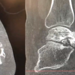
When orthopedic surgeons attempt to evaluate and manage osteochondral lesions of the talus (OLTs) using conventional computed tomography (CT), they may overestimate their size. This, in turn, influences treatment strategies. Therefore, OLTs often warrant advanced imaging studies, especially in revision or cases with cystic defects. To determine the effectiveness of these techniques, a study, “Value of 3D Reconstructions of CT Scans for Calcaneal Fracture Assessment,” was performed by Foot & Ankle International (FAI).

Understanding the True Anatomy and Size of OLTs May Help Guide Treatment

OLTs are defined as localized irregularities in the articular chondral surface also involving the subchondral bone. The exact cause of these lesions is unclear, but they are likely the result of a rotational ankle injury, such as a ligamentous sprain or bony fracture. OLTs are a common issue in orthopedic medicine; however, treatment remains a clinical challenge. Most OLTs can initially be managed with nonoperative treatment. When this approach fails, surgery is often indicated. Surgical intervention depends on the chronicity of the lesion, size of the lesion, presence of cystic lesions, and the stability of the cartilage. Many procedures for larger lesions require autograft or allograft tissue to fill the void, but these procedures can be very costly and do not always address the problem effectively. Understanding the true anatomy and size of these lesions may help better guide treatment, allowing for accurate preoperative planning and potential cost savings.

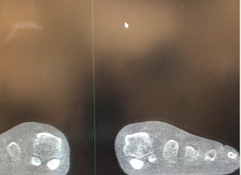
Conventional CT Scans Overestimate OLT Size

The results of the FAI study showed that conventional measurements of OLTs with CT scans grossly overestimated the size of the lesion. The average approximate OLT volumes from 10 independent observer CT measurements comprised of a rectangular prism (Vrec), a right cylinder (Vcyl), and right cone (Vcone) were compared to morphometric true-volume (MTV) via 3D reconstruction. On average, the Vrec, Vcyl, and Vcone estimates were 303%, 864%, and 285% greater, respectively, than the MTV. The FAI study demonstrates that volume measurements of OLTs using conventional CT typically overestimate true-volume approximations when compared to OLTs created with 3D reconstructions.

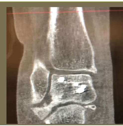
CurveBeam Imaging Provides Valuable Insights to Orthopedic Surgeons

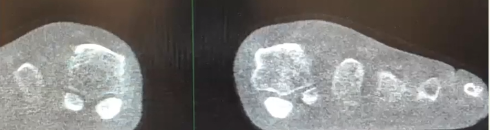
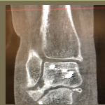
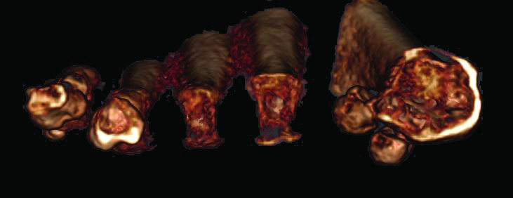
3D reconstructions of CT scans are used frequently in orthopedic surgery; however, the usefulness of this technique in OLTs has not been reported in the literature. 3D CT scans, such as those created using CurveBeam Cone Beam CT imaging technology, provide insight previously unavailable through either 2D and plain radiography. These detailed images, like those seen below, provide more precise measurements that can assist surgeons in both accurate diagnosing and preoperative planning.

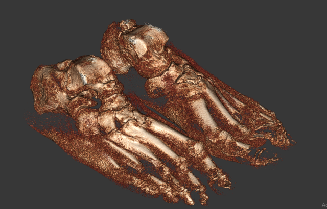
Bilateral – Talar Head Exposed Sharp Filter: CubeVue, CurveBeam’s custom visualization software, can segment a volume to reveal joint surfaces, such as the talar head.

bilateral

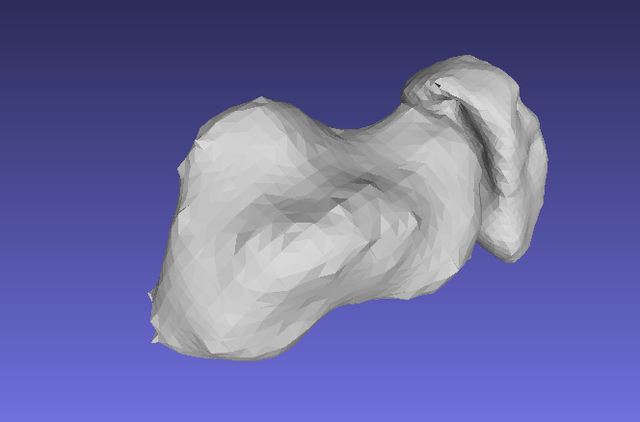
Talus Navicular Cluster: CurveBeam’s proprietary segmentation tools (under development) are demonstrated here using third-party viewing software.

talus

CurveBeam designs and manufactures Cone Beam CT imaging equipment for the orthopedic and podiatric specialties. CurveBeam’s pedCAT system and CubeVue custom visualization software have been installed in facilities across the United States, Europe, Australia, and China. More recently, CurveBeam’s InReach system, a multi-extremity CT optimized for hand, wrist, and elbow imaging, and LineUP system, a weight-bearing multi-extremity CT for imaging of the foot, ankle, and knee, have been added to the product line. Today, CurveBeam Cone Beam CT scans are setting new standards in orthopedic 3D imaging worldwide.

To find out more, visit https://www.curvebeam.com/about/about-curvebeam/.


[1] https://www.researchgate.net/publication/324808166_The_Role_of_3D_Reconstruction_True-Volume_Analysis_in_Osteochondral_Lesions_of_the_Talus_A_Case_Series