CurveBeam Announces Development of Weight Bearing CT Imaging System that will Scan the Hip and Pelvis

CurveBeam is proud to introduce the next level of weight bearing CT imaging, which will have the unique capability of scanning the hip and pelvis in weight bearing position, at the 2019 RSNA Annual Meeting.

With the HiRise, musculoskeletal radiologists and orthopedic specialists will be able to assess alignment of the total leg in three dimensions.

The HiRise is investigational only and is not available for sale in the United States.

The HiRise will permit visualization of the femoral head within the acetabulum. These scans could be used in pre-operative planning for knee replacement surgery, as surgeons will be able to assess alignment of the femoral head to the knee joint in three-dimensional weight bearing position.

Learn more about the HiRise at the RSNA Innovation Theater on Wednesday, Dec. 4 at 11 a.m.

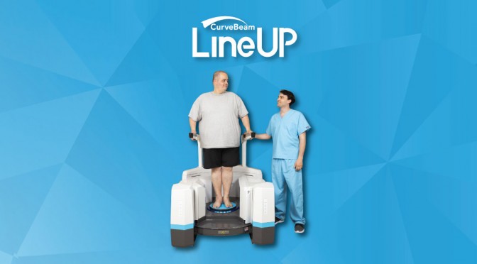
CurveBeam’s weight bearing systems boast the largest patient platform and field-of-view in their class. Patients can stand naturally with both feet side-by-side. Both the left and right limb are captured in a single scan.

“The weight bearing position allows surgeons to make better decisions regarding alignment during pre-surgical planning,” said Dr. Robert Santrock, MD, an associate professor at West Virginia University Health System. “Bilateral weight bearing imaging enables concomitant deformities to be assessed.”

The wide bore will allow for continuous scanning along the lower limb via multiple orbits, with patient remaining still in one position.

The HiRise gantry will raise and lower along a vertical track for lower extremity scanning. The gantry will tilt 90 degrees for upper extremity scans as well as non-weight bearing lower extremity scans. An optional table will allow patients to be fully supine during non-weight bearing scanning if necessary.

“Extremity cone beam CT systems are utilized as a point-of-care modality to improve workflow and have been widely accepted by the orthopedic community for lower extremity applications to provide an accurate weight bearing assessment of alignment,” said Dr. John Carrino, MD, MPH, Vice Chairman of Radiology for the Hospital of Special Surgery.

CurveBeam utilizes cone beam CT technology (CBCT) in its systems. CBCT images are initially acquired as two-dimensional projections using a rotating gantry with a relatively low-power X-Ray source, a pulsed X-Ray beam and a flat panel detector. The projections are reconstructed into a volumetric dataset. Cone beam CT scans are optimized for trabecular detail.

With an approximate footprint of 60″ x 73″, the HiRise will be a practical solution for outpatient settings. In addition, the HiRise will plug into a standard 120V outlet and is anticipated to require minimal external shielding.

Radiation dose of a cone beam CT scan of the distal extremities is typically a fraction of a comparable conventional CT scan of the same region.

To learn more and see the HiRise on display, visit CurveBeam at RSNA in South Hall – 1404. Visit www.curvbeam.com, call 267-483-8081 or email info@curvebeam.com to set up a demo.

FOOTInnovate Webinar Preview: Dr. Albert Armstrong

Don’t miss Dr. Albert Armstrong’s forthcoming webinar titled “Podiatric Radiology – Weight Bearing CT Imaging as an Essential Tool for Diagnosis.”

FOOTInnovate will host Dr. Barry’s webinar on Thursday, October 24 at 9 p.m. EST. Click here to register. (A FOOTInnovate account is required to attend.)

Dr. Armstrong is a Professor of Radiology at Barry University. He is a board-certified Diplomat with the American Board of Podiatric Medicine and the American Board of Wound Management. He attended Barry University School of Podiatric Medicine and completed his residency with Mount Sinai Medical Center in Miami, FL.

Dr. Albert Armstong, DPM, Professor of Radiology at Barry University School of Podiatric Medicine, in Miami Shores, FL, poses with a group of podiatry residents.

Dr. Albert Armstong, DPM, Professor of Radiology at Barry University School of Podiatric Medicine, in Miami Shores, FL, poses with a group of podiatry residents.


AFFAS 2019: Gaining Knowledge, Sharing Expertise

CurveBeam will be exhibiting at the 7th Triennial Asian Federation of Foot and Ankle Surgeons (AFFAS) Scientific Meeting in Bangkok, Thailand. Scheduled for October 11 – 12, 2019 at the Centara Grand and Bangkok Convention Center at CentralWorld in the heart of Bangkok, AFFAS 2019 provides a unique opportunity for orthopedic foot and ankle professionals from around the world to come together to exchange knowledge and innovation in the clinical and research issues currently shaping the specialty.

CurveBeam Lunch Symposium

Join CurveBeam on Thursday, October 11, for a Lunch Symposium  in which Dr. Alexej Barg, MD, of University of Utah Orthopedics, will be presenting a Clinical Case Review on WBCT. The Symposium will take place 12:15 p.m. to 1:15 p.m. in Ballroom C.

WBCT Society Symposium

Then, on Friday, October 12 in Ballroom A&B, the International WBCT Society will be hosting a special WBCT Symposium. Dr. Barg’s Presidential address will kick this event off at 11:20am with these highly anticipated presentations to follow:

Time Presentation


Presenter
11:25am-11:35am Experience on 11000 Cone Beam WBCT


Martinus Richter, MD, PhD
11:35am-11:45am Comparative Study of the Alignment of the Hindfoot and Position of the First Metatarsal and Sesamoid of Hallux Valgus with WBCT and WB X-Ray.


Jian-Zhang Zhang, MD, PhD
11:45am-11:55am Cartilage Imaging with Low Dose WBCT


Oliver Michelsson, MD
11:55am-12:05pm Comparison Between X-Ray and Weight Bearing Simulation CT on Varus-Type Stage B Osteoarthritis of the Ankle


Kiyonari Tomiwa, MD
12:05pm-12:10pm Closing Remarks: The Future of WBCT


Francois Lintz, MD, FEBOT
12:10pm-12:20pm Discussion Alexej Barg; Francois Lintz,


Please join CurveBeam at our AFFAS 2019 Lunch Symposium on October 11,  and be sure to attend the International WBCT Society’s Symposium on October 12. Both events feature renowned orthopedic foot and ankle specialists who will be sharing their expertise on industry-relevant topics.


Press Release: CurveBeam Announces New Validated Protocols for LineUP™ Weight Bearing CT Imaging System

LineUP System Now Compatible with Wright Medical’s PROPHECY™ Preoperative Navigation System and Portfolio of Total Ankle Replacement Implants

HATFIELD, PENN. — Sept. 10, 2019 – CurveBeam, a leader in extremity CT imaging, announced it has developed and validated protocols for the LineUP weight bearing CT imaging system that are compatible with Wright Medical Group’s PROPHECY™ Preoperative Navigation System and portfolio of total ankle replacement implants. Hospitals and orthopedic practices selected to participate in a controlled release will be able to submit LineUP scans to be used in PROPHECY’s virtual preoperative alignment process to create patient-specific surgical guides, with the potential for the protocol to be available to all LineUP users.

CurveBeam will showcase examples of how weight bearing computed tomography (WBCT) scans produced on the LineUP were utilized in Wright’s proprietary technology to model patients’ unique anatomy to determine implant component sizes and alignment at the American Orthopaedic Foot & Ankle Society (AOFAS) Annual Meeting in Chicago from September 12 – 15, 2019.

Dr. Robert Santrock, an orthopedic surgeon at the West Virginia University Health System, will deliver a lecture at CurveBeam’s exhibit (#932) on Friday, September 13 to discuss how he used the LineUP composite knee-foot scan protocol with the PROPHECY system for his patients who are undergoing a total ankle arthroplasty procedure.

“WVU elected to place the LineUP in the Orthopedic Department, which makes scheduling and performing scans for patients more convenient,” Dr. Santrock said. “I am delighted I am now able to use LineUP technology in conjunction with the PROPHECY Preoperative Navigation System.”

“The application of WBCT imaging to patient-specific surgical modeling holds tremendous potential to enhance the accuracy of preoperative plans and assessment of post-operative results. It has a tremendous potential to improve outcomes,” said Arun Singh, President & CEO of CurveBeam.

The Wright Medical PROPHECY system is used with Wright’s INFINITY™ and INBONE™ Total Ankle Replacement Systems and its INVISION™ Total Ankle Revision System.

The CurveBeam LineUP is a weight bearing Extremity CT imaging system. Its innovative design allows for a patient to be scanned while he/she is standing naturally on both feet. The AOFAS recommends weight bearing imaging when possible to get the most accurate assessment. The LineUP offers the largest field-of-view in its class, which allows for both distal limbs to be captured in a single scan.

The LineUP is compact, self-shielded and plugs into a standard wall outlet, making it ideal for point-of-care orthopedic settings. A study published in the International Journal of Diagnostic Imaging concluded, “[due to extremely low levels] radiation should not be a concern when considering the use of WBCT imaging for foot & ankle examination.” For more information about CurveBeam’s extremity CT imaging solutions, visit www.curvebeam.com.

About CurveBeam

CurveBeam researches, designs and manufactures cone beam CT imaging systems for the orthopedic specialties. CurveBeam’s corporate headquarters is located in Hatfield, Pennsylvania. The company was founded in 2009 and is privately owned and operated.

CurveBeam Connect: The Humanitarian Effort with a Growing Global Footprint

When we sprain our ankle or break a toe, it’s assumed that proper care is rarely far from reach. This isn’t the case for everyone, especially those who live in rural corners of the world. But there is significant hope for these patients with debilitating foot and ankle issues.

summary screenshot myersonClick here to download the executive summary podcast for this episode of CurveBeam Connect, featuring Dr. Mark Myerson, founder of the Steps2Walk Foundation.

The philanthropic organization, Steps2Walk, offers life-changing treatments to patients all over the world. In many cases, the program’s surgeon’s have given permanently disabled patients the ability to walk again. We explored this forward-thinking organization on CurveBeam Connect.

Our guest was the esteemed doctor himself, founder and executive director of Steps2Walk, Dr. Mark Myerson. Regarded as one of the premier foot and ankle surgeons in the world, Dr. Myerson explored the operation of his organization, and how its reach is only expected to grow in the coming years.

“We are ‘Doctors Without Borders’ for the foot and ankle,” Dr. Myerson said when explaining the organization in brief; and if Dr. Myerson had his way, in coming years Steps2Walk would be just as much of a household name. With over 200 surgeons volunteering to participate in Steps2Walk, the organization is a force to be reckoned with. But the humanitarian program doesn’t accept just any surgeon.

Participants must be excellent educators, as well as highly skilled surgeons accustomed to deformities not often found in the Western world, like Cleft Foot. Steps2Walk aims to not only treat patients in need but to educate them on their foot and ankle health.

“Probably 80% of work that we’re doing is teaching.” Dr. Myerson said.

Steps2Walk is continuing to change patient lives and the way care is administered in a variety of countries with different health care systems. For doctors interested in participating in the program, or interested listeners wanting to know more, visit www.Steps2Walk.org.