Weight Bearing CT Helps Visualize OCD Lesions

Osteochondral lesion of the talus is a broad term that describes injury or abnormality to the talar articular cartilage and adjacent bone.  Researchers have shown that radiographs alone miss osteochondral lesions of the talus in up to 50 percent of patients (1) .

Dr. Albert Armstrong, DPM, MS, BSRS,  Professor of Radiology and Medical Director of Advanced Imaging at the Barry University School of Podiatric Medicine, shared such a case in a recent FOOTInnovate lecture.

A 40-year-old male patient presented with pain in his left ankle. He was a self-described tough guy who said he has been tolerating the pain for years, but his new job required him to be on his feet all day.

The patient’s X-Rays showed a previous avulsion fracture and some radiolucency in the talar dome, but were otherwise inconclusive:

This 40-year-old male patient’s X-Ray revealed a previous avulsion fracture and some radiolucency around the talus, but was otherwise inconclusive as to why the patient was suffering ankle pain.

A weight bearing CT study was ordered, and it clearly showed two osteochondral lesions in the talar dome, as well as an osteophyte formation on the anterior ankle. Both of these findings pointed to osteoarthritis in the ankle.

The CurveBeam weight bearing CT scan revealed two osteochondral lesions as well as
The CurveBeam weight bearing CT scan revealed two osteochondral lesions as well as an osteophyte formation on the anterior ankle.

Large lesions and bipolar lesions associated with advanced degenerative joint disease do not respond to simple debridement or cartilage resurfacing techniques.  In a case featured in Podiatry Today, a weight bearing CT scan taken after standard X-Rays of a 52-year-old patient with chronic right ankle pain revealed large subchondral cystic changes and bipolar lesions. After reviewing the X-Ray and weight bearing CT. doctors determined the patient to be a good candidate for a total ankle replacement.

ACFAS Scientific Conference

Will you be in San Antonio for the ACFAS Scientific Conference Feb. 19 – 22? Visit CurveBeam at exhibit #407 to see several more examples of how weight bearing CT can provide more detailed diagnostic information than X-Ray alone.

(1) Badekas T, Takvorian M, Souras N. Treatment principles for osteochondral lesions in the foot and ankle. Int Orthoped. 2013; 37(9):1697-1706.

FOOTInnovate Webinar Recap: “Seeing 2020: My First 6 Months with Weight Bearing CT” with Dr. Blake E. Moore, MD, FAAOS

Dr. Blake E. Moore, MD, FAAOS, recently delivered a FOOTInnovate webinar titled “Seeing 2020: My First 6 Months with Weight Bearing CT,” diving into specialty views provided by CurveBeam’s LineUp weight bearing CT solution.

Dr. Moore is a board-certified orthopedic surgeon and a member of Atlantic Orthopaedic Specialists in Virginia Beach, VA. Atlantic Orthopaedic Specalists is a private orthopedic group comprised of 21 orthopedic surgeons, including two foot and ankle surgeons, and three hand and upper extremity surgeons.

Leveraging Specialty Views

While the LineUP unit provides 0.3mm multi-planar reconstructions and vivid 3D renderings of patient scans, it’s the machine’s ability to create simulated X-Rays of specialty views with ease that most impresses Dr. Moore.

CubeVue Insta'X tab
CubeVue Insta’X tab

“You can get multiple X-Ray views and specialty views that, sometimes, radiation techs that you have in the office may not be necessarily proficient in,” he said. “The image quality is phenomenal there.”

In particular, Dr. Moore has begun utilizing pedoscopic views, which were difficult to obtain prior to the introduction of weight bearing CT.


moore webinar screenshot 1

“I didn’t really utilize (the pedoscopic view), until we had access to weight bearing CT scanning. … You can really see the distribution of weight along the plantar aspect of the foot,” Dr. Moore said. “That can sort of help you guide your treatment in terms of if patients are having medial or lateral column overload.”


Putting Weight Bearing CT into Practice

Dr. Moore presented several case studies that highlight the effectiveness of weight bearing CT and the impact it’s already having on his practice.

In one case, the bilateral 3D rendering showed how the fibula was impaled into the calcaneus and the lateral aspect of the posterior facet of the subtalar joint, and how the subtalar joint had no congruity at all with the talus.

In this bilateral 3D rendering that Dr. Moore presented on FOOTInnovate, the degree of hindfoot deformity is clearly visible.

“You can really see the difference between the contralateral side and the affected side and as well look at the contribution of physiologic weight bearing to the overall deformity of the foot,” Dr. Moore said. “Seeing the normal side can really help you visiualize where you need to get to to get back to neutral alignment.”

Subtle sub-fibular impingement

Dr. Moore also shared a case in which a subtle subfibular impingement was revealed.

In another case, Dr. Moore reviewed a patient who had undergone total ankle arthroplasty. While some loosening may be observable through traditional scanning, weight bearing CT allowed Dr. Moore to get a more accurate picture of just how significant that loosening was.

Dr. Blake Moore explained that traditional radiographs obtained for post-operative evaluation may obscure loosening around the implant.
Dr. Blake Moore explained that traditional radiographs obtained for post-operative evaluation
suggested some early loosening around the tibia on the left side.


Dr. Blake Moore explained a weight bearing CT imaging reveled the cystic changes were more significant than the X-Ray suggested.
However, Dr. Blake Moore explained a weight bearing CT imaging reveled the cystic changes were more significant than the X-Ray suggested, and may need bone grafts.

Finally, Dr. Moore was able to thoroughly evaluate osteochondral lesion of the talus in a patient through CT scanning.

Dr. Blake Moore presented a case with an osteochondral lesion in his FOOTInnovate webinar.
Dr. Blake Moore presented a case with an osteochondral lesion in the talus in his FOOTInnovate webinar.

“I usually get, in addition to an MRI, a CT scan for most of the osteochondral lesions that I treat in order to really get a true idea of the size,” he said. “I think MRI really overestimates the size of these osteochondral lesions and you need to know what the cystic component of that is to pre-operatively plan for these appropriately.”

Preop Templating for TAA

Dr. Moore said weight bearing CT is going to play an enormous role in pre-op templating for total ankle arthroplasty moving forward.

“I think pre-operative templating for total ankle arthroplasty, which is just now starting with the CurveBeam LineUP, is going to be a game-changer for looking at complex deformity and correcting that with patient-specific cut guides. It increases the reliability and reproducibility of the procedure.”

We’re just starting to understand the role that rotation plays when placing these implants, Dr. Moore said.

“When the engineers look at the pre-op templating and make the plans, they really base the rotation off the tibia, and I think that that talar rotation is sometimes difficult to figure out in terms of where that talar implant should be rotated,” he said. “When the templating is currently done, the contralateral affected side is really subtracted immediately. I think that there’s a lot of critical information that can be gained from looking at the normal rotational axis of the unaffected tibiotalar joint.”

Key Financials

Dr. Moore highlighted the immediate financial impact of completing 150 weight bearing scans, which is as follows:

  • Scans per month: About 35 (90% foot and ankle)
    • Breakeven: 11 – 12 scans/ month
  • Reimbursement range: $50 to $750
  • Average reimbursement: $266
  • Breaks down to about $39,000 in the first three months

Mobile Weight Bearing CT

Dr. Moore had to decide which location of his practice would house the CurveBeam LineUP machine he utilizes, he said that, were he offered another crack at initial selection, he would have preferred a mobile solution.

CurveBeam offers such a solution, as CurveBeam’s Mobile CT Scanning solutions can bring industry-leading CT capabilities directly to your practice.

“If I could do it all over again, I would have put the unit on a truck so that we could have moved it from office to office,” Dr. Moore said. “But the experience for the patients is first-class.”

Moving Forward

In summary, Dr. Moore said that weight bearing CT has been the most critical addition to his practice since he began it, adding that it “greatly enhances the level of care and sophistication of preoperative planning.”

To learn more about CurveBeam’s LineUp, visit curvebeam.com/products/lineup/.

3D Printing Custom Surgical Guides with Dr. Kristian Buedts, MD

Custom surgical guides help improve placement and surgical success. Dr. Kristian Buedts, MD, a foot and ankle surgeon at ZNA Middleheim in Antwerp, Belgium recently shared how he uses weight bearing CT scans to create his own surgical guides, and then how he 3D prints those guides at his hospital.

Dr. Buedts highlighted the challenges associated with arthrosis in younger patients, noting that osteo-arthritis typically develops in younger patients as a post-traumatic symptom. More than half the time, the condition presents as asymmetrical wear of the tibio-talar joint (Witteveen 2013), and realignment surgery is often considered to avoid the implications of ankle replacement and joint fusion in younger patients (Krahenbuhl 2017).

These surgeries have been made easier by the accurate 3D representations made possible with weight bearing CT scans, providing professionals like Dr. Buedts with a more consistent pre-operative planning process that included the following steps:

  • Computer simulation software aids in generation of 3D models
  • Displacement is calculated by comparing the affected bone to the normal side through superimposition
  • Virtual osteotomies are conducted
  • Commercial software helps translate these simulations into custom-made guides
  • These guides are 3D printed to assist with the actual surgery

In one case, a patient with ankle instability in the ankle joint as a result of ligamentous laxity and a complex cavo-varus deformity with congruent varus deformity in the ankle joint, among other problems, underwent a corrective dome osteotomy.

While cases such as the patient’s are often hard to reproduce and are difficult to operate on, Dr. Buedts and his team utilized weight bearing CT imaging and the 3D printing of pre-operative guides that were used during the procedure to achieve an extremely positive outcome.

To learn more about the possibilities of weight bearing CT imaging, visit curvebeam.com/contact/.

FOOTInnovate Webinar Recap – Soomekh: How I Have Integrated Weight Bearing CT into My Practice

David J. Soomekh, DPM, is a Diplomate of the American Board of Foot and Ankle Surgery and a Fellow of the American College of Foot and Ankle Surgeons.

In this CurveBeam-sponsored FOOTInnovate webinar, Dr. Soomekh delivers a presentation about how exactly weight bearing CT technology has been integrated into his practice.

Click here to watch the FOOTInnovate webinar.  A FOOTInnovate account is required to access.

How Dr. Soomekh Leverages the pedCAT

To begin, let’s dive into how Dr. Soomekh uses the CurveBeam pedCAT system in his practice.

“In a lot of cases, I will go straight to a WBCT scan and not do an X-Ray because I know I am looking for subtleties and I don’t want to give the patient a double dose, ” Dr. Soomekh said. “In some cases you might say I have sort of replaced the plain film machine.”

Dr. Soomekh said that the pedCAT is instrumental in his ability to:

  • diagnose fractures that are difficult to appreciate on plain film due to bone overlap;
  • detect stress fractures;
  • appreciate joint relationships in dislocations more easily;
  • and get an accurate 3D view of joint changes and subchondral lesions associated with arthritis.

In addition, the pedCAT assists Dr. Soomekh with pre-operative planning and an in early detection of osteomyelitis.

Dr. David Soomekh, DPM, shares patients' pedCAT scans with them via a tablet. He said the datasets wirelessly upload to the viewer in less than 10 minutes."
Dr. David Soomekh, DPM, shares patients’ pedCAT scans with them via a tablet. He said the datasets wirelessly upload to the viewer in less than 10 minutes.”

“I [view images on] a mobile device. … It’s wireless, and the image still comes up in about six to eight minutes after it’s processed,” Dr. Soomekh said. “I’ll bring it into the room and show the patient the views and the pathology.”

pedCAT Reimbursements and Authorization

Though the rates presented by Dr. Soomekh are specific to Southern California, they provide valuable insight into the operational benefits of a pedCAT system.

For Medicare, Dr. Soomekh said his reimbursement rate is $130 for a scan. With PPO insurance, the rate can range from $180 to $280, and as high as $350. Dr. Soomekh’s cash-pay rate is $200.

While Medicare doesn’t require authorization, Dr. Soomekh’s practice has implemented some best practices for securing authorization for PPO insurance.

His staff will make a pre-authorization call while the patient is in-office, and billing is held until authorization is received. Occasionally, a peer-to-peer call is required. The patient signs a consent form he or she will be responsible for the cost of the CT if it is not covered by insurance.

“In most cases, I don’t wait until I get the authorization before I go ahead and take the scan,” Dr. Soomekh said. “Some patients ask me to wait until they get the authorization, and we’ll do that. … Otherwise, the staff will be doing an authorization call while we’re doing the scan. If we get authorization right then, we’ll let the patient know.”

Results of Weight Bearing CT in Action

Though a thorough watching of Dr. Soomekh’s webinar will reveal many cases in which weight bearing CT is beneficial, let’s take a look at how the pedCAT helps Dr. Soomekh assess Hallux Valgus.

The system helps Dr. Soomekh in pre-operative planning as he assesses the degree of frontal plate rotation of the first metatarsal and its association with the sesamoids. He can also appreciate the degree of transverse plane angulation at the first metatarsal cuneiform joint, among other benefits.

The image below, for example, provided Dr. Soomekh with a look at the internal rotation of the first metatarsal in a younger patient.

metatarsal internally rotated

The image below shows the result of corrective surgery for the same patient.

sesamoids al“The sesamoids are underneath, now, in the appropriate position, and the angulation is closed,” Dr. Soomekh said.

pedCAT Benefits

To conclude the webinar, Dr. Soomekh outlined the major benefits of adopting the use of a pedCAT machine at his practice.

These include:

  • Excellent image quality
  • Instant results
  • Immediate and focused treatment for the patient
  • Patients benefit from “one-stop shop” experience
  • Increased practice revenue
  • Images can be sent to radiologist for an official report

National Standing CT Conference Set for March 19 in Manchester

UPDATE: The Standing CT Conference has been re-scheduled to Thursday, June 11, 2020. Please contact kirsty.collins@standingct.com. 

On March 19, CurveBeam will again proudly serve as the primary sponsor for “Bipedalism: Defining Humanity and Foot & Ankle Surgery,” the National Standing CT Conference.

The event, which provides a one-day course aimed at surgeons, podiatrists, specialist physiotherapists and radiologists interested in the latest weight bearing imaging technology for the foot and ankle, is set to take place in Manchester, England’s City of Manchester Stadium, also known as the Etihad Stadium.

The conference is organized by Standing CT Company, which provides weight bearing CT services via mobile imaging centers to hospitals and orthopedic practices in the United Kingdom and Europe. Standing CT Company exclusively utilizes CurveBeam’s weight bearing CT systems.

The conference will feature a variety of speakers and courses for attendees.

Course organizers include Stephen P. Bendall, MBBS, FRCS, FRSC (Orth), an orthopedic foot and ankle surgeon at Princess Royal Hospital in Haywards Heath, Sajid Butt, MB BS, FCPS, FCPR, a radiologist at Royal National Orthopedic Hospitals in London and Stanmore, Andrew Goldberg, MD, and Matthew Solan, BSc, MB, BS, FRCS (Tr & Orth), Medical Director, Chair of MAB and consultant orthopedic surgeon.

Other faculty include CurveBeam President & CEO Arun Singh, Matthew Welck, MBChB. BSc (hons). MSc. FRCS (Orth), Nicholas Cullen, MBBS, BSc(Hons), FRCS, FRCS(Tr&Orth), Lee Parker, BM MRCS FRCS (Tr. & Orth.) Paul O’Donnell, MBBS, MRCP, FRCR, and Francois Lintz, MD .

StandingCTBlogBody

Question-and-answer sessions will follow each of the four main discussions.

To book your place at this critical conference, call 0800-047-1010 and provide your contact details or email Kirsty.Collins@standingct.com.

FOOTInnovate Webinar Recap: Weight-Bearing CT as a Diagnosis Tool in Podiatric Radiology, Dr. Albert Armstrong DPM

Albert Armstrong, DPM, MS, BSRS, Professor of Radiology and Medical Director of Advanced Imaging at the Barry University School of Podiatric Medicine, is an important voice in the field of podiatry. He recently delivered a FOOTinnovate webinar titled, “Podiatric Radiology: Weight Bearing CT Imaging as an Essential Tool for Diagnosis.”

The Barry University Foot & Ankle Institute is comprised of three hospital-based podiatry clinics in greater Miami. Barry University podiatry students spend a portion of their third year rotating through these clinics. Barry University acquired a pedCAT system for both clinical and research applications in 2018.

In the webinar, Dr. Armstrong focused on how the CurveBeam pedCAT system improves diagnosis and workflow in clinic.

Click here to watch the FOOTInnovate webinar. A FOOTInnovate account is required.

pedCAT’s Efficiency

Dr. Armstrong explained a pedCAT study takes about the same amount of time and produces about the same amount of radiation as three traditional radiographs of the foot.

“The 3D, weight-bearing CT machine that we have is the pedCAT. … It’s small, compact, and it fits in our X-ray room,” Dr. Armstrong said. “It’s fast and easy, and we get instant results.” And patients often comment on the state-of-the-art technology.

pedCAT datasets can be displayed as Multiplanar Reformats (MPR), providing coronal, sagittal and transverse slices of the anatomy.

pedCAT users can also manually get “slices in virtually any plane,” Dr. Armstrong said.

Dr. Armstrong reviewed several cases from clinic in the webinar. Three of those cases are summarized below.

Case 1

Dr. Armstrong presented a case of a 52-year-old female who had undergone a bunionectomy three decades prior. She presented in clinic with pain in the left foot great toe. The pain was preventing the patient from walking, running and wearing heels.

Traditional X-Ray imaging didn’t provide an adequate look at the patient’s foot due to superimposition of the sesamoids and metatarsals.
However, scrolling through the coronal plane of a weight bearing CT exam revealed a large osteophyte plantar of the first metatarsal head that was fused to the tibial sesamoid.

While traditional imagine didn’t provide a clear diagnosis, a coronal view created from the 3D volume revealed a large osteophyte and other factors that were hidden by the superimposition of the sesamoids and metatarsals in the X-Ray exam.

The more complete diagnosis, Dr. Armstrong said, led to a much better surgery plan for the patient.

The pedCAT and 3D volume imaging allows for detailed looks on several layers, as doctors can essentially “peel off,” as Dr. Armstrong puts it, layers ranging from shoes worn during weight-bearing scans to skin and soft tissue, etc.

Case 2

Another case, Dr. Armstrong said, highlights how critical weight-bearing imaging can be.

A 37-year-old male came to the Barry University Foot & Ankle Institute after a work injury was still painful two months later. The patient had fallen at his construction job. He went to the emergency room, where an X-Ray exam was read as negative, and he was diagnosed with an ankle sprain. The treatment prescribed was an ace wrap and a brace.

Two months later, the patient came to the Barry University Foot & Ankle Institute.

A sagittal slice of the weight bearing CT scan showed an intra-articular calcaneal fracture to the subtalar joint, as well as patchy osteoporosis throughout the foot.

The fracture had been completely missed on the initial X-Ray. Patchy osteoporosis can be a sign of complex regional pain syndrome, and is typically radiographically indistinguishable as well.

Case 3

A 70-year-old male with a history of osteoarthritis and Type 2 Diabetes had synthetic cartilage implanted into his 1st MPJ joint 9 weeks prior. The patient had severe pain when standing and walking and had to walk on the lateral edge of his foot to compensate. Plain X-Rays came back negative.

With weight-bearing imaging, the coronal MPR images highlighted first metatarsal head was everted to such a degree that the patient was bearing weight on the tibial sesamoid when standing. That was causing his pain.

Without weight-bearing CT imaging, the rotation of the first metatarsal head in this patient’s foot would not have been revealed.

In summary, Dr. Armstrong said that pedCAT provides fast and efficient weight-bearing CT imaging, highlights the value of 3D volume and MPR images, and helps translate critical weight-bearing images into improved results for patients.

CurveBeam’s Automatic IMA Angle Measurement Tool, Presented by Dr. Francois Lintz, MD, FEBOT, PhD

At the 2019 AOFAS  Annual Meeting in Chicago, Dr. Francois Lintz, MD, FEBOT, PhD showcased CurveBeam’s efforts to develop an automatic measurement tool, Smart M1-M2. This tool automatically calculates the intermetatarsal (IMA) angle on a 3D foot & ankle CT scan in a matter of seconds. The validation effort for the Smart M1-M2 tool is being led by Dr. Jonathan Deland, MD at the Hospital for Special Surgery.

Watch the video below to learn more.


FOOTInnovate Webinar Recap: Dr. Alessio Bernasconi, “3D, Weight-Bearing CT’s Role in Assessing Pes Cavovarus”

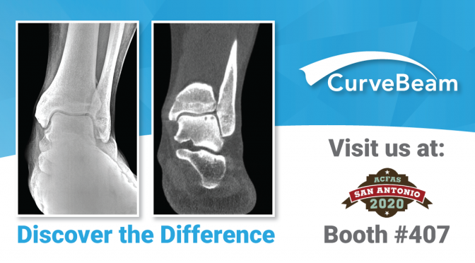
Dr. Alessio Bernasconi, MD, FEBOT and  his colleagues at the Royal National Orthopaedic Hospital in London have produced critical and salient findings about the role of weight-bearing CT imaging (WBCT) in assessing pes cavovarus.

Dr. Bernasconi recently shared these findings in a FOOTinnovate webinar, and his presentation is outlined below.

Click here to watch the FOOTInnovate webinar. A FOOTInnovate account is required.

A Refresher on Pes Cavovarus Diagnosing Procedures

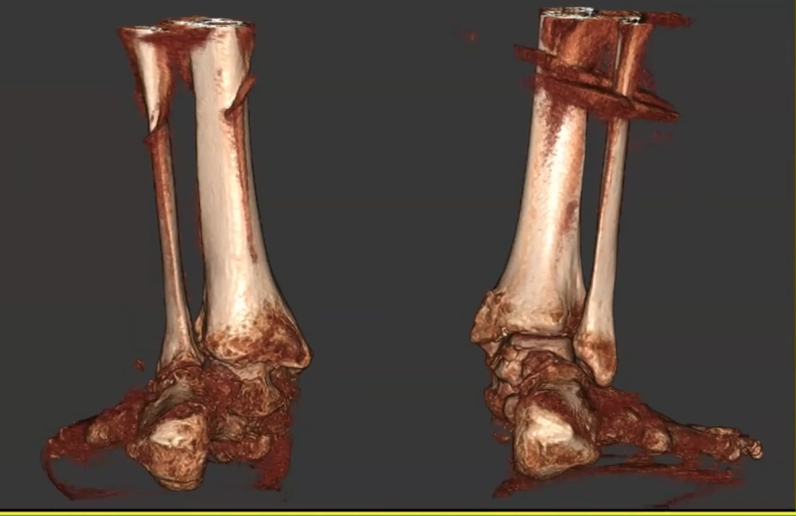
Pes cavovarus is considered a three-dimensional deformity and is characterized by a high-arched, varus hindfoot alignment with a forefoot that may be abducted or plantarflexed depending on the individual case.

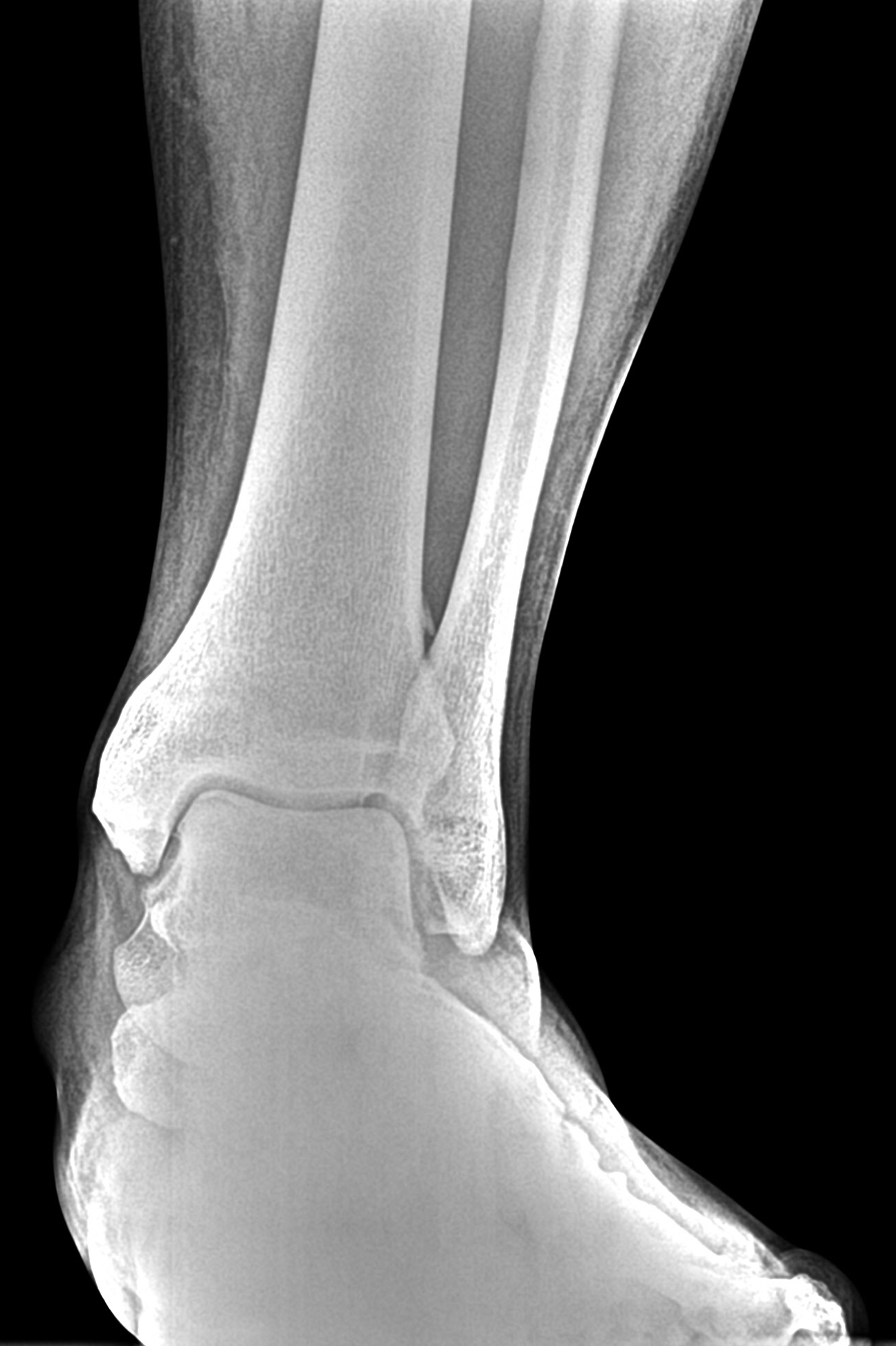
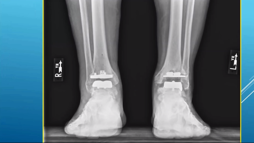
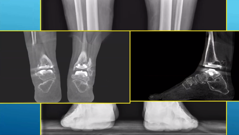
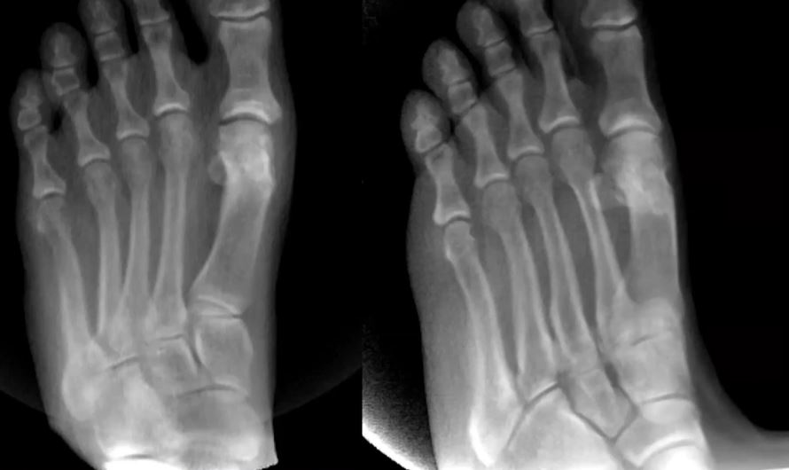
Diagnostic work for pes cavovarus is based on a thorough clinical assessment. However, additional imaging is needed, and X-Ray imaging is the current standard. Additional CT or MRI imaging can be ordered to further characterize the deformity and assess the status of joints and soft tissues.

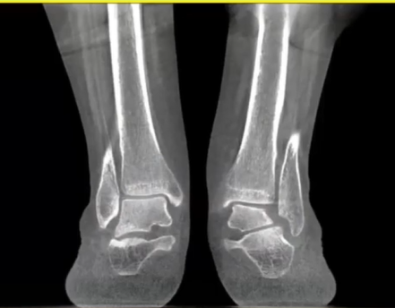
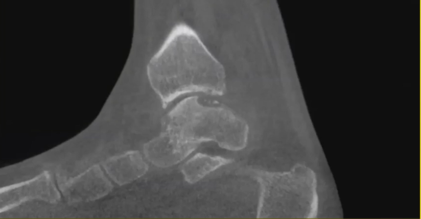
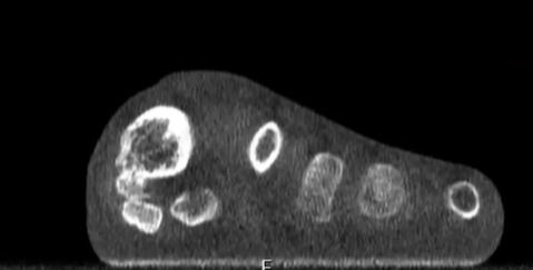
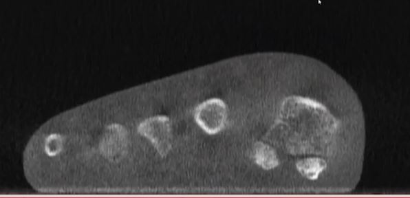
However, common dorsoplantar, lateral views of bilateral cavovarus deformity can bring along with them difficulty in precisely identifying joint planes and status. Recent studies, including published papers from PubMed suggest that radiographs have inherent bias as beam position, operators, foot position, and more can change what doctors see and identify.

Is there any objective way to identify hindfoot alignment?

Improving Identification of Hindfoot Alignment

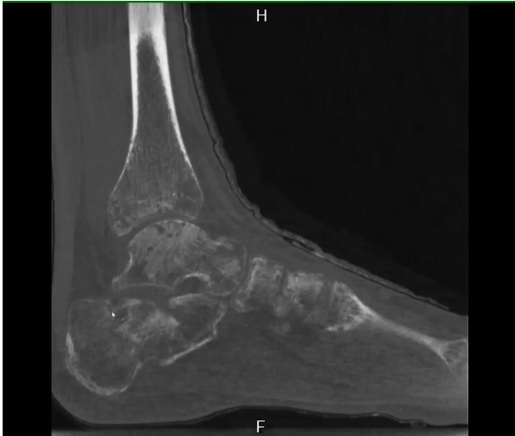
The Saltzman view – developed by Charles Saltzman, MD and Georges El-Khoury, MD – is designed to find the main axis of the tibia and the main axis of the calcaneus, then to define the angle between those two lines.

However, this view can still be limited by rotation of the foot.

That’s why authors like Jonathan Kaplan, MD, who outlined a solution in Foot & Ankle International, propose the use of weight-bearing CT scanning as a better alternative when diagnosing malalignment associated with pes cavovarus.

The Implications of Weight-Bearing CT Scanning

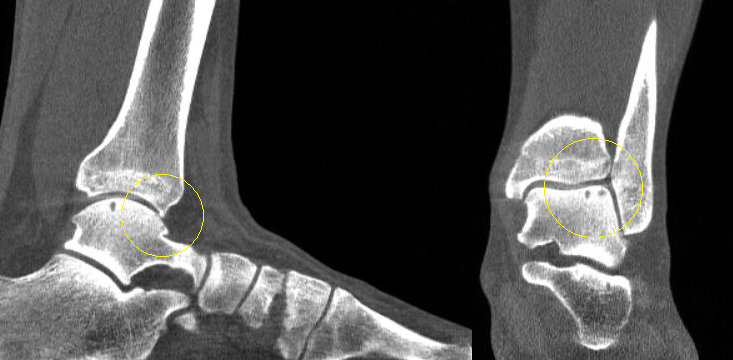
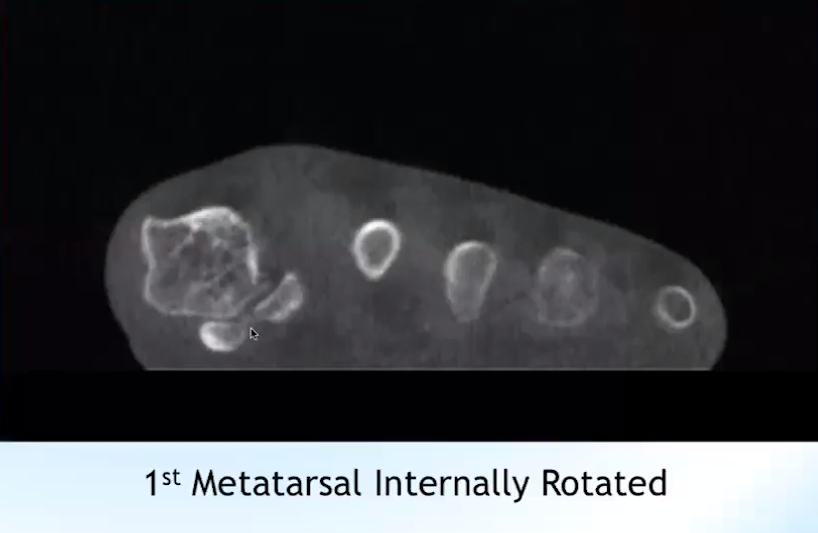
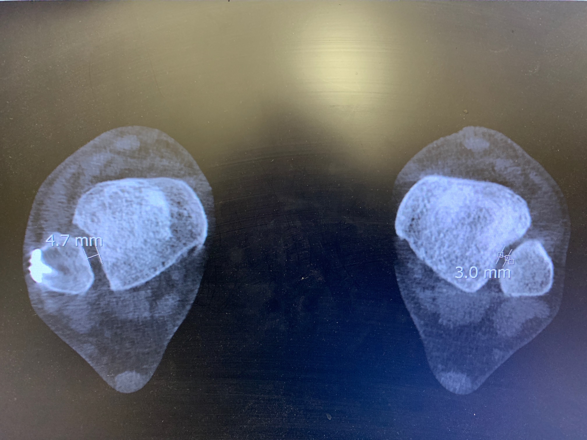
Two studies published in 2017 and 2018 introduced the idea of using three-dimensional measurements on weight-bearing CT images, allowing the measurement of malalignment to move away from a single plane and toward a volume-based view that produces a more reliable and true view of the bones in physical space.

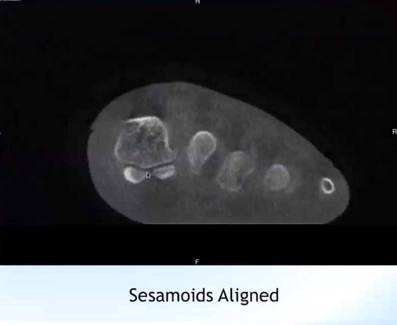
The three measurements proposed are foot and ankle offsets, the calcaneal offset and the hindfoot alignment angle.

In 2017, a study assessing 135 datasets including normally aligned feet, varus hindfeet and valgus hindfeet demonstrated excellent inter and intra-observer reliability using this new measurement.

The true revolution in utilizing 3D measurements is that, in gathering the three measurements, you can add a fourth point to represent volume that provides that additional beneficial depth.

“What you need is a cone-beam, weight-bearing CT device,” Dr. Bernasconi said. “In our institution, we use the PedCAT one. You need a software to elaborate data – CubeVue – and you need a TALAS plug-in.”

Essentially, Dr. Bernasconi said, this landmark software solution can provide a simpler way to obtain values relative to the normative values that highlight varus or valgus malalignment.

Critical Questions

Dr. Bernasconi highlighted three critical questions regarding the reliability of this new solution:

  • How reliable are 3D measurements when diagnosing pes cavovarus?
  • Does etiology affect reliability?
  • Does the severity of the deformity affect reliability?

To find the answers, Dr. Bernasconi and the staff at the Royal National Orthopaedic Hospital completed a retrospective study based on STROBE guidelines of patients with a pes cavovarus diagnosis from their hospital between 2015 and 2017.

Eventually settling on 17 patients, the team matched those patients with an equal number of normally aligned feet. Three observers with different levels of expertise and different knowledge of the weight-bearing CT imaging process produced excellent inter-observer reliability. Further, there was little variance between the observers, suggesting that the data is reliable regardless of previous expertise, etiology and deformity severity.

“The reason that we have studied foot morphology, thanks to the help, obviously, of this new technology, (is that) standing CT allows you to reproduce in a more faithful way the true geometry of the bone and the joints in the space,” Dr. Bernasconi said. “We think that this may potentially help us better understand the etiology.”

Though Dr. Bernasconi said his findings were far from enough to fully corroborate this positive outcome, they are certainly a starting point.

The takeaway, he said, is that this new, 3D approach will allow for reliable assessment of pes cavovarus, both in the diagnosing stage and in monitoring patients after procedures to determine how successful the correction was.

FOOTInnovate Lecture Preview: Dr. Blake Moore

Be sure to register for Dr. Blake Moore, MD, FAOOS’ upcoming webinar on FOOTInnovate, titled “Seeing for the First Time: Integration of Weight-Bearing CT into Your Practice.” The webinar will broadcast on January 8, 2020 at 9 p.m. EST.

FOOTInnovate-DrMoore

Dr. Moore specializes in the diagnosis, treatment and surgery of the foot and ankle and foot and ankle reconstruction and general orthopedics. He joined Atlantic Orthopaedic Specialists in Virginia Beach, Virginia in 2014. Dr. Moore will highlight several cases where weight bearing CT imaging made a difference in his clinical decision making, including the post-operative syndesmosis case illustrated below.

In this post-operative syndesmosis case, an interposed medial fragment is pushing the syndesmosis open. This syndesmosis widening could only be appreciated on a bilateral weight bearing CT exam, which provides a comparison to the contralateral side. (acquired via cell phone camera)


Dr. Moore has a special interest in treating athletes like himself. He specializes in lower extremity trauma, complex hindfoot and ankle reconstruction, and total ankle replacement surgery.


International WBCT Society Announces Book Release

The first textbook on weight bearing CT imaging, written by the board of the International WBCT Society, will be released in February 2020. The textbook is a scientific, technical and clinical guide to Weight Bearing Cone Beam Computed Tomography (WBCT), presenting all of the relevant content to date on the development, implementation, interpretation and clinical application of WBCT for the foot and ankle.

Pre-order the hardcover text from Springer here.

978-3-030-31948-9_Cover_PrintPDF indd

“At the beginning of 2019, the board of the society discussed how to further spread the knowledge of WBCT,” said Prof. Dr. Martinus Richter, past president of the International WBCT Society. “In contrast to standard textbooks, the scientific content is emphasized and placed at the beginning to reflect the initial scientific approach of WBCT ISG and International WBCT Society.

“We wish that this book serves all current and upcoming users of WBCT in foot and ankle to answer all scientific and technical questions, to educate and instruct the clinical use including measurements.”

Weight Bearing Cone Beam Computed Tomography (WBCT) in the Foot and Ankle: A Scientific, Technical and Clinical Guide is separated into three informative sections.

  • Part One describes the history of, development of, and need for WBCT as an imaging option, and features a scientific overview of the procedure.
  • Part Two is an exhaustive scientific background comprised of 16 landmark studies, describing its advantages for selected foot and ankle injuries and deformities (both congenital and acquired).
  • Part Three uses this science as context, and includes chapters that:
    • Explain the technical aspects and necessary background for WBCT
    • Introduce the different devices used for WBCT
    • Provide insight into the actual measurement possibilities, including the initial software solutions for automatic measurements
    • Feature current clinical applications via case material illustrated in an atlas-like fashion
    • Explore future developments and further applications of WBCT, such as dynamic scans and measurements or hologram-like visualization

“In the early stages of the scientific work-up in different institutions, different methodologies, especially for angle measurements, were inaugurated. No standard for the methodology for the image acquisition and measurement was defined, and different methods have been published,” Dr. Richter explained.

“Since then, the society is further growing and the member number as well as the number of Pubmed-listed publications by members will both reach 100 by the end of 2019.  The International WBCT Society can be considered as one of the most scientifically active societies,” Dr. Richter said.

Weight Bearing Cone Beam Computed Tomography (WBCT) in the Foot and Ankle will be available online and in stores beginning February 2020, providing an excellent resource for orthopedic and foot and ankle surgeons, radiologists, and allied medical professionals working in this clinical area.