The Face of Customer Service at CurveBeam

Satisfied customers are not just the result of well-made products, but the result of continuing support over the lifetime of those products as well. Tami Alexander is the Director of Worldwide Customer Relations at CurveBeam, based out of company headquarters in Warrington, Pennsylvania, United States. She joined the CurveBeam family in October of 2015, moving from the West Coast to take over the position.

Tami Alexander serves as CurveBeam's Director of Worldwide Customer Relations.

Tami has more than 30 years in the medical imaging/radiology world. This includes insurance contracting & credentialing, reimbursement, accreditation and radiation regulations. Prior to joining CurveBeam, she worked at the California and Arizona locations of HealthWest Partners for 13 years, as Director of Operations for five PET/CT imaging centers, and a radiation oncology center.

“I enjoy working with customers in my new role and giving them the support they need long after the sale,” Tami said. “In my previous role,  operations was such an integral part of  the overall experience our customers received  while visiting our centers that I have carried over those same quality standards to meet or exceed our customer’s expectations.”

Tami works closely with Andrew Kochanasz, who is the Technical Support Manager, to ensure that any problems requiring service are quickly addressed. She also works closely with the CurveBeam sales team to coordinate site preparation, installations, and training. If an annual maintenance or any other service is needed at the customer’s site, Tami schedules CurveBeam’s service technicians. If you have warranty questions, or if you would like to purchase additional licenses for CubeVue software, schedule your annual maintenance, or request additional training, Tami is the best person to contact.

Tami would like to let all pedCAT customers know, “Don’t hesitate to give us a call. We are here to help in any way we can. From tech support & customer service, to helping our customers find resources the resources they need for their CBCT systems.”

You can contact Tami at 267-483-8089 or tami.alexander@curvebeam.com.

Webinar Recap: “Using Weight Bearing CT to Guide Clinical Decisions”

Dr. Steven K Neufeld, MD, presented “Using Weight Bearing CT to Guide Clinical Decisions” on FOOTInnovate™  in late 2015. In one of his opening statements, Dr. Neufeld described why he decided to give the webcast: “This talk was put together really out of my enthusiasm and excitement and interest in this technology.”

The pedCAT saves time

“… (It’s) quicker for me to get a cat scan in my office, and show the results to my patient,” Dr. Neufeld said.  By the time the patient leaves the X-Ray room and gets back to the exam room, the images are ready.

Also, it helps streamline his pre-op planning. “I don’t have to send the patient to the hospital for a CT. I don’t have to schedule another appointment to review the results,” he said. “All the information is right there. It also saves the patient money. The cost of a CT scan in my office is much less than a big hospital or institution.”

Immediate feedback to the patient also helps put the patient at ease. “The patient feels comfortable with me, that I have a plan and that I know what we were going to do, and I go in there and I execute it,” Dr. Neufeld said.

The in-office workflow has improved accuracy and consistency in imaging for him as well, “Instead of getting a standard Saltzman or heel view … the weight bearing CT scan is much easier for the technician,” he said. “You don’t have to position the foot in any particular way, you don’t have to angle your beam in a particular way. You just have them stand on the machine and you then push the button, and you can determine alignment very easily.”

The pedCAT aids in definitive diagnosis and evaluation

In his webcast, Dr Neufeld proposed the following: “Based on our knowledge, it’s pretty much understood that the standard of care for any Orthopaedic foot and ankle or Podiatric evaluation of a foot and ankle problem is a weight-bearing radiograph – at least currently. The big question again is: does this X-Ray give enough?”

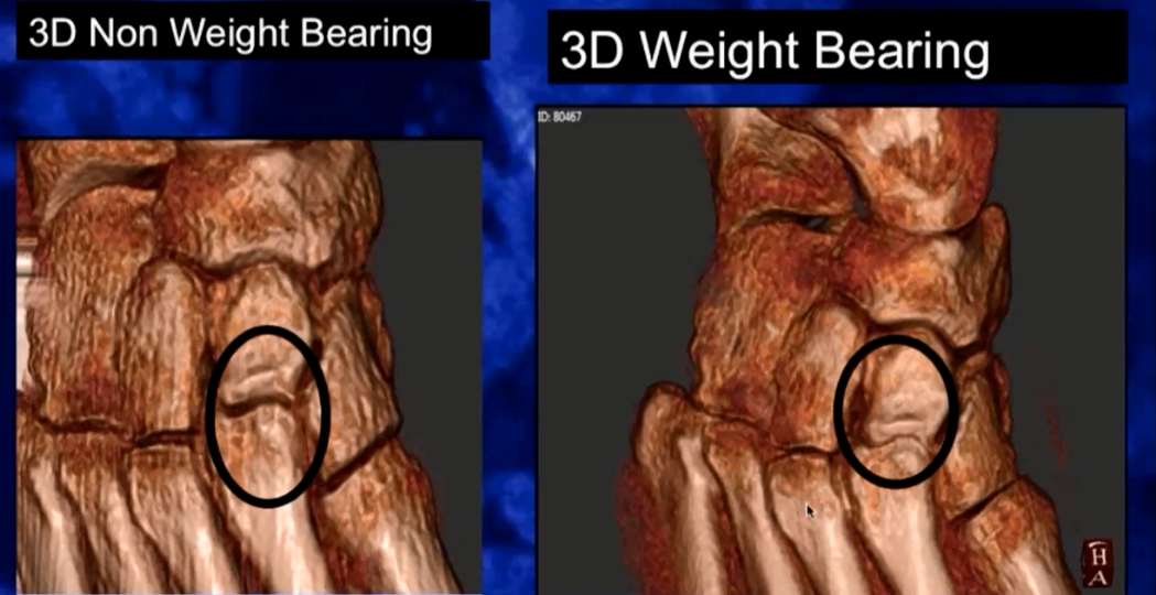
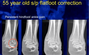
When it comes to alignment, weight-bearing is deemed necessary to properly evaluate the ankle under load. This is especially true when superimposition of structures does not allow you to properly determine position, rotation, translation, and joint space. In the case shown below, only weight-bearing CT allows you to see the impingement in the subtalar region; a 2D x-ray would not allow you to properly evaluate the joint spaces.

neufeldpost1

neufeldpost2

A Total Ankle Replacement (TAR) is another surgical procedure where weight-bearing CT is a must, according to Dr Neufeld. “A non weight-bearing CT scan would really not give me information about the implant position… (The PedCAT is) a great modality for post-op,” he said. The convenience of having 3D imaging on hand for follow-up exams, even if they are non-weight-bearing, makes it easier to follow the progress of healing post-op. Failed hardware and non-unions are more readily diagnosed this way.

Dr Neufeld also regularly prescribes weight-bearing CT scans for unknown pain. “The other way that I use weight-bearing CT scan is … sometimes a patient comes in the office, and they just hurt! And you just can’t figure it out!” he said. “You know – X-Rays are normal, the exam is very vague … we’re all faced with this patient. You just can’t figure out what is going on sometimes.”

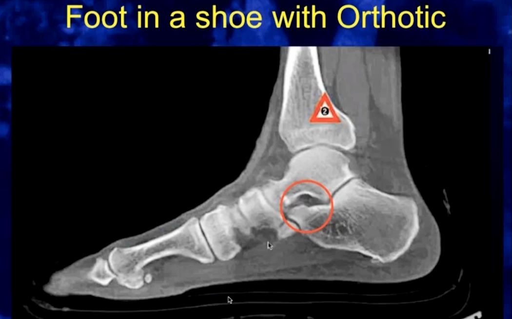
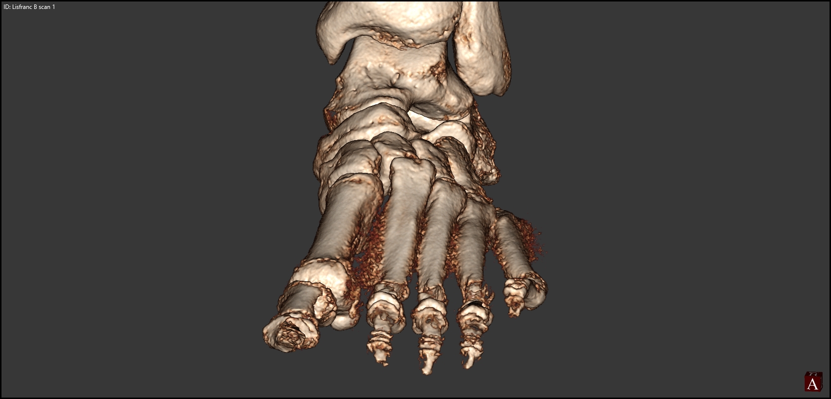
Other uses for his weight-bearing CT include Lisfanc, navicular & other hard to evaluate fractures, sesamoid problems (especially when coupled with Metatarsalgia), and arthritis. The fact that you can scan a foot in a shoe with an Orthotic also makes it easier to determine if the Orthotic is accomplishing what it is supposed to do, as shown in the image below.

neufeldpost3

An investment worth the regulatory hurdles

Dr Neufeld, Director of the Orthopedic Foot and Ankle Center in Falls Church, Virginia, began using his pedCAT in August of 2015. The state of Virginia requires medical facilities to obtain a Certificate of Need to purchase CT systems. The approval process can be challenging. However, Dr Neufeld was both persistent and patient in his determination to bring the new weight-bearing technology into his office.

“It is brand new,” he said. “There is nothing like it out there in the state. If you can prove and show that this technology is different than what they have out there- you’re not competing with the hospitals, you are not competing with the radiology centers – and that it is unique and essential for your practice, then you can put up a fight. And if you have perseverance and some patience, you can get it, and that’s what we did.”

So why persist? Dr Neufeld’s answerer is simple.“The PedCAT has been a game-changer for my practice and one of the best investments,” he said. “It has really become a crucial part of my practice, and I would say it’s probably one of the most profitable, influential investments in modalities that I’ve added.”

pedCAT: Weight Bearing CT Views of Lisfranc conditions

Lisfranc injuries can easily be missed on traditional radiographs.

One research study tried to determine the level of accuracy when using plain radiographs to assess midfoot conditions. Their results confirmed a high incidence of missed diagnosis even by experienced observers.¹

“Radiographic evaluation is crucial in the diagnosis…Weight bearing views are more sensitive since tarso-metatarsal instability may be revealed…The threshold for cross sectional imaging such as multi-detector CT should be low,” according to the study.

With the pedCAT, it is possible to get a weight bearing, three dimensional scan at the point of care. The tarso-metatarsal joint can be viewed without any superimposition from surrounding anatomy.

Delaying a 3D scan can be detrimental to recovery.

“The identification of Lisfranc joint injury can be difficult and is often not detected on initial presentation to the accident department. This is important because of the correlation between delay in treatment, particularly more than six months, and poor functional outcome,” the study reported.

Midfoot dislocation, fracture, pedCAT

In this pedCAT scan, the 3D rendering shows the 2nd metatarsal is pushing on the 3rd metatarsal. The multi-planar slices reveal the fracture that is causing the dislocation. The sagittal and axial slices show a bone fragment as well. It would be impossible to see this information on a plain radiograph.

lisfranc AP

This pedCAT scan clearly depicts the gap between the first and second metatarsals.

¹ Sherief, Tamer. “Lisfranc injury: How frequently does it get missed? And how can we improve?” International Journal of the Care of the Injured. Elsevier Ltd. 10.106/j.injury.2006.10.002

pedCAT: Will it Fit?

The pedCAT has been deployed to a range of medical facilities – from large hospitals to single physician practices. Each site has its own unique requirements, and CurveBeam works with each customer to ensure the first weight bearing CT scanner dedicated to the foot & ankle meets all safety requirements. Because the pedCAT is an ultra low dose device, the shielding infrastructure required is similar to that of a plain X-Ray device. The pedCAT does not need to be in a lead lined room.

Because of its compact size and relatively minimal required shielding, the pedCAT can easily fit into a practice with limited space. Take a look at these examples:

This pedCAT was placed next to a treadmill used for gait analysis.
This pedCAT was placed in a patient examination room in this New York City podiatrist’s office.
This pedCAT fit into a tight corner next to an X-Ray machine in this California podiatrist’s office.