pedCAT Shown to be Most Effective Imaging Option for Mülller-Weiss Disease

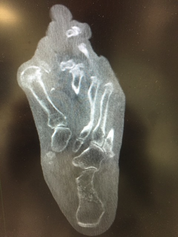
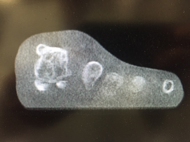
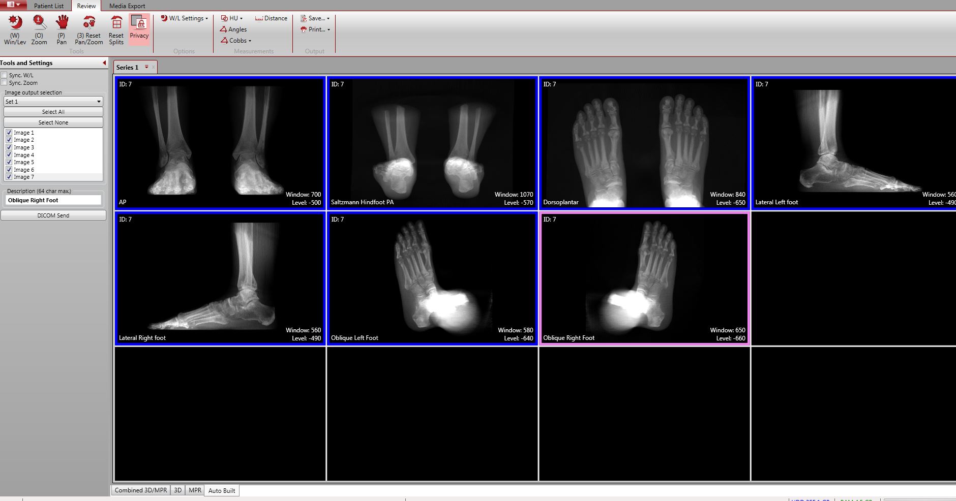
The CurveBeam pedCAT imaging system was recently recommended as an incredibly beneficial option for the treatment and analysis of Mülller-Weiss disease. While the disease can be detected in its early stages with magnetic resonance imaging and radiographs, there is a considerable advantage to using weight-bearing computed tomography (WBCT) for diagnosis and evaluation.

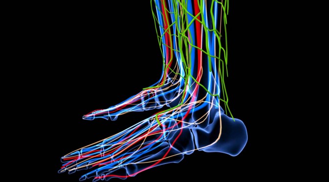
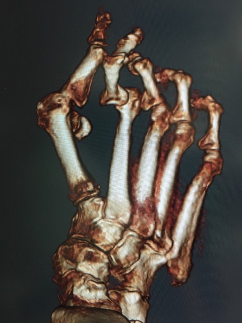
Mülller-Weiss involves a painful deformity caused by osteonecrosisKöhler_I 
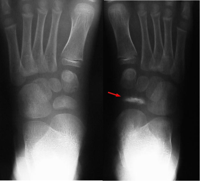
and fragmentation of the navicular, most commonly occurring in adults and disproportionately in female patients. The pedCAT, a cone beam CT imaging system, provides an opportunity for early diagnosis and better preoperative analysis, resulting in improved treatment. Standard computed tomography (CT) in itself beneficial for several reasons.

First, it allows for easier examination of the extent of the deformity and for evaluation of any surrounding arthritis. With CT data, surgeons can evaluate and plan for any future attempts at remedying the deformity.

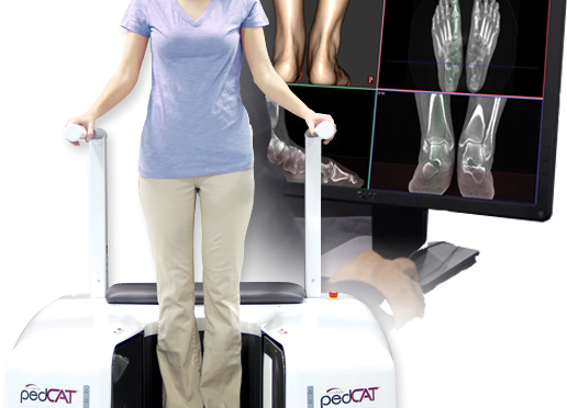
However, because standard CT scans are not weight bearing, they do not provide an accurate view of the relationship between the hindfoot and midfoot deformity. pedCAT WBCT scans allow doctors to go one step further than standard CT scans.

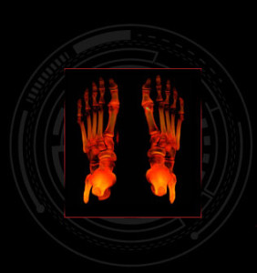
pedCAT 3D datasets can be fully manipulated in order to provide an opportunity for in-depth analysis before proceeding with surgery on a complex deformity. In addition, the pedCAT provides for a better patient experience as the procedure only takes between 19 and 48 seconds, depending on the extent of the deformity.

Weight Bearing CT scan. (Top Left) straight subtalar alignment. (Top right) Anteroposterior view shows navicular bone stock. (Bottom left) Sagittal slice shows features of subtalar varus, superior talonavicular arthritis. (Bottom right) straight subtalar alignment. Source: Foot & Ankle Specialist.

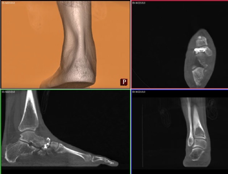
The pedCAT also cuts down on radiation, with a low dose of 1.4 μSv, an amount that is not significantly higher than the 0.7 μSv dosage of the traditional three radiographs of a foot taken to identify Mülller-Weiss. Overall. the pedCAT allows for an accurate and thorough analysis that cannot be achieved through normal X-rays, MRIs nor bone scans.

When creating a treatment plan for Mülller-Weiss disease, the goal is to reestablish a balanced foot, which may require realignment surgery or surgery involving a structural graft.

Due to the complexity inherent in any attempt at treatment, pedCAT imaging is ideal, as it allows doctors to accurately gauge the amount of medial column shortening as well as determine the navicular bone stock and density.

Identifying these key details as accurately as possible is imperative for operations such as calcaneal osteotomy to properly restore alignment or repairing the medial column length.

muller weiss1
Weight bearing CT scan. (Top left) Subtle subtalar varus. (Top right) Anteroposterior view with increased cut thickness allows accurate determination of lateral navicular compression in weight bearing position. (Bottom left) Sagittal slice allows accurate determination of talonavicular and potential subtalar and naviculo-cuneiform arthritis. (Bottom right) Thus demonstrates amount of subtalar varus in weight bearing position. Source: Foot & Ankle Specialist

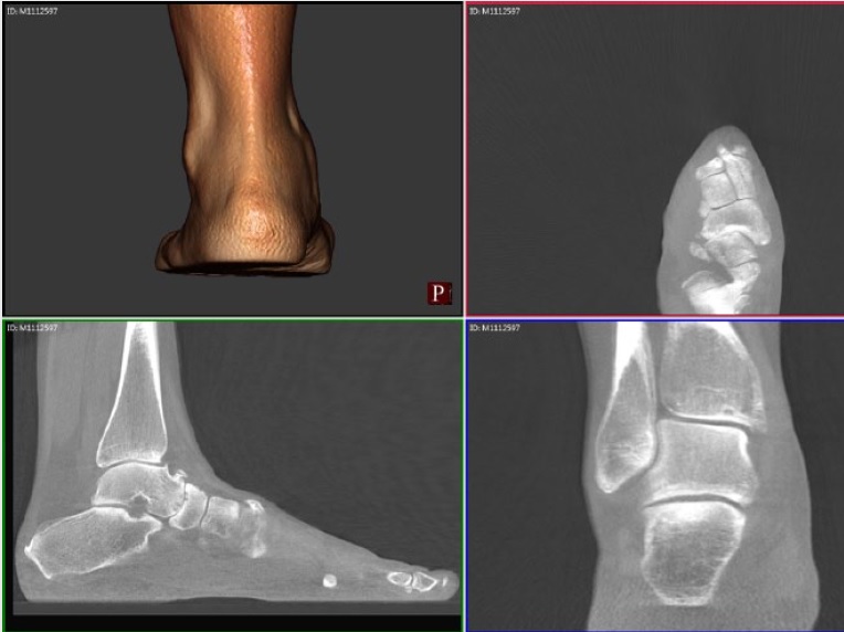
While a combination of weight bearing X-Ray and unloaded CT scanning are sometimes suggested as an alternative, the degree of the deformity caused by Mülller-Weiss will not be accurately represented.

It is only through WBCT scanning that a true picture of the dynamics within the foot will be understood. pedCAT is a compact and ultra low-dose system, which aims to make such 3D scanning possible for clinics.

Weight Bearing CT Advances Planning for Forefoot Reconstructive Surgery

Think of a surgical procedure like a construction project. No contractor would build a building without a full set of plans, cross-sectional diagrams and 3D renderings. Dr. Bob Baravarian, DPM, feels foot surgeons should be just as prepared.

As he explained in Podiatry Today, Dr. Baravarian has always been interested in architecture and real estate, yet was drawn to 159682158975132.Zfw2RIZiU5Lh04gznDby_height640medicine.

Dr. Baravarian sometimes works next to a hip & knee surgeon in the OR. One day, he observed the hip & knee surgeon had a digital tool that laid out an exact surgical plan for the osteotomy he was performing. The plan rendered leg length and acetabular rotation a non-issue.

Soon after, Dr. Baravarian incorporated a pedCAT weight bearing CT imaging system into his podiatric practice. He found he could plan angular corrections to the degree, calculate the exact size of a graft for an Evans procedure, and could even plan the amount of rotation necessary for anatomic alignment of the hindfoot.


Overtime, he began to apply the same methods to forefoot procedures as well.

  • For hallux valgus cases, he scans the patient in the post-corrected position so he can determine if a hindfoot alignment correction is also needed.
  • For hallux rigidus cases, he uses weight bearing CT to evaluate the level of articular damage and the possibility of cartilage graft procedures. “This is impossible to do at such high levels with plain radiographs,” Dr. Baravarian said in the Podiatry Today article.

pt07treatment1 (1)Bilateral, weight bearing three dimensional views of the foot and ankle give specialists the data they need to create optimal treatment plans. The pedCAT, with a scan time of one minute, is a compact, ultra-low dose CT imaging system that is ideal for orthopedic and podiatric clinics. Patients benefit from the convenience of point-of-care advanced diagnostic imaging, and the pedCAT automatically generates all standard X-Ray views in addition to the full CT volume.

As software programs become more advanced, Dr. Baravarian said, surgeons may be able to perform osteotomies and shift the bones in preoperative planning, which will allow even more advanced planning along with far less operative time and less risk to the pt07treatment3 (1)patient.

Dr. Baravarian is the Chief of Podiatric Foot and Ankle Surgery at the Santa Monica UCLA Medical Center and Orthopedic Hospital, and Director of the University Foot and Ankle Institute in Los Angeles.

Read the entire article here.

Exclusive Video From The First pedCAT Weight Bearing CT Scientific Users’ Meeting in Berlin

meeting-83519_960_720 (1)The first pedCAT Weight Bearing CT Scientific User’s Meeting recently convened at the 2016 Foot International (EFAS, DAF, I-FAB) in Berlin. The meeting focused on weight bearing cone beam CT (WBCT) technology and the potential applications and challenges it presents.

Fill out this form to access the video.

Look for several interesting items as you watch:

  • Professor Martinus Richter’s work included a time workflow study on the use of X-Ray,  conventional medical CT, and the pedCAT; the results will surprise you. His study also showed how weight-bearing  CT measurements are more accurate than X-Ray measurements.
  • Dr. Arne Burssens’ study on hindfoot alignment answered critical questions: How do we identify varus and valgus, how do we accurately measure them, and how can we be sure that the measurements are clinically useful and reproducible?
  • Dr. Francois Lintz gives us a sneak peek at a new weight-bearing measurement CT tool that will be available exclusively in CubeVue, pedCAT’s software. It will enable users to measure alignment in three dimensions, and distinguish between normal and pathological cases.
  • Dr. Cesar Netto’s study focused on adult acquired flatfoot deformity, comparing  measurements on weight bearing CT vs non-weight bearing CT.
  • Dr. Michael Wachowsky’s study focused on the use of weight bearing CT  with pediatric patients, and asks: With the new weight bearing CT technology, what will be the exact definitions of the measurements?

The challenge created by this new technology is clear: How do we now define what we are measuring, since the current X-Ray methods have just been rendered obsolete?

“We can’t have non-weight bearing CTs and say anything about alignment – that’s over,” concluded Dr. Charles Saltzman. “What do we want to measure, and how do we know what we’re measuring represents reality?”

Fill out this form to access the video.

pedCAT: A Positive User Experience


Baravarian Western

“After using the pedCAT for a year, I am very impressed with how much I need it, and how many different things I use it for,” Dr. Bob Baravarian said at the 2015 Western Foot and Ankle Conference held in Anaheim, Cali. The California Podiatric Medical Association hosted the meeting at the end of June.

Dr. Baravarian cited Hallux Rigidus as one example where pedCAT imaging has been helpful because he is able to properly assess the sesamoids.

Dr. Baravarian confirmed his practice is realizing positive revenue streams as a result of the device.

pedCAT CubeVue: Automatic X-Ray Views

CurveBeam pedCAT Weight Bearing CT

It takes less than a minute to scan a patient in the pedCAT, but that’s enough time for the pedCAT system to collect enough data to create a 3D reconstruction of the foot and ankle, as well as .3 mm slices in all three planes.

COMING SOON: A new feature will let you take that data one step further – pedCAT’s CubeVue software will automatically generate the data into common X-Ray views, including the Saltzman hindfoot view, the dorsoplantar view, and left and right lateral views.

The automatic X-Ray view feature will save the physicians valuable time when analyzing their patient data.

To learn more about this and other CubeVue features,  contact your CurveBeam representative!

New Study: pedCAT Allows for More Accurate Bone Angle Measurements than Radiographs or CT

A study published in Foot and Ankle Surgery compared three common angle measurements of the foot taken from non-weight bearing CT, weight bearing digital radiographs, and weight bearing pedCAT scans. The study concludes, “The angles differed between radiographs, CT and pedCAT, indicating that only pedCAT is able to detect the correct angles… pedCAT prevents inaccuracies of projection and foot orientation in contrast to radiographs due to the 3D dataset which is principally independent from projection and foot orientation.”

The study also found the average image acquisition time for the pedCAT (average 270 seconds) was 70 percent faster than with radiographs and 35 percent faster than with CT.

The study was authored by Martinus Richter, Bernd Seidl, Zech Stefan, and Sarah Hahn.

pedCAT: Improved Outcomes

pedCAT: Improved Outcomes

The message at RSNA Annual Meeting this past November was clear: new US healthcare laws mean the practice of radiology is no longer about volume, but value.

“We are focusing on quality metrics. It’s becoming important for us to become champions of quality in our institutions,” said Dr. Vijay Rao, MD, in a course at the meeting.

How could a pedCAT add value to your practice?

We might get an idea by looking at a comparable new technology. Breast tomosynthesis mammography provides 3D imaging for breast cancer screening, similar to the way the pedCAT provides 3D imaging for the foot and ankle. Breast tomosynthesis technology can detect breast cancers earlier than traditional 2D mammography, and can more accurately pinpoint the size, shape and location of abnormalities, according to the Massachusetts General Hospital Imaging Department.

Dr. Liane Philpotts, professor of diagnostic radiology at the Yale School of Medicine, called tomosynthesis a “game changer” and a “win-win.”

In the same way, the pedCAT eliminates variability, helps lead to better diagnoses, and makes both you and your patients more confident that treatment will result in better outcomes.

Dr. Erik Nilssen, MD, said the pedCAT helps him determine exactly when to allow patients to ambulate, “based on our ability to monitor fracture healing and fusion rates.”

Also, pedCAT scans can take the guesswork out of hindfoot alignment, said Dr. Martin O’Malley, MD, because they allow for reproducible measurements.

“We’ve never had a reproducible measurement,” until now, he said.

To see an example of a pedCAT scan that led to a more accurate diagnosis, click on the blog post title.

Continue reading pedCAT: Improved Outcomes

It’s Time to Embrace New Technology

The deal made headlines in the orthopedic community when Stryker, one of the world’s largest medical technology companies, acquired MAKO Surgical Corp., in December 2013. MAKO pioneered the advancement of the robotic arm to assist in hip and knee orthopedic surgeries.

A post-script of an Orthopedics This Week podcast on the subject, the author noted:

“One re-occurring questions from surgeons, who are all dealing with the realities of an increasingly cost conscious health care system, is why would Stryker acquire a company which sells million dollar robots in this period of health care cost reduction?”

We often hear similar feedback from physicians who say they cannot justify a pedCAT purchase when major changes to healthcare loom ahead. Our response is that now more than ever, definitive diagnoses, accurate pre-surgical plans, and comprehensive post-surgical evaluations are crucial. The new medical landscape demands more efficient care.

It seems Stryker agrees. It’s response, as reported by Orthopedics This Week:

“Stryker’s answer is that MAKO will simplify joint reconstruction procedures, reduce variability, and enhance the surgeon and patient experience. And in dollar terms, Stryker’s implants comprise about 80 percent the total cost of large joint replacement. The rest of the cost of surgery is everything else including the surgery itself, rehab, and any problems that come up. Small gains in the non-implant portion of surgery, like fewer errors, more consistent outcomes, can have a huge effect on overall costs. At a time when insurance companies are asking hospitals, surgeons and implant suppliers to virtually guarantee outcomes and eliminate variability, Stryker’s purchase of MAKO is a $1.65 billion bet that robots are the answer.”

Made in the USA

At CurveBeam, we are proud that the pedCAT is manufactured entirely in the United States. Our factory headquarters are located in Warrington, Penn., just north of Philadelphia. The factory floor is housed in the same building as our administrative, sales, and research & development offices. We are committed to creating opportunities in our community. Most of the pedCAT’s components are sourced from local manufacturers as well.

CurveBeam’s manufacturing process is certified by ISO, a developer of voluntary industry standards. Each pedCAT goes through a rigorous testing process to ensure it produces high quality images.

An engineer surveys inventory at CurveBeam headquarters.

Engineers on the factory floor follow the principles of lean manufacturing, developed by Toyota. The process is easily scalable, meaning CurveBeam can react quickly to large orders or orders that require a fast turnaround time.

We would like to extend an open invitation to all foot and ankle specialists to visit us. We’d be happy to give you a tour and show you the pedCAT “behind the scenes.”

Tweet: Check out this example of high tech medical device manufacturing on American soil: http://ctt.ec/HPlnc+

Welcome to the CurveBeam Blog!

Welcome to the CurveBeam Blog!

2foot-with-platform1.jpg

It’s an exciting time at CurveBeam. Almost two years ago  we received FDA-approval for our first product, the pedCAT. The in-office 3D weight bearing imaging system for the foot and ankle has been integrated into orthopedic and podiatric practices around the world.  Foot and ankle specialists, considered leaders in their field, have told us that the pedCAT will become the standard of care for lower extremity imaging in the near future. In fact, in some of their practices, it already is.

We’re dedicated to continuing the improvement of foot & ankle imaging and are excited to share with you the technology advancements, right here on this blog in the upcoming months. So check back often!