CurveBeam Selected to Join The OrthoForum

2017 is in full swing, and we at CurveBeam are excited about the prospects the year has in store for both our company and the field of orthopedics. At only eight years old, we are relatively young, but in that short time we have emerged as a leading manufacturer of imaging equipment for orthopedic and podiatric specialties around the globe. Our imaging equipment helps provide better patient outcomes, and we are continuing to innovate new solutions for leading specialty practices. It is with great pleasure we announce we have recently been added as an approved vendor to The OrthoForum Purchasing Alliance.

CurveBeam participated in OrthoForum’s Annual Conference in Laguna Niguel, Cali., last week, and will also be exhibiting at the regional conferences throughout the rest of the year.

The OrthoForum is a national physician specialty organization whose membership includes over 3,090 physicians and some of the largest private orthopedic practices in the country – and growing. Members are individually selected to help meet the unique challenges that integrated orthopedic group practices face in today’s healthcare environment. The top priority of the organization is to protect the ability of orthopedic practices to remain independent and provide the highest quality patient care. Advocacy, engagement between members and elected officials, and collaborations with other orthopedic and medical associations are methods The OrthoForum employs to fulfill its mission.[1] CurveBeam is thrilled to join The OrthoForum Purchasing Alliance and is ready to demonstrate our commitment to helping fellow members and all orthopedic practitioners thrive.

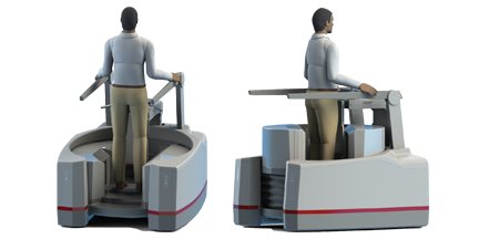
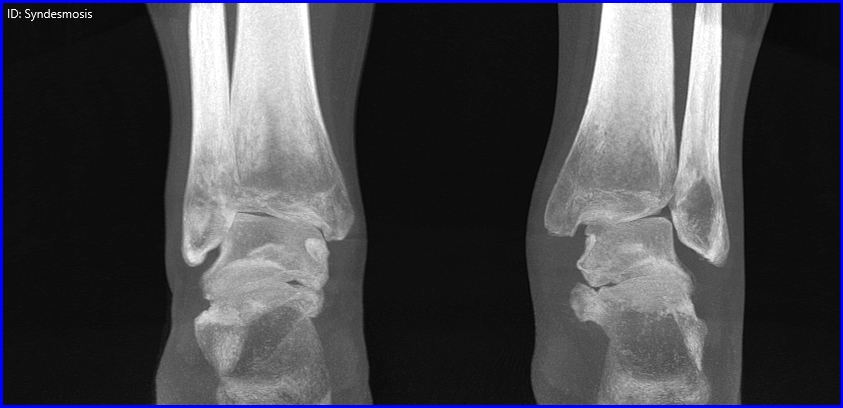
Our dedication to orthopedics is exemplified by our revolutionary pedCAT system, which utilizes an innovative design and compact footprint to bring point-of-care advanced imaging into the foot and ankle specialty. Practices all across the United States, Europe and Australia rely on the pedCAT system, and its custom visualization software, CubeVue, for 3D imaging that is essential to formulating comprehensive treatment plans. Research has shown pedCAT is ten times faster than X-rays and CT scans, resulting in fewer patient trips to the hospital and significantly reduced exposure to radiation.[2]

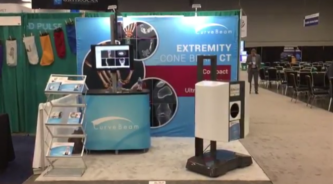
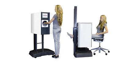
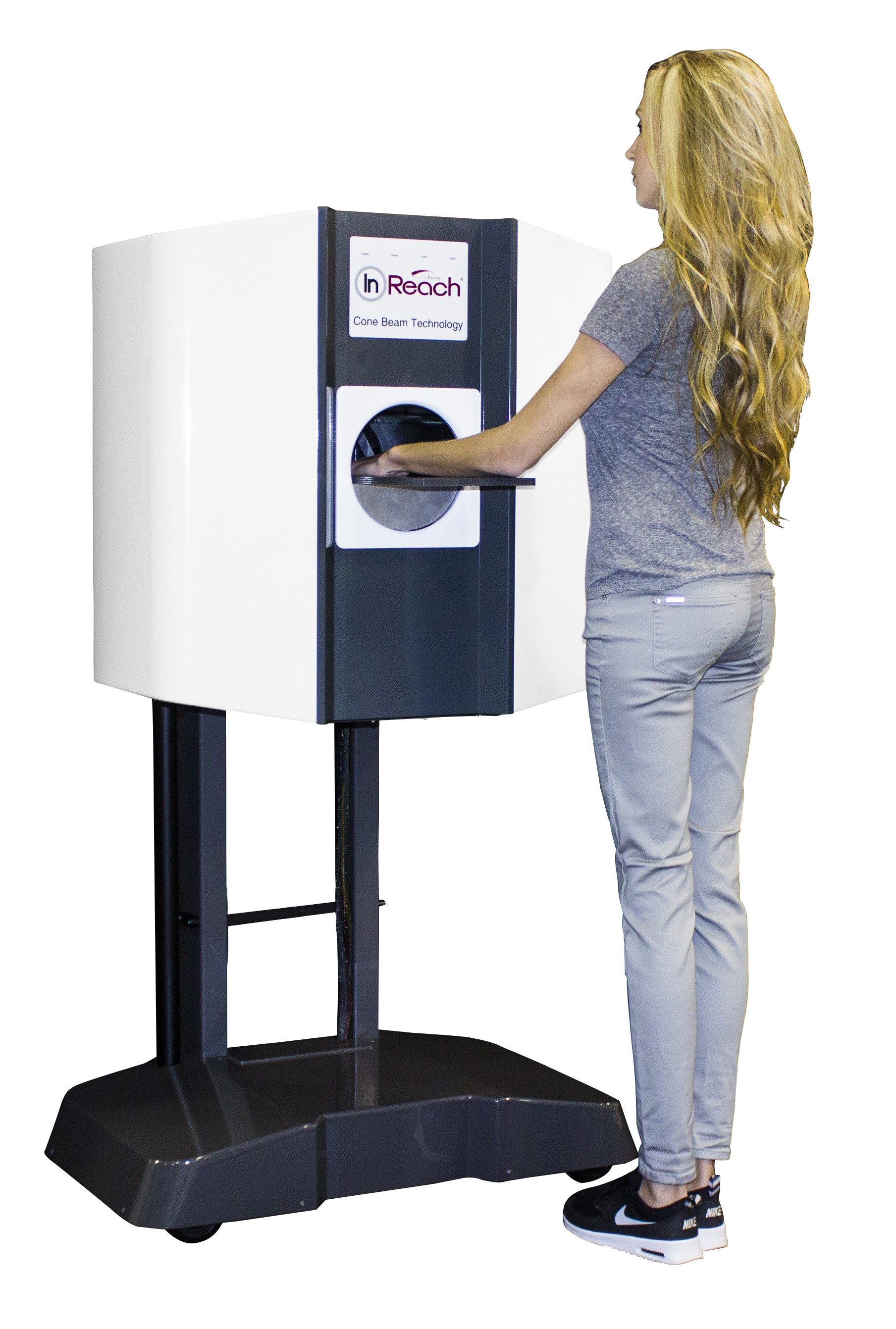
As innovators, we are constantly developing new technologies to improve patient care. This year we’re looking forward to launching InReach, an extremity cone beam CT scanner that images the hand, wrist, forearm, elbow and the lower extremities. At 20 seconds per scan, InReach provides fast and easy imaging. Our professionals are also hard at work designing a revolutionary, complete lower extremity imaging solution for orthopedic and podiatric specialists that we hope to bring to market in the months ahead.

At CurveBeam, we’re passionate about engineering technology that advances better patient outcomes, and streamlines the workflow of clinicians within the orthopedic specialty. The OrthoForum advocates for operators within the entire orthopedic field, and we are honored to participate in these efforts as a new member of this respected organization.

[1] http://www.theorthoforum.com/about-us/
[2] https://marketscaleblogs.com/curvebeam/join-the-talas-revolution-in-foot-and-ankle-diagnosis/

The Opportunity for Private Office Imaging

Changes in healthcare insurance, regulations, and other factors have shifted the bulk of imaging services to hospitals, but the opportunity for private radiologists is still very promising.

“Hospital outpatient imaging has been migrating from private offices since about 2010,” says Dr. David C. Levin of Thomas Jefferson University, as reported by Aunt Minnie. “But that doesn’t mean that radiologists still can’t make private office ventures work.”

Dr. Levin goes further than just his diagnosis of the medical imaging service industry by prescribing a solution to private radiology offices interested in increase their imaging business.  His business model suggestions include a mix of customer service, marketing, and cost competitive advice that can turn around a struggling radiology office and build a successful and sustainable private practice.

Implement a Cost-Effective Payment Schedule for Patients with High Deductibles

The increasing number of high deductible healthcare plans has created a patient incentive to shop around for low price imaging services. Understanding these plans will help your office develop a fee schedule that is attractive from a cost perspective.

“Patients are going to have higher and higher deductibles, and that’s going to make them shop around for healthcare,” says Dr. Levin. “If you can offer, say, an MRI of the knee for $600, while the local hospital has it at $4,000, you can be sure patients will come to you.”

Marketing – It Really Works

Make prospective patients aware of your services by advertising an elevated quality of care and customer service.

“Hospitals generally don’t market their radiology services, but private practices certainly can,” says Dr. Levin. A well-designed advertising campaign can make your practice stand out and drive new customers to your location.

Don’t Neglect Customer Service

There is so much more that goes into the patient experience besides the treatment itself. Examine the way patients are treated, from the initial contact all the way through follow up, to identify opportunities to improve the experience. Patient frustration can arise at any step in the process. Time saving processes as well as friendly and helpful engagement go a long way to improving the overall patient experience.

Efficiency.  Efficiency.  Efficiency.

Dr. Levin stresses the importance of efficiency throughout the business.

“The more efficient your practice, the lower your costs will be,” he says. Fully utilizing assets and people in a streamlined operation brings down overhead and operating costs, and makes for a lean, cost competitive business with higher margins.

At the end of the day, Dr. Levin emphasizes private radiology practices have an amazing opportunity to improve their imaging services and overall business. Taking the time to evaluate the situation, develop an action plan, and implement constructive changes will certainly pay dividends now and for years to come.

CurveBeam’s Volunteer Day Warms Community

The holiday season is an excellent time for any company to celebrate and share its values. The team at CurveBeam believes in giving back, just as much as it believes in innovation. To reach out with this spirit, a group of CurveBeam volunteers recently connected with an amazing organization.

Everyone had a simple goal in mind: to make sure kids in the community have the supplies they need for this season and beyond.

Cradles to Crayons serves as a resource for communities in need and a hub of civic engagement in Greater Philadelphia. This organization provides children from birth through age 12 living in homeless or low-income situations with the essential items they need to thrive – at home, at school and at play – free of charge.

Ensuring kids are safe, warm and ready to learn throughout the year

Cradles to Crayons was founded in 2002 to get basic necessities to the kids who need them, including clothing, school supplies, toys, and more. The organization provides all the physical goods that children need to be safe, protected from the elements, and ready to learn.234585951194320-834hbvhp2y7forv0xdvp_height640

By supplying kids with high-quality used or new goods, Cradles to Crayons and its partners make children feel valued.

The organization partners with social service organizations to connect groups that have surplus resources with the communities and families that need them. Cradles to Crayons makes it simple for children, families, and corporations to participate by organizing a clothing collection, hosting a fundraiser, or working in one of its warehouses.

Charity Navigator – an independent group that rates charities on both their organization capacity and efficiency – recently awarded the Cradles to Crayons a four-star rating: their highest ranking. In receiving this designation, Cradles to Crayons was recognized for exceeding industry standards and outperforming most charities in its cause.

In addition, Cradles to Crayons Philadelphia was included in Main Line Today‘s list of local charities to support.

A CurveBeam volunteer party

In lieu of a traditional Christmas party, a group of CurveBeam volunteers spent half a day at Cradles to Crayons, helping prepare items to go out to kids in need in time for the holidays.

The team enjoyed the experience. It was the perfect expression of the company’s ongoing commitment to giving back, and one the team looks forward to repeating next year.

To learn more about CurveBeam’s work and its talented and warm-hearted team, visit CurveBeam.com. Happy New Year!

Curvebeam’s Booth Is a Must-See at RSNA 2016!

In just a little over a week, many of the greatest minds and innovators in radiology will convene in Chicago for RSNA 2016, and we at CurveBeam are proud to have our talented and skilled team there representing us. Over the course of the conference, there will be groundbreaking research presented, technological advances displayed and educational classes offered. In the heart of it all, our booth will be featuring some incredible innovations – proving once again that CurveBeam is continuing to find new ways to push the boundaries of our field.

Our core team pioneered Cone Beam CT imaging technology for the dental and ENT specialties. The ability to provide point-of-care imaging revolutionized these industries and allowed not only for improved care, but for the possibility of custom dental implants. Now we are taking that same breakthrough technology and adapting it to the orthopedic field, and we are proud to introduce the results of our efforts at this year’s RSNA. Here are our top three reasons why you can’t afford to miss the CurveBeam booth this year:

  1. See the LineUp

We will  unveil the prototype for the LineUP, a bilateral weight-bearing Cone Beam CT imaging system for the knees and lower extremities. It will not only plugs into a standard wall outlet, but will also be self-shielded and will complete a scan in under a minute. While the technology is still investigational only, it is PACS/ DICOM compliant and is sure to revolutionize the way orthopedic clinics approach radiology.


  1. Experience InReach

While the LineUp is certainly fast, the InReach completes scans of the hand wrist, forearm, elbow and extremities in under 20 seconds flat. We designed InReach technology to bring the idea of point-of-care extremity CT imaging  to upper extremity specialists. While it is also still investigational technology, the progress we’ve made toward this goal is worth coming to see.


  1. Discover TALAS

One of the most crucial elements of orthopedic surgical planning is the precise pre-operative measurement of hindfoot realignment. TALAS is a tool that makes the process not only smoother, but more accurate and precise. It is a semi-automated hindfoot alignment measurement device that has been adapted for weight-bearing CT. Come check it out for yourself, and discover the potential TALAS has to one day be a true game-changer in the field.

“We are tremendously excited to share our recent research and development efforts with the global radiology community,” says CurveBeam President and CEO Arun Singh. RSNA is an opportunity to share and learn together as a community and we are thrilled to be a part of that experience. Feel free to come by and discover our innovative new technology. If you want to learn more before seeing our products in person, visit CurveBeam.com. We hope to see you soon at RSNA 2016!

Using The InReach to Detect Scaphoid Fractures

Scaphoid fractures are often difficult to diagnose. The scaphoid may be difficult to visualize on X-Rays due to superimposition.  A delayed or incorrect diagnosis of a scaphoid fracture could result in grave outcomes such as nonunion, delayed union, decreased grip strength, reduced range of motion, and/or osteoarthritis of the radiocarpal joint.

X-ray is currently the standard method of diagnosis, but according to Vinti Singh, an Advanced Medical Imaging Specialist at CurveBeam, “…that particular part of the wrist is really hard to see on X-ray.  CT scans show scaphoid fractures clearly, but traditionally, patients must travel to a hospital or imaging center to access this service.” Singh adds that CurveBeam is developing the InReach, a point-of-care cone beam CT imaging system for the upper extremities. The InReach is investigational only and is not available for sale in the United States. The CurveBeam R&D team is striving to  “provide faster, more accurate detection and as a result, better patient care.”

Since scaphoid fractures rarely appear on X-rays right away, doctors often send a patient home in a splint or cast for two to three weeks if they suspect a scaphoid fracture. Typically, the patient will then return for another X-ray.  With the InReach, a patient could get a CT scan, at the physician’s discretion, for a definitive diagnosis.

The CurveBeam InReach will provide point-of-care CT imaging of the upper extremities. The InReach is investigational only and is not available for sale in the United States.

CurveBeam’s InReach system will scan the wrist structures in nineteen seconds. MRIs take much longer, as do bone scintigraphy processes, which also require injections of imaging dye or radioactive material. The InReach delivers high resolution images with 0.3mm slices, accounting for their improved diagnostic accuracy over the X-ray. The unit will have a small footprint and uses a regular wall outlet, unlike a traditional CT unit, which requires  more space, power and additional cooling needs of a traditional CT. Patients need only place a hand in the machine for a few seconds, as opposed to having to lie down on a CT bed. So not only does the InReach system reduce patient frustration at scheduling and waiting for multiple appointments, but it also maximizes patient comfort.

With complicated injuries like the scaphoid fracture, timely diagnosis and treatment are critical in avoiding the potential long-term wrist dysfunction resulting from misdiagnosis. Compared to the previous standards of X-rays, MRIs, bone scintigraphy, and traditional CT scans, the InReach would offer superior speed and accuracy while delivering a high level of patient care and comfort. Learn more about CurveBeam and the InReach imaging technology here.

From RADAR to pedCAT, Amrita Kar is Making Her Mark

The strength of our company originates with engaged and driven employees. The staff at CurveBeam incorporate their ambitious and innovative ideas into the quality services you can expect when seeking extremity imaging solutions. Our team of engineers have made it possible to bring the same point-of-care Cone Beam CT imaging used in the dental industry to the field of foot and ankle diagnostics.

The CurveBeam team created the pedCAT, a compact and ultra-low dose CT imaging system for orthopedic and podiatric clinics. They then went one step further, building an imaging system that provides bilateral, weight bearing 3D views of the foot and ankle within one minute. This revolutionary technology has become the standard level of quality for our team of engineers.

One of those engineers is Amrita Kar. In less than two years with CurveBeam, Amrita has carved out her place as a talented and creative expert in her field. In 2006 she started training at the Indian Defence Research & Development Organization (DRDO), a department of the Indian Ministry of Defence. Amrita contributed to building Radar control and signal processing systems, and others in the industry soon recognized her talents. She was quickly recruited by the private sector to oversee end-to-end development on many classified projects. Then in 2011, she discovered some Computed Tomography images, which sparked her interest in medical imaging. Hoping to explore her new passion, she earned a full scholarship to Villanova University, where she pursued her Master’s degree in Electrical Engineering.

After receiving her graduate degree with a specialization in Biomedical Signal Processing, the search to utilize her new skills began.

“A startup medical device company was a natural professional choice based upon my past experiences working effectively with cross-functional teams at a fast-paced environment and dealing with evolving customer requirements,” says Amrita.

Thus, she began serving as an intern in September of 2014 for our team, and by December she had advanced to the position of Senior Development Engineer. She serves as a mentor for other CurveBeam scientists, physicians, and engineers, and together they have achieved innovations in the world of medical imaging, including the pedCat system.

“It’s inspiring as a developer,” she says, to work with others to help physicians provider faster and more accurate diagnoses. Further, she admits to feeling privileged to “work here among very sharp, intelligent, and competent team [members]” toward the goal of “fast implementation of changing customer needs.”

This system is in fact helping change the methods used by medical professionals in their approach to foot and ankle care. We are proud to be making a difference and proud to employ dedicated people like Amrita Kar on our team.

Enjoyed this post? Meet another member of the CurveBeam team.

Healthcare costs have skyrocketed in recent years

This trend has bewildered patients, who see their medical bills ballooning with no commensurate improvement in care. As a result, medical professionals and administrators are on the hunt for the cause of this spike in costs. One common scapegoat is diagnostic imaging, such as Computerized Tomography (CT) scans and x-rays. These technologies have been painted as expensive, unnecessary, and overused just to line the pockets of greedy doctors and hospitals. But is that really the case?

This question was investigated by Molly T. Beinfeld and G. Scott Gazelle in their paper “Diagnostic Imaging Costs: Are They Driving Up the Costs of Hospital Care?” for the Radiological Society of North America. Benfield and Gazelle gathered and analyzed billing data for 17,139 patients admitted to Massachusetts General Hospital between 1996 and 2002. This data clearly showed an obvious increase in patient costs, but the causes of this increase, and the conclusions that can be drawn from them, are less obvious.

On average, total patient costs increased about 8% per year, and imaging costs also increased 8% per year. This means that medical imaging costs did not drive up overall costs any more than any of the other services that the hospital provides. Additionally, this 8% increase in imaging costs is even more benign than it seems. Between 1996 and 2002, the number of CT and MR images obtained at Massachusetts General more than doubled, but the cost per image actually went down thanks to technological improvements. The underlying causes behind these two facts help to illustrate the difficulty in pinning the blame for the rise in medical costs on any one factor.

One large reason for the increased use of medical imaging is because medical imaging technology has improved, making it a more powerful tool that can be applied to conditions that it previously wasn’t. During the range of the study, the usage of CT scans for patients undergoing appendectomies increased nearly 800% and the usage for patients with stroke and transient ischemic attack more than doubled. The use of these images allowed doctors to better treat their patients, and its cost was likely defrayed by the resultant decrease in use of other methods. The cost per CT fell by nearly half over the course of the study, in part due to the increase in number of CT scans performed and the resultant decrease in per-scan fixed indirect costs. By looking at a cost benefit analysis, the rise in use of CT scans makes perfect sense.

Average CT imaging costs vary widely from a few hundred dollars to several thousand, depending on a patient’s location, healthcare provider network and insurance status. According to a recent piece in Health magazine, prices range from $410 to $2,334. For customers who are uninsured and pay with cash within 60 days, there may be a discount: the average CT scan price without insurance ranges from $1,016 to $1,440. Medicare Part B covers CT scans when your provider orders them, but deductible and copayment may apply. For other only covered under Medicare Part A, the cash price for a CT scan varies. Despite the costs, the CT scan is considered cost effective in many circumstances and is well-studied.

Identifying the underlying reasons for why healthcare has continuously become more expensive is the first step in tackling the rising costs problem. As the work of Beinfeld and Gazelle shows, there is more to the rise in medical costs than meets the eye. Usage of CT imaging has increased greatly, but that is merely because it has become more effective at helping doctors to diagnose and treat their patients, and it has also become more affordable to use as a result. The increased use of CT imaging has in fact likely served to temper the rise in healthcare costs while also helping to minimize morbidity and mortality among the patients it serves. Understanding that CT imaging is not as expensive as commonly believed is important because it helps healthcare professionals to make better decisions about patient care.

CurveBeam Announces Development of Extremity CT System for Knee

A new orthopedic CT system promises to improve the standard of care for knee imaging. On the CurveBeam LineUp, patients are scanned while standing upright and fully weight bearing.

CurveBeam LineUP
CurveBeam LineUP provides bilataral weight bearing CT imaging of the knees and lower extremities.

Traditional CT and MR images are acquired in a non-weight bearing position, leading to “missed diagnoses of meniscal damage,” according to Dr. Neil Segal, who has been overseeing research efforts using a LineUp prototype, first at the University of Iowa and currently at the University of Kansas.

Neil Segal, MD
Neil Segal, MD

Although plain radiographs can be acquired while the patient is in a full weight-bearing position, the optimal degree of knee flexion and X-Ray beam tilt to best visualize the joint surface is person specific.

“Difficulty in reproducing the same view of the joint over time impairs ability to detect joint disease, and the 2D nature of radiographs makes these images of overlapping bony anatomy very insensitive for detecting abnormalities until there is advanced joint damage,” Dr. Segal said.

The LineUp was developed by CurveBeam, a Pennsylvania-based company that specializes in extremity cone beam CT systems for orthopedics. CurveBeam anticipates it will submit an application for and receive FDA 510(k) clearance for the LineUp in 2017. The LineUp will be on display at RSNA at Booth #8008 in the North Hall.

CurveBeam introduced the pedCAT, a bilateral weight bearing CT system dedicated to the foot and ankle, in 2012. Since then, the device has been added to the imaging services of hospital foot & ankle sections, orthopedic clinics and podiatry offices.

Like the pedCAT, the LineUp will provide isotropic, three-dimensional volumes of the anatomy with a high resolution output of between 0.2 mm and 0.3 mm slices. The LineUp will be the only cone beam CT system that can provide bilateral, weight bearing scans.

A study led by Dr. Segal focused on osteophytes, one structure

linked to pain in people with knee osteoarthritis. Knees of community-dwelling adults with knee OA were imaged with MRI (reference), fixed-flexion radiographs, and weight bearing CT. The sensitivity and accuracy for detecting osteophytes and subchondral cysts were higher with weight bearing CT imaging in comparison to fixed-flexion radiographs. The study was published in the August 2016 issue of the Journal of Orthopedic Research.

“Clinically, this is a highly meaningful improvement,” Dr. Segal said. “It suggests that weight-bearing CT could replace radiographs as the recommended means of assessing knee OA. This advancement is even more significant given that it was made without significantly increasing the radiation dose (0.01 mSv for SCT vs. 0.005–0.102 mSv for a series of knee radiographs).”

Another research effort led by Dr. Segal indicates weight-bearing CT arthrography studies can provide outstanding delineation of articular cartilage with better differentiation between cartilage and subchondral bone then MRI studies, while also visualizing the cruciate ligaments. In knees with osteoarthritis, meniscal tears not visualized on MRI were detectable on weight-bearing CT.

“Thus, we found that some potential advantages of weight-bearing CT over non-weight-bearing MRI/MRA include 3D measures of meniscal position, detection of pathology not detected in unloaded positions, and ability to bear weight in a functional position, thus better recreating the magnitude of body weight and muscle forces acting around the knee during usual standing,” Dr. Segal said.

CurveBeam Announces First pedCAT Installation at an Accredited College of Podiatric Medicine

CurveBeam is proud to announce the installation of a pedCAT cone beam CT imaging system at Kent State University College of Podiatric Medicine.

Kent State is the first member of the American Association of Colleges of Podiatric Medicine to acquire weight bearing CT imaging technology.

“Kent State University is proud to be the first facility in the Northeast Ohio region to provide this ‘state- of-the-art” imaging modality,” said Dr. Allan Boike, Dean & Professor of Foot & Ankle Surgery at KSUCPM. “The CurveBeam pedCAT will allow the college to improve the foot and ankle health of the community while providing the highest quality education for our students and research opportunities for our faculty and residents.”

The pedCAT is the only cone beam CT imaging system that allows for bilateral, weight bearing CT images of the foot & ankle. Due to its compact size and low radiation exposure, the pedCAT is an an ideal solution for CT imaging at the podiatric point-of-care. Recent articles in orthopedic journals demonstrate weight bearing CT imaging is a valuable research tool that is shedding new light on even our basic understanding of foot & ankle biomechanics and deformities.

“Our initial euphoria in acquiring the pedCAT weight-bearing cone beam CT has only been eclipsed by actually getting to use it first-hand,” said Dr. Lawrence Osher, Director of the Radiology Department. “Simply put, this is an utterly amazing tool in the podiatric diagnostic and research armamentarium.  The ability to do 3D and multi-planar reconstructions on a weight-bearing foot and ankle, coupled with the prospect of marrying structure and function, opens up a seemingly endless array of research opportunities.  Bounded only by our creativity, we at KSUCPM look forward to adding significantly to the pool of knowledge in the medical literature.”

The pedCAT was funded through a research grant from the Ohio College of Podiatric Medicine (OCPM) Foundation, which was established to promote podiatric medical education and research.

“Dean Boike is to be commended for his foresight in procuring this advanced apparatus which further enables the College to maintain and expand its competitive edge in podiatric medical education,” said Dr. David Nicolanti, Executive Director of the OCPM Foundation. “In addition, this weight bearing CT imaging system provides a basis for enhancing collaboration between the College and foot and ankle specialists, from all medical stratums throughout the state of Ohio and the encompassing region.”

The College has a number of weight bearing CT research initiatives planned.

The pedCAT is located at the Cleveland Foot and Ankle Clinic’s midtown office at 7000 Euclid Ave in Cleveland. Weight bearing CT services will be offered to the clinic’s patients.

Weight Bearing CT Sheds Light on Understanding of Rotational Dynamics in Syndesmosis

As the orthopaedic and podiatric specialties continue to advance, there is great potential for technology like CurveBeam’s pedCAT system to revolutionize care.

Recent research highlighted the importance of weight bearing scans to the understanding of foot and ankle anatomy, suggesting a role for pedCAT in both a research and clinical setting. “Rotational Dynamics of the Normal Distal Tibiofibular Joint with Weight-Bearing.

Computed Tomography” a 2016 study published in Volume 37 of
syndesmosisFoot & Ankle International sought to determine the normal range of motion for uninjured distal tibiofibular joints. Researchers hoped this reference would be useful as a comparison when assessing injured and repaired ankles.

Until recently, all measurements of this motion had been conducted on cadavers or through non-weight bearing scans. In contrast, this study used a weight-bearing CT (WBCT) system to survey the ankles of 32 subjects as they stood on one foot, then the other.

They found a “total movement of 1.5 mm and rotation of 3 degrees” in the syndesmosis as the average across subjects. However, the study also found that intersubject variation was extremely high, meaning different people had vastly different ranges of motion despite similar orthopaedic histories.

These differences were not correlated with sex or age. Intrasubject variation, or the difference in movement between a person’s right and left foot, was significantly smaller and more consistent, less than 1 mm on average.

The study therefore concluded “the contralateral ankle should be used as a reference when dynamic alignment of the distal tibiofibular joint is studied.”

In other words, surveying a person’s uninjured ankle will give a better idea of what is normal movement for that particular individual than comparing the injured ankle to a standardized range, like the one this study endeavored to produce.

CurveBeam’s pedCAT technology could have helped researchers eliminate possible errors in these findings. In the article, researchers admitted “it is possible that we were unable to optimize posture and rotation identically on both sides “ as a result of the limited field of view of the imaging equipment.

The device used in the study could only scan a partial foot in a scan. Test subjects had to stand on one foot, then twist and then switch and twist again. Researchers were unable to measure the force with which the subjects moved on each side.

The pedCAT’s field of view accommodates bilateral imaging, which would have allowed researchers to survey both weight bearing feet at the same time, providing helpful insights. The research indicates the importance of weight bearing measurements of a person’s right and left ankle to determine their normal range of motion.

Being able to accurately assess their syndesmosis on one side will help physicians more accurately assess and repair damages to the other.

CurveBeam’s pedCAT technology is the ideal imaging solution because it allows physicians to scan and survey ankles side-by-side for increased accuracy and ease.

CurveBeam and its technical solutions have the potential to revolutionize orthopaedic and podiatric research and care. Visit us at curvebeam.com to learn how pedCAT and other CurveBeam technologies can make a difference in your practice!