Webinar: Pitfalls In Hindfoot and Ankle deformities tackled by weightbearing CT

Arne_BurssensYou are invited to join FOOTinnovateTM and Dr. Arne Burrssens for the webinar “Pitfalls In Hindfoot and Ankle deformities tackled by weightbearing CT” sponsored by Curvebeam.

Click here to register.

Dr. Burrssens is an orthopedic surgery resident at Ghent University in Flanders, Belgium. During his one-hour presentation on Monday, June 5, 2017 9:00 PM – 10:00 PM (W. Europe Standard Time), he will discuss his personal experience with weight bearing CT and reveal how it has improved his patient care. He’ll also share a few specific cases to provide a deeper look into his work.

Dr. Burrssens is co-author of the article “Weightbearing CT in normal hindfoot alignment — Presence of a constitutional valgus?” published in February 2017 by the journal Foot and Ankle Surgery. The purpose of the study was to assess hindfoot alignment using weightbearing CT.

The research concluded that weightbearing CT provides an objective assessment of hindfoot alignment with reproducible results, which can aid a surgeon during a calcaneal osteotomy.

PLEASE NOTE: You must register for this session at least 15 minutes prior to the start time. Click here to create a free account.

CurveBeam and Carestream Health Announce Collaboration to Promote Weight-Bearing CT Awareness and Research

Warrington, Penn. – June 6, 2017 – Medical device companies CurveBeam and Carestream Health announced a collaboration to support and facilitate education and research for weight-bearing computed tomography imaging.

The ability to capture weight-bearing CT images allows foot and ankle specialists to study foot biomechanics in three dimensions. Significant clinical research has already demonstrated weight-bearing CT has the potential to be the new standard of care for an initial diagnosis. However, much work remains to be done to increase awareness among orthopaedic specialists,” said Vinti Singh, marketing manager for CurveBeam. “Through collaboration, CurveBeam and Carestream can best support these efforts.

Before the introduction of weight-bearing CT imaging in 2012, patients could get either a weight-bearing radiographic 2D X-ray or a non-weight bearing medical CT scan. X-rays can at times obscure and distort anatomical details, while non-weight bearing CT scans may inaccurately represent joint space and alignment. The American Orthopaedic Foot & Ankle Society recommends weight-bearing imaging when possible to get the most accurate assessment.

The image quality and detail captured with 3D weight-bearing CT exams allow the surgeon to view and measure anatomical abnormalities of lower extremities under natural load conditions,” said Helen Titus, Carestream’s Worldwide Marketing Director for Ultrasound & CT. “The goal of this collaboration is to spread the word about this new imaging modality among orthopaedic specialists.”

The companies will work together to support the Weight-Bearing CT International Study Group, which is comprised of orthopaedic researchers from leading institutions in the U.S. and Europe. The group is working to create standardized protocols for weight-bearing CT measurements and analysis.

The companies’ first joint initiative is to co-sponsor an evening scientific session during the American Orthopaedic Foot & Ankle Society (AOFAS) annual meeting in Seattle, WA. Speakers at this session include Dr. Mark Easley of Duke Orthopaedics in Durham, NC, and Dr. Lew Schon of MedStar Union Memorial Hospital in Baltimore, MD. For more information about the Study Group, and how to RSVP for the session, please visit www.wbctstudygroup.com.
Both companies offer weight-bearing solutions for extremity CT imaging. The devices employ cone beam CT technology and are designed for point-of-care settings, as well as hospitals and imaging centers.

To view Carestream’s latest news announcements, please visit www.carestream.com/news.

CARESTREAM is a trademark of Carestream Health.

Keep Your Orthopedic Practice Alive Through Diversification

Medical providers are always looking for ways to create optimal patient outcomes while lowering costs. Dwindling reimbursement can dissuade people from pursuing medical careers—or their chosen specialty. Medical Student Education published a study finding 15 percent of fourth-year medical school students reported payment as the primary factor driving their career choices.

These concerns pose unique challenges for orthopedic surgeons. If we fail to improve revenues for orthopedic practices, then many of these practices will fail. If that happens—at the same time baby-boomers are reaching the age of needing more musculoskeletal care—our nation will experience a true healthcare crisis.

Not long ago, developing a financially successful practice hinged on a physician’s clinical expertise. But expertise alone will no longer serve to attract patients and generate revenue. Our long-term goal should be to generate revenue from multiple sources, diversifying practices to ensure practice viability.

The American Academy of Orthopedic Surgeons, AAOS, created a 45-page guide, Enhancing Your Practice’s Revenues: Pearls and Pitfalls (A Primer for Orthopedic Surgeons), recommending orthopedic practices offer in-office imaging.

Imaging has become an integral part of many orthopaedic offices,” the report says. “In-house imaging provides an ancillary revenue stream, improves practice efficiency, and may play a role in overall patient satisfaction. The types of imaging services offered vary depending on a variety of factors, which include but are not limited to practice size, geographical location, political considerations, and orthopaedic specialties represented in the office.

Revenues from imaging services can add significantly to your bottom line. A reasonably busy MRI, for example, may generate more revenue and profit for the practice than an orthopaedic surgeon, while X-ray often generates an additional $50,000-$100,000 or more of operating profit per orthopaedic surgeon.”

For orthopaedists, the history and physical has long been key to the successful diagnosis of patients with musculoskeletal patients. Traditional film X-rays provided them an additional diagnosis tool. Imaging technology continues to evolve, allowing orthopaedists to diagnose diseases, disorders and conditions more quickly and with even greater accuracy. The result is a win-win situation for patients and doctors alike.

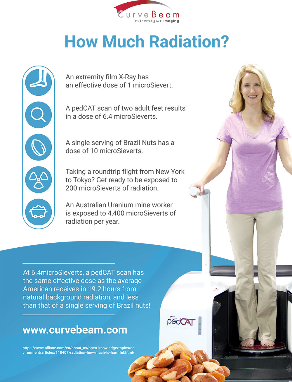
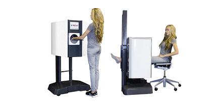
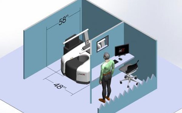
The CurveBeam pedCAT is designed to fit into your practice and workflow, with a footprint of about 4’ x 5’. The unit plugs into a standard wall outlet and gives you the bilateral, weight-bearing 3-dimentional views of the foot and ankle you need to create a comprehensive treatment plan.

The pedCAT scans are reimbursed through CPT code 73700 – CT lower extremity without contrast.

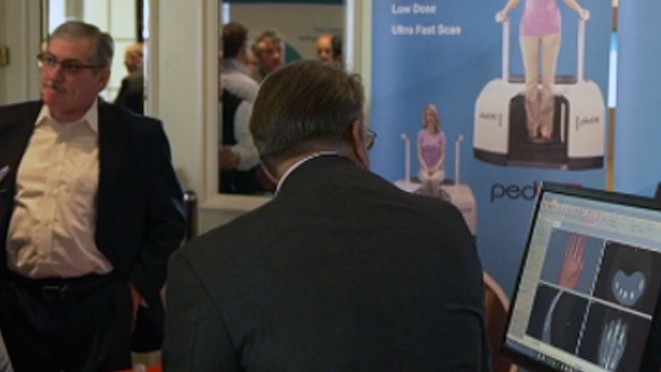
The pedCAT is a compact, ultra-low dose CT imaging system that is ideal for orthopedic and podiatric clinics. Patients benefit from the convenience of point-of-care advanced diagnostic imaging, scan time is one minute, and the pedCAT automatically generates all standard X-ray views in addition to the full CT volume. All of this contributes both to improved patient care and the bottom line.

To learn more about how the pedCAT can enhance your practice, visit CurveBeam.com today!

A pedCAT Scan: Less Radiation Than A Serving of Brazil Nuts

Most people may not realize it, but humans are exposed to natural background radiation all the time. The source is naturally occurring radioactive materials and cosmic radiation from outer space. Geography plays a huge factor in how much radiation a person is exposed to, but the average  American adult receives an effective dose of about 3000 microSieverts a year.

So how does the Curvebeam pedCAT scan compare to radiation from other activities? See below!

A-pedCAT-Scan-Less-Radiation-Than-A-Serving-of-Brazil-Nuts-infographic

The pedCAT Weight-Bearing CT Scan Put CurveBeam a Step Ahead of the Competition

It’s hard to forget about your feet—socks and shoes, not to mention pain at the end of a long workday. As the foundation of your mobility, foot health is vital to overall health. But foot health involves more than just keeping them clean and dry. It is vital to wear properly fitted shoes—custom insoles (avoid flimsy over-the-counter insoles) and supportive sandals are also a good idea. In fact, feet are so important to us that they even get their own specialists.

An expert in the field of podiatry, Dr. Kyle Kinmon of Boca Raton, FL recently shared some advice on WPTV for maintaining optimal foot health. Kinmon used part of his television segment to introduce viewers to the latest pedCAT technology from CurveBeam: a weight-bearing CT scan for diagnosing foot and ankle issues.

In fact, Kinmon’s office is the first in South Florida to offer a 3D foot scanner, the pedCAT technology from CurveBeam. Introducing the tool to TV viewers on WPTV, Kinmon said, “This is called the pedCAT. It’s the newest technology in the world of foot and ankle. It’s a weight-bearing CT scanner that, in 19 seconds, will provide us with every possible image you could want to see in the foot and ankle.”

To learn more about Dr. Kinmon’s practice, please visit www.certifiedfoot.com.

Dr. Kinmon, a fellow of the American College of Foot and Ankle Surgeons, has contributed to research in the field of podiatry with many peer-reviewed publications, including an award-winning article in the Journal of the American Podiatric Medical Association and an article on pediatric flat foot for New Balance shoe company’s health line. Kinmon is also the Founding Director of Residency Training for Bethesda Memorial Hospital’s three-year Podiatric Surgical Residency Program, further supporting residents in surgery at three area residency training programs.

While Kinmon is highly trained in foot and ankle surgery, he always considers surgery the last resort. In the vast majority of cases, surgery is not required, and the condition is resolved using conservative treatment alone. Still, even for someone as skilled as Dr. Kinmon, diagnosing foot and ankle issues can be challenging.

The pedCAT’s bilateral, weight-bearing 3D views of the foot and ankle give specialists the information they need to create comprehensive treatment plans. Its compact (4’ x 5’), ultra-low dose (2-5 micro Sieverts per scan) CT imagine system plugs into the wall and is ideal for orthopedic and podiatric clinics. Scan time is one minute, and the pedCAT automatically generates all standard X-ray views in addition to the full CT volume. The pedCAT is PACS/DICOM compatible and supports DICOM modality worklist (HIS/RIS) and PACS integration.

One can definitely understand why Kinmon was excited to share the news about pedCAT with television viewers. With the pedCAT from CurveBeam, his job is now a lot easier and his diagnoses more accurate than ever.

To learn more about the exciting new pedCat from CurveBeam, visit http://curvebeam.com/products/pedcat today!

CurveBeam Announces FDA 510(k) Clearance for InReach Cone Beam CT Imaging System for the Upper Extremities

InReachMay 8, 2017 – Warrington, Penn. – CurveBeam announced it has received FDA 510(k) clearance for the InReach, a Cone Beam CT imaging system primarily designed for the hand, wrist &elbow; & lower extremities in non-weight bearing position.

The InReach is an ultra-compact CT scanner that provides high-contrast 3D datasets of bony anatomy, which could potentially replace radiographs as a first line of diagnosis.                                                                                              

The InReach is ideal for the point-of-care because of its small footprint, its self-shielded design, and standard power requirements. Point-of-care 3D imaging allows for faster diagnosis and more accurate treatment plans.

curvebeaminreach

“The InReach will revolutionize the speed and accuracy of assessment of upper extremity conditions that specialists have traditionally found challenging to diagnose with plain X-Ray, such as scaphoid fractures,” said CurveBeam President & CEO, Arun Singh. “The InReach continues the company’s mission to elevate advanced diagnostic imaging capabilities to enhance orthopedic care.”

The InReach is designed with patient comfort in mind. Patients’ hand, wrist or elbow is positioned in a height-adjustable bore while in standing or sitting position. The unit can also accommodate non-weight bearing, lower limb imaging. Scan times are less than 30 seconds.

The InReach device is supplemented by CubeVue, CurveBeam’s custom visualization software.  CubeVue gives orthopedic specialists unprecedented access to multi-planar slices and vivid 3D renderings of the anatomy previously not easily accessible to specialists. CubeVue’s Insta-X feature provides Digitally Reconstructed Radiographs, potentially eliminating the need for radiographic exams altogether. 

The InReach is the second extremity CT imaging system CurveBeam has introduced to the market.

CurveBeam is the leader in Weight-Bearing extremity CT imaging, starting with the introduction of its pioneer product, the pedCAT, in 2012. The pedCAT is the only CT system that allows for bilateral, true weight bearing imaging of the lower extremities. Since 2012, the pedCAT has been integrated into leading foot & ankle orthopedic and podiatric practices around the world.

CurveBeam is currently developing its next generation multi-extremity device, the LineUP, which will provide bilateral Weight-Bearing images of the knees in addition to feet, as well as hand, wrist & elbow. CurveBeam anticipates the LineUP will be submitted for FDA review by July, 2017.

CurveBeam Is the Innovator in 3D Orthopedic Imaging Technology

Cone beam computed topography, or CBCT, has become the standard of care in orthopedic imaging. This technology has revolutionized the practices of doctors in the orthopedic and podiatric industry. Automatic measurement tools lead to better patient outcomes with the added convenience of point-of-care advanced diagnostics. CurveBeam’s goal is to lead the way with cutting-edge innovations in orthopedic imaging. CurveBeam not only adopts new technology quickly, but is increasingly setting the standard.

CurveBeam imaging products are designed specifically to analyze extremities, especially knees, ankles, and feet. These compact, affordable units give specialists high-quality, weight-bearing 3D views of a patient’s foot and ankle. Scans are low-dose, so units don’t require shielding or dedicated facilities. Units are lightweight, mobile, and require no special power sources, so they can easily become an essential tool in virtually any practice.

In the past, doctors had to interpret images manually. CurveBeam’s software uses artificial intelligence to predict a patient’s future prognosis and recommend a comprehensive treatment plan. Providers will find this advanced capability directly impacts their practice, resulting in improved diagnostics, streamlined workflow, and greater productivity.

CurveBeam is an innovator setting the standard of care in orthopedic imaging. Check out our video below to learn more about this exciting technology!

Jon-David Deeson Testimonial on pedCAT

During OrthoForum’s Annual Conference in early March, OrthoTennessee CEO Jon-David Deeson discussed his experiences with the pedCAT, our low-dose, in-office bilateral weight- bearing foot and ankle CT scanner. His clinic in eastern Tennessee has been using the pedCAT for just about a year.

[The pedCAT] allows our providers the opportunity to obtain a CT image in-house and get immediate results. It’s a quick scan that takes about the same amount of time as an X-ray,” said Deeson. The pedCAT scans a patient in about one minute.

Deeson’s staff appreciates that the pedCAT “offers CT imaging under true physiological weight-bearing loads.” Plus, Deeson says, the pedCAT is “not only a great diagnostic tool but it’s also great for patient education.” Orthopedists and podiatrists can interpret the images themselves in-house and inform patients of their pain point(s) using CubeVue, our custom visualization software.

The pedCAT’s foot print is about 4′ x 5′, which allows for installation in small spaces. “Ours is in an old X-ray file room with very little buildout required,” he said.

According to Deeson, the CurveBeam staff made it simple to get started. “The implementation and training was easy. The CurveBeam staff was very knowledgeable and friendly. And, they’ve remained available to us for questions that our staff and physicians may have.”

Read more about our sponsorship with OrthoForum and watch the video here. Visit curvebeam.com to learn more about the pedCAT.

CurveBeam’s pedCAT Imaging System Exhibited at CIRMS Annual Meeting

The Council on Ionizing Radiation Measurements and Standards (CIRMS) 25th Annual Meeting in Gaithersburg, MD will showcase new cutting edge technologies touching on the fundamental aspects of radiation measurements and focusing on the theme of ‘Past, Present, and Future’. The dynamic and diverse aspects of the importance of measurements and standards in this area will be addressed by international experts from academia, industry, and government. These experts will examine radiation protection, industrial applications and radiation effects, medical applications, homeland security, and other related areas.

CurveBeam is pleased to announce we will be participating. A CurveBeam engineer will be presenting on Tuesday, March 28 during “Breakout Session III: Real Time Imaging for Orthopedic Applications.” Her talk is titled “Why Cone Beam CT Can Make 3D the Standard of Care in Extremity Imaging.” With Cone Beam CT imaging, CurveBeam is revolutionizing the way specialists diagnose and create comprehensive treatment plans for podiatric and orthopedic issues.

The core team behind CurveBeam pioneered Cone Beam CT imaging technology for the dental specialties. The introduction of point-of-care Cone Beam CT imaging revolutionized the industry and ushered in the advent of custom dental implants and improved practices in orthodontics and oral surgery. Today, Cone Beam CT scans are virtually the standard of care for advanced oral surgery treatment planning. In the same way, CurveBeam hopes to contribute to the improvement of the orthopedic and podiatric specialties worldwide through their new product pedCAT.

One of the goals of the CIRMS Annual Meeting is to get input from audience participants on the need for developing a 3D real-time imaging tool for evaluating orthotics inside shoes with the patient in a weight bearing position. Once the orthotic has been made, an image of the patient can be taken in the weight bearing position, with the patient standing in his/her shoes with the new orthotic installed in the shoe. This type of analysis could be used to evaluate if the orthotic does what it is meant to do. The 3D weight bearing images can provide information about specific bone alignment issues using the new orthotics. The images can also reveal if the new orthotic is providing the expected amount of correction without compromising other foot anatomical issues, and if the spacing between the major foot joints is affected by the new orthotic.

CurveBeams’s pedCAT system allows specialists a unique bilateral, weight bearing 3-dimensional view of the foot and ankle to fully diagnose and create comprehensive treatment plans. The pedCAT is a compact, ultra-low dose CT imaging system ideal for orthopedic and podiatric clinics. Patients benefit from the convenience of point-of-care advanced diagnostic imaging. Scan time is one minute, and the pedCAT automatically generates all standard X-Ray views in addition to the full CT volume. Depending on the scan protocol, the patient is exposed to 2 – 5 micro Sieverts per scan. That’s about the same as a plain X-Ray study of the foot and ankle, and less than the average daily background radiation a U.S. resident is exposed to.
.
To learn more about the CurveBeam pedCAT Imaging System and how it is revolutionizing the orthopedic and podiatric fields, visit curvebeam.com, or talk with our team at the CIRMS 25th Annual Meeting, March 27th to March 29th in Gaithersburg.

The Hospital for Special Surgery Utilization of pedCAT

The Hospital for Special Surgery (HSS) located in Manhattan is ranked as the #1 hospital for orthopedics by the prestigious US News and World Report (2016-2017). And when the surgeons and clinicians at this elite medical institution need to make a proper foot and ankle diagnosis, they rely on CurveBeam’s revolutionary pedCAT system for fast and accurate 3D imaging.

Assessing the root cause of a patient’s pain is essential for developing a comprehensive treatment plan. At HSS, this evaluation process begins with an interview so that specialists can learn a patient’s unique history and obtain information regarding the primary care physician’s prognosis. Then the patient steps into the pedCAT scanner, where expert radiologists can conduct foot and ankle imaging faster than X-rays and CT scans, resulting in fewer hospital trips and significantly reduced exposure to radiation. The pedCAT system delivers the highest quality images and robust data, allowing physicians to make the most accurate diagnosis of the malady, and guiding the surgeon in the operating room for a superior treatment outcome. Watch below and visit curvebeam.com to learn more about the pedCAT.