Orthopedic Cone Beam CT: A Primer for Radiologists

Two foot CBCT illustrationIn the past five years, cone beam CT technology has been incorporated into an increasing number of orthopedic clinics and hospital orthopedic departments. When a radiologist reviews a CT volume captured from an orthopedic cone beam CT device, he or she will likely observe that trabecular bony detail is comparable to or even superior to those acquired from a conventional medical CT system.

What a radiologist may not realize is that filters and kernels applied during reconstruction are set to accentuate hard tissue and bone, and are not changeable. Therefore, soft tissue windowing is limited. A radiologist may also not realize that the effective dose of cone beam CT scans is typically significantly lower than a scan of the same body part on the low dose settings of a conventional medical CT.

CurveBeam has put together an introductory primer for radiologists that details the differences between cone beam and traditional CT volumes.

Enter your email address below to email a copy to yourself.

Name *

Email *

In Lecture, Dr. De Cesar Netto Calls WBCT an Essential Diagnostic Tool

In-Lecture,-Dr.-De-Cesar-Netto-Calls-WBCT-an-Essential-Diagnostic-Tool

The field of foot and ankle pathology has seen recent technological advancements that allow specialists to diagnose and treat ailments with far greater accuracy and effectiveness than was previously possible. Dr. Cesar de Cesar Netto, a board member of the Weight-Bearing CT (WBCT) International Study Group, recently outlined in a lecture on FOOTInnovate the leap we have made by moving from traditional X-Ray scans to the modern weight-bearing CT scans, such as the pedCAT developed by CurveBeam.

“Comparing the X-Ray with weight-bearing CT technology is like comparing a game of FIFA on the Sega [Genesis] with the PS4. “There’s no comparison,” De Cesar Netto says. “You’re seeing the same team, the same game. The concept is the same but the visualization is completely different.”

The pedCAT uses is cone-beam CT technology, which means that a cone shaped X-Ray beam is used to gather volumetric data from all sides of the foot or ankle in just a single rotation. What makes this technology particularly significant, according to De Cesar Netto, is the ability to see how the bones and tendons in the foot interact differently when weight is placed on them. Not only that, but cone-beam CT scans only put out around 3.8 micro Sieverts of radiation, compared to the 25 micro Sieverts of a traditional CT scan.

In his lecture, De Cesar Netto lists standardization as one of the central goals of the WBCT Study Group. By establishing automatic alignment parameters, doctors will be able to more easily detect anomalies in the scans. To show this he walks the audience through several case studies.

The first involves a 54-year-old patient who is suffering from a bad ankle sprain. Through the use of WBCT scan software that provides 3D biometrics, De Cesar Netto shows how it is possible to use alignment analysis to determine where the patient’s talus is compared with where is should be if all bones in the foot were properly aligned. The second and third case studies, involving patients suffering from progressively flattening feet, also used WBCT imaging to show issues with the ligaments and inflamed tendons contributing to the problem.

By utilizing WBCT scans and CurveBeam’s latest software tools, De Cesar Netto says doctors can implement more effective solutions through complete analysis than if they simply relied on conjecture based on traditional X-Rays. The more complete the picture, the better the treatment.

De Cesar Netto is anticipatory of CurveBeam’s LineUP system, which will provide doctors with a bilateral data set image of the foot, ankle, and knee at the same time, allowing them to have a complete picture of deformity in the lower extremities, leading to a better evaluation.

CurveBeam is constantly seeking to move the field forward and provide doctors with the tools they need to care for their patients. It’s why Dr. De Cesar Netto mentions in his lecture that he believes CurveBeam is one of the best available options on the market.  View Dr. De Cesar Netto’s webinar on FOOTInnovate here. Membership is required to access the webinar, but is free for foot and ankle specialists.

A Recent Study Demonstrates the Need for CT Scans in Complex Ankle Fractures 

CT scans were shown to be better at detecting fractures over plain radiographs, according to a recent study by the American Orthopedic Foot and Ankle Society (AOFAS).[1] The study set out to determine the effects of computed tomography (CT) on both the detection of fractures as well as its effects in changing a surgeon’s overall operative plan.

Ankle fractures rank among the most common of injuries, with 150 out of 100,000 people experiencing a fracture every year. Of those injuries, up to half of them involve the posterior malleolus, a bone near the lower back of the tibia. Due to its position as well as its tendency to break irregularly, fractures in the posterior malleolus are particularly difficult for orthopedists to diagnose.[2] The AOFAS study noted that multiple past studies show that plain radiographs are unreliable for determining and diagnosing fractures. It remains imperative to have powerful imaging like CT in order to determine with complete confidence the presence ankle fractures of this kind as well as others.

The AOFAS study’s methods focused on finding patients with record of ankle fractures treated both operatively and nonoperatively. Patients were drawn entirely from the registry of the study’s home institution. Using this pool of more than three thousand patients, the study surveyed three orthopedic surgeons, asking them to determine if the radiograph of the fracture presented was complex or simple. Then, based on these scans, they asked these surgeons for their operative plan moving forward. In the study’s second part, the same surgeons were presented with a randomized set of CT scans from the same patients, and they were asked the same set of questions. Those in charge of the study then compared changes in operative approach, operative indications, and positioning after the CT scan was reviewed.

The results showed that complex fractures were less likely to be identified using a radiograph as compared to a CT scan. The writers of the study made suggestions based on these results to use CT scans more regularly in the diagnosis of ankle fractures. These results and this conclusion are consistent with prior studies, which generally agree that CT scans are more accurate, and are therefore more useful for orthopedic surgeons.

When it comes to complex fractures and fractures in the problematic posterior malleolus, this study affirms that CT scans are more reliable for developing operative plans. If the annual occurrence of ankle fractures continues to be so common, it’s important for orthopedic surgeons to take note of these results and adapt their methods.

To learn more about CT scanners that are ideal for orthopedic clients, click here.

[1] https://app.asana.com/app/asana/-/download_asset?asset_id=491852374049556

[2] https://www.verywell.com/posterior-malleolus-fractures-2549438

CurveBeam Shares Some Key Takeaways from the 2017 RSNA Annual Meeting

chicago-1804479_1920

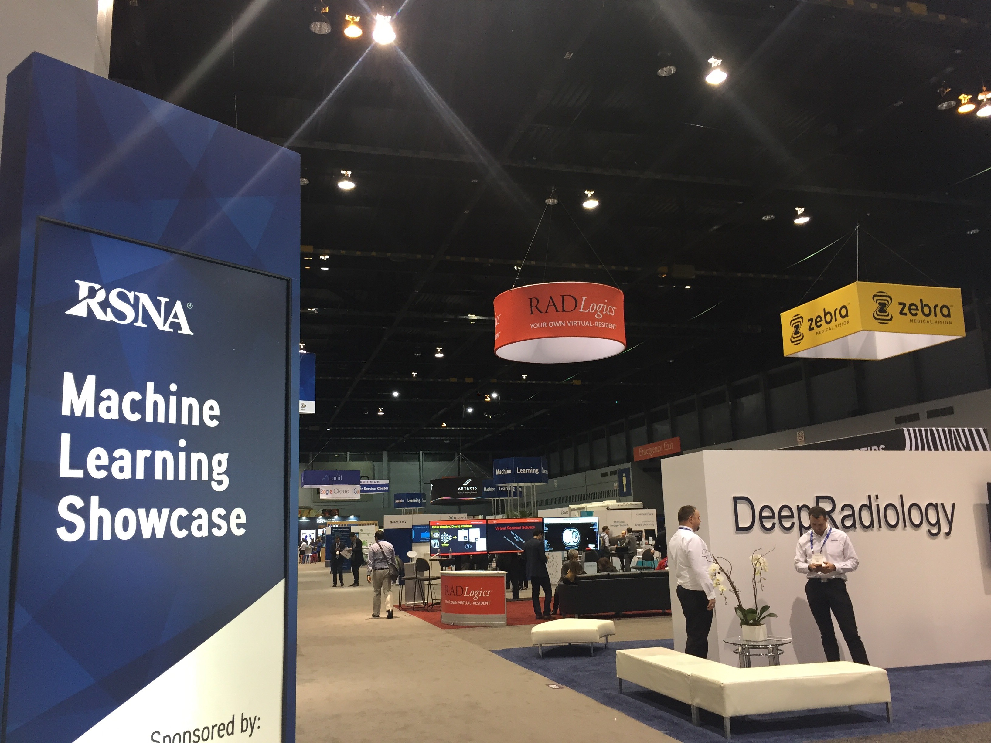
CurveBeam recently had the pleasure of attending and participating in the 103rd Annual Meeting of the Radiological Society of North America (RSNA), held in Chicago from November 26 to December 1. The RSNA Annual Meeting is a unique opportunity for attendees from around the world to gather and meet with thought leaders and innovators to learn about the latest advances in imaging. Post-event, we wanted to share our experiences including some key takeaways.

Rising Injuries in Youth Sports
Diego Jaramillo, MD, MPH, from Miami Children’s Hospital in Miami, delivered a presentation titled “The Perfect Storm for Athletic Injuries: Youth, Growth, and Hormones”. Dr. Jaramillo shared how childhood sports have become increasingly intense and competitive. As young athletes dedicate more time and energy to one specific sport and even specialize in specific functions within that sport, the type of injuries, particularly repetitive injuries, are intensifying. Increased bone porosity of a young skeleton coupled with an increase in muscle strength can stress a skeleton and predispose it to damage. Puberty, including the hormonal influences on the growth plate, also increases the vulnerability of the skeleton to injury. Following x-rays, most young athletes are diagnosed using CT, especially for injuries that involve the head or that are primarily osseous such as the triplane fracture.

Effect of Weight Loss on Knee Cartilage Degeneration
Alexandra S. Gersing, MD, from the University of California, San Francisco, shared the results of a study on “How Weight is Lost Can Slow Knee Cartilage Degeneration”. The study examined which types of weight loss are most beneficial for patients who need to lose a significant amount of weight to slow the progression of knee cartilage degeneration. The study looked at 760 male and female patients with a body mass index greater than 25 who were enrolled in the Osteoarthritis Initiative (OAI), a U.S.-based study focused on the prevention and treatment of knee osteoarthritis. Patients were categorized into groups according to the amount of weight they were asked to lose over a 48-month period, as well as the weight loss method. Changes in the right knee were assessed at baseline, 48-, and 96-months using 3T MRI. Patients who lost weight showed significantly less T2-value increase in the bone layer of all compartments compared to those with stable weight, suggesting less cartilage degeneration over 96 months.

Patients Prefer Immediate Test Results
Radiology resident David Mihal, MD, of the University of Cincinnati College of Medicine and the Cincinnati Children’s Hospital (CCH), shared a “Survey that Showed Patients Prefer to Get Immediate Test Results”. Dr. Mihal began the four-phase study after learning that 20 percent of patients were uncertain about how they were going to get their results and had expressed anxiety to the technologist doing the exam. In phase one, techs screened outpatients to identify those who were nervous about their results. In phase two, the front desk staff checked for patients who didn’t have a follow-up appointment and offered them the option of immediate results. In phase three, all radiography outpatients were offered results as part of a questionnaire at check-in after a 10 to 20 minute wait. In phase four, the wait-time notification was eliminated from the questionnaire. Each successive phase tripled the rate at which patients used the service. Perhaps more important, 97 percent of patients understood that immediate results were available, and 92 percent of patient comments on the availability of the service were positive.

CurveBeam designs and manufactures Cone Beam CT imaging equipment specifically designed for the orthopedic and podiatric specialties, including the pedCAT, a compact, ultra-low dose CT imaging system. This technology allows doctors to make a better diagnosis the first time, eliminating the need for additional scans and, therefore, reducing low-level radiology exposure to patients. Best of all, the practice has access to the results right away. CurveBeam is proud to have participated in the 2017 RSNA Annual Meeting and is looking forward to RSNA 2018!

To learn more, visit CurveBeam.com today.
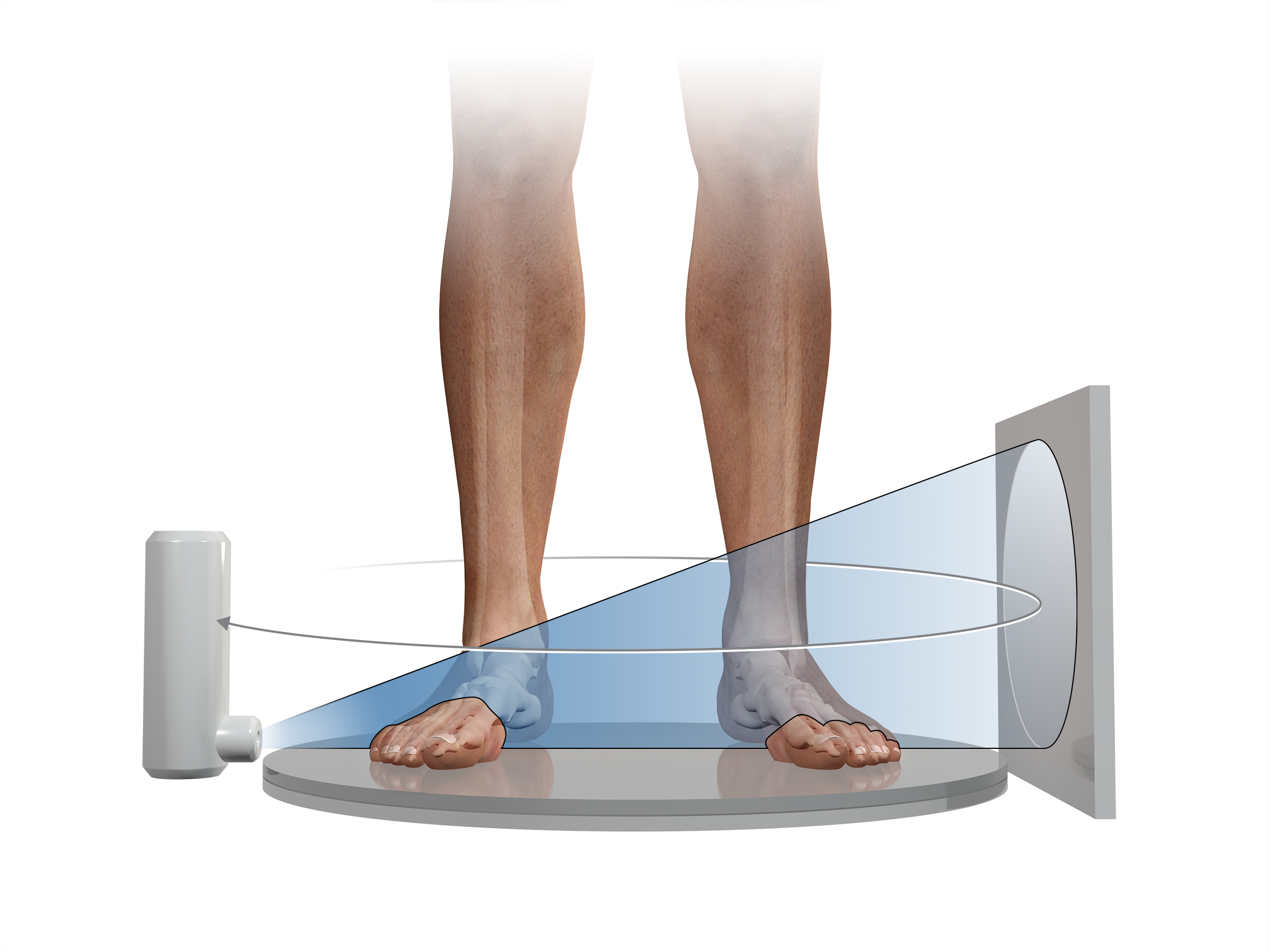
CurveBeam Systems Offered on the Stratum Med GPO to Save Practices Money

CurveBeam, the designer and manufacturer of Cone Beam CT imaging equipment for orthopedic and podiatric specialties, is pleased to announce a new partnership with Stratum Med, a collaborative alliance of physician-owned and health system-employed physician groups. Stratum leverages the collective “Groups” consolidated purchasing power to achieve higher discounts and reduce costs. CurveBeam systems are now offered through an exclusive contract with Stratum Med. Stratum saves its shareholders and clients money on their medical/surgical supply, equipment, laboratory, pharmacy and office supply purchases.

Stratum Med, Inc. is a physician owned and governed  company, founded in 1996 headquartered in Urbana, Illinois with relationships with 60+ physician groups and integrated health systems across the country  consisting of over 10,000 physicians. Stratum strives

to advance the quality of care, improve financial performance and optimize operational efficiency by using a  collaborative alliance model to leverage the collective groups’ consolidated purchasing power and yet preserve independent purchasing practices. At the same time, its dedicated support teams ensure consistent, first-class service. This model also provides a “watch dog” to ensure pricing integrity and consistency based on contract terms, as well as networking opportunities where materials managers, lab directors, pharmacy directors and imaging directors can challenge and learn from one another.

Stratum has achieved savings ranging from 8-20% and has developed a non-acute, integrated purchasing program providing Stratum Shareholders and Clients access to pricing, services, and features typically available only to large hospitals and healthcare systems.

CurveBeam is proud to be a part of such a useful purchasing model and looks forward to the opportunities this new partnership will provide. To learn more about the benefits of purchasing CurveBeam imaging machines for your practice, visit curvebeam.com/products/pedcat/ today.

RSNA Daily Dispatch – A New Focus on Machine Learning

For the first time ever, the technical exhibits halls at the RSNA Annual Meeting include a machine learning showcase, where a variety of researchers and industry ventures have the opportunity to share cutting edge work in artificial intelligence and deep learning in healthcare and radiology.

rsna 3

One company collected 9 million radiology data sets over three years to train computers to automatically recognize and diagnose a wide range of injuries and pathologies. Other research involved using parameters of patients’ demographic information and medical history records  to predict a prognosis. From managing massive amounts of patient data to quickly assessing complex medical images, machine learning can assist radiologists in their current tasks and allow for a level of planning and diagnosis that would not be possible without the trove of information stored in big data.

RSNA Daily Dispatch

The Monday, Nov. 27 schedule of the 2017 RSNA Annual Meeting included a variety of scientific sessions discussing technical and clinical developments in Cone Beam CT systems. From improving image quality to correlating with other modalities, researchers are validating the efficacy of CBCT in diagnosis and treatment of musculoskeletal injuries and pathologies.

@Ingakoerte tweeted a photo of the crowds at the RSNA Annual Meeting.

In a poster presentation for “High Resolution Extremity Cone-Beam CT with a CMOS X-Ray Detector: System Design and Applications in Quantitative Assessment of Bone Health” Biomedical Engineer Qian Cao evaluated the ideal Cesium Iodide scintillator thickness to visualize trabecular bone detail for applications such as early detection of osteoarthritis. He compared image quality characteristics of an optimized CMOS detector to an amorphous silicon detector and a micro CT scanner. The optimized CMOS detector had superior trabecular detail compared to the Amorphous Silicon detector and comparable detail to the Micro-CT, with the advantage of a much larger field of view than Micro-CT.

In “Effect of Motion Compensation on the Image Quality of Cone Beam CT Scans in Musculoskeletal Setting” Guarav K. Thawait, MD, research associate at Johns Hopkins shared the results of a study where involuntary patient motion in CBCT scans was corrected using an iterative reconstruction algorithm. The algorithm improved motion artefacts significantly in bone structures.

In “Evaluation of Bone Erosions in Rheumatoid Arthritis Patients using CBCT and MRI,” Dr. Thawait discussed a study in which a radiologist reviewed CBCT, MRI, and Ultrasound datasets for signs of rheumatoid arthritis. The correlation between CBCT and MRI was moderate while the correlation between CBCT and US was a bit higher. Though the correlations were only moderate to good between modalities, test-retest reproducibility for CBCT scans was excellent and the modality shows promise as a useful tool for RA diagnostic evaluation.

RSNA Daily Dispatch

The Radiological Society of North America (RSNA) commenced its 103rd Annual Meeting in Chicago on Sunday, Nov. 26, 2017.

@judywawira tweeted a photo of  a standing room only session at the RSNA Annual Meeting on Nov. 26, 2017.
@judywawira tweeted a photo of a standing room only session at the RSNA Annual Meeting on Nov. 26, 2017.

Attendees from around the world gathered to learn about the latest advances in imaging.

In an afternoon session titled “Open Gantry Systems: Advances, Challenges, and New Applications,” Dr. Jeffrey H. Siewerdsen, PhD, of Johns Hopkins Biomedical Engineering, discussed the vast applications of cone beam CT for interventional and diagnostic imaging. The devices presented included C-arms for image guided surgery, Cone Beam CT guided radiation therapy systems, and diagnostic imaging devices for breasts, dental/ENT, and orthopedic extremities.  Siewerdsen emphasized the  revolutionary potential of weight-bearing extremity devices in orthopedic specialties, and mentioned the CurveBeam pedCAT and LineUP systems.  He also discussed the image quality parameters of Cone Beam CT and artifacts specific to this type of imaging. He offered suggestions on how to quantify image performance of such systems, many of which are relatively new and do not have established quality metrics in the imaging community. Finally, he presented some of the newest Cone Beam CT devices to come to market, including the extremely compact CurveBeam InReach made primarily for hand and wrist imaging.