Webinar Summary: Advanced CT Imaging in Foot and Ankle Surgical Considerations

We all know that X-rays and MRIs aren’t the be-all, end-all of diagnostic imagery.

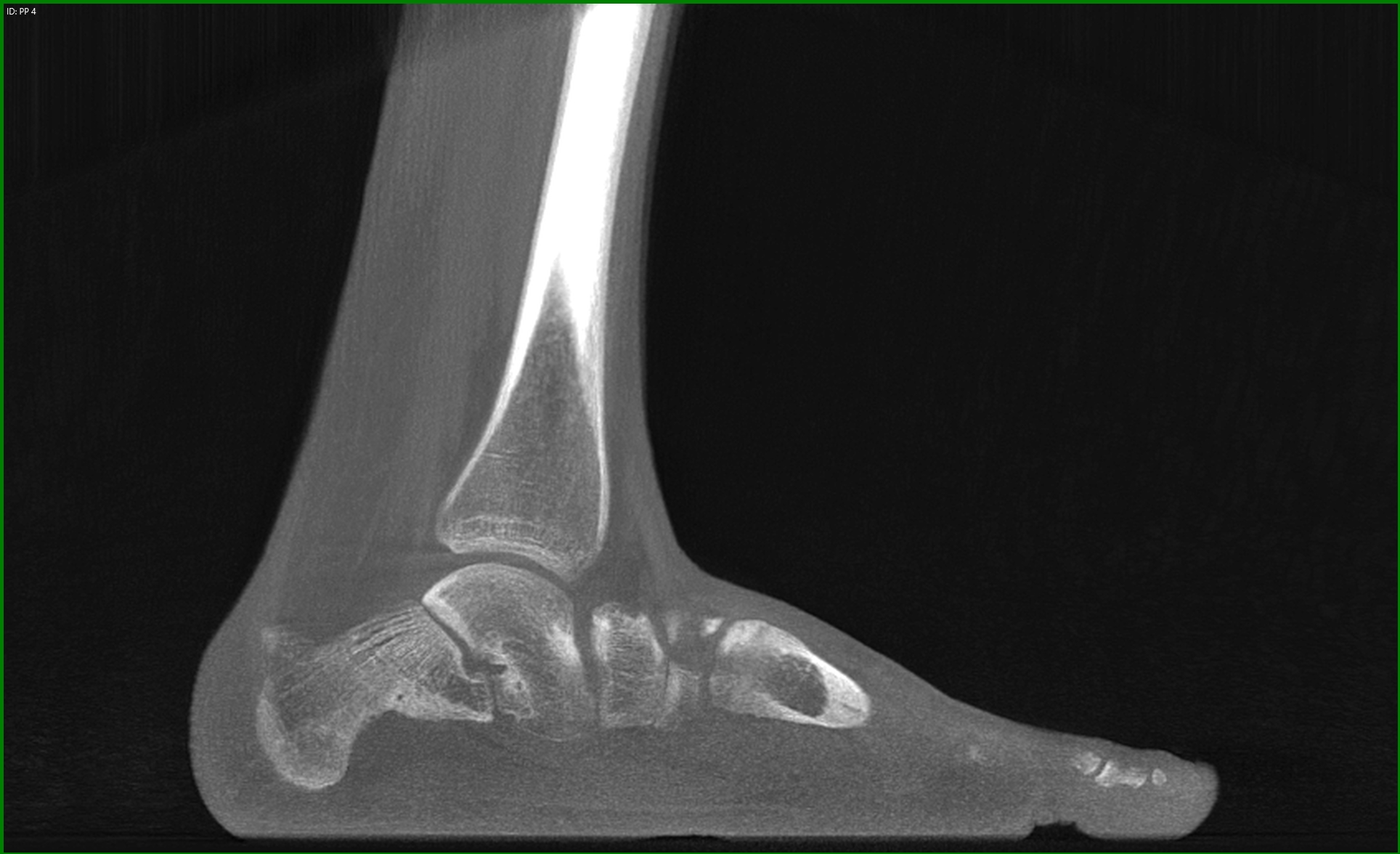
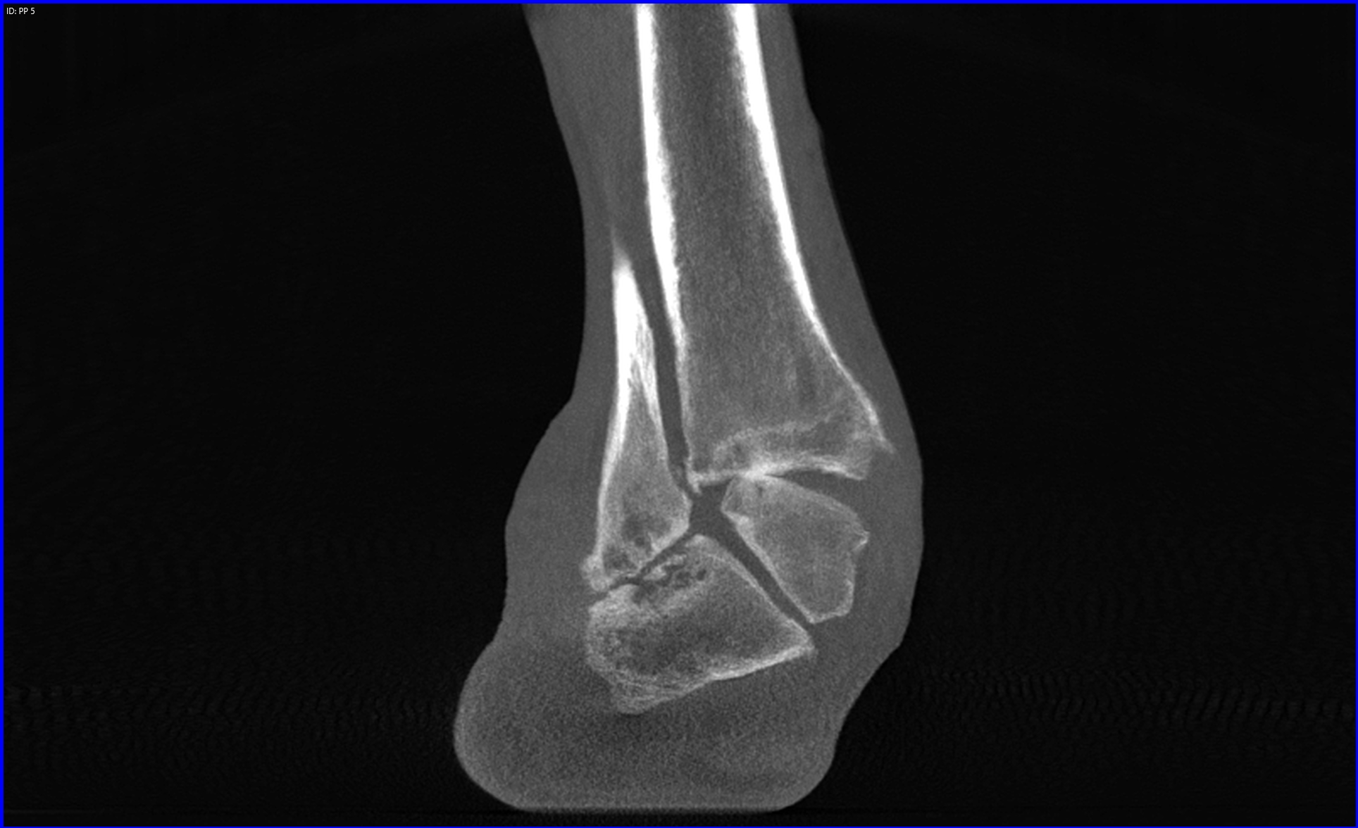
Try determining the frontal plane rotation of the sesamoid and first metatarsal with an X-ray. It isn’t possible. Correction of the hallux valgus rotation in bunion surgery depends largely on the repositioning of the sesamoidal apparatus which is impossible to assess without an axial view, and X-rays fall completely short when it comes to assessing these relational details from the vantage point of a single plane.

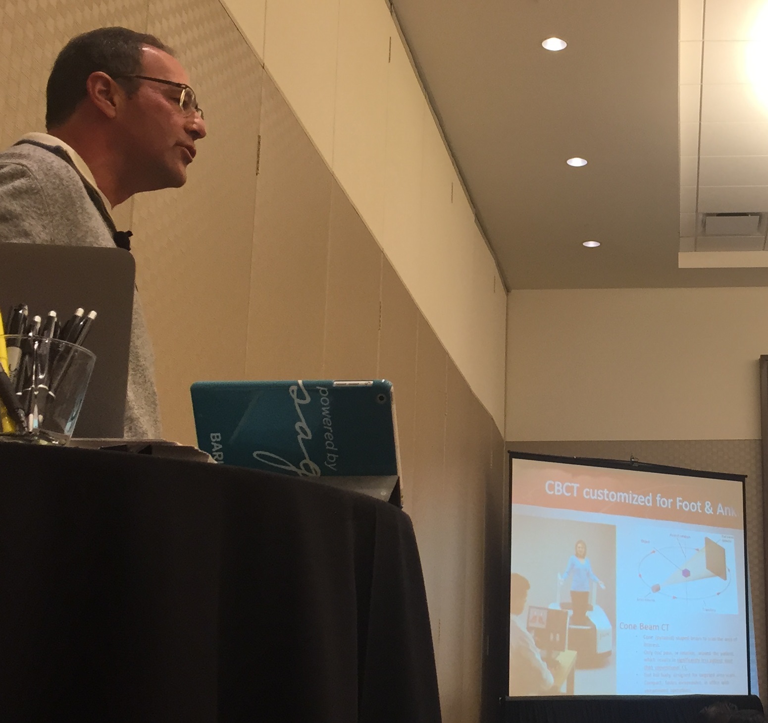
Such was the topic of discussion during the latest Curvebeam webinar led by Dr. Bob Baravarian, Director of University Foot and Ankle Institute in Southern California. He explained how weight-bearing 3-dimensional CT scans are changing the game of podiatric diagnostics.

Throughout the webinar, titled “Advanced CT Imaging in Foot and Ankle Surgical Considerations,” Dr. Baravarian offered a very straightforward presentation of the often-not-so-straightforward complexities of foot and ankle deformities and how advanced imaging technology can improve both surgical planning and surgical outcomes. He cited the example of hallux valgus among many others as “impossible to imagine treating without 3-D imaging technology” these days – given the results he’s seen with his patients and scope webinar 3
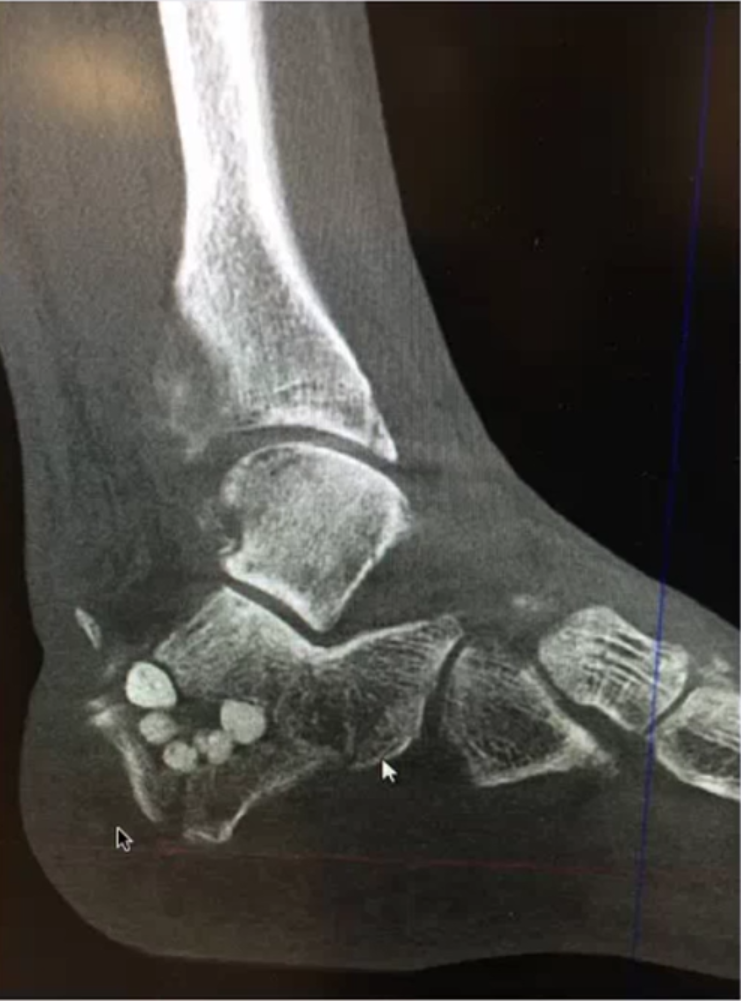
of its applications.

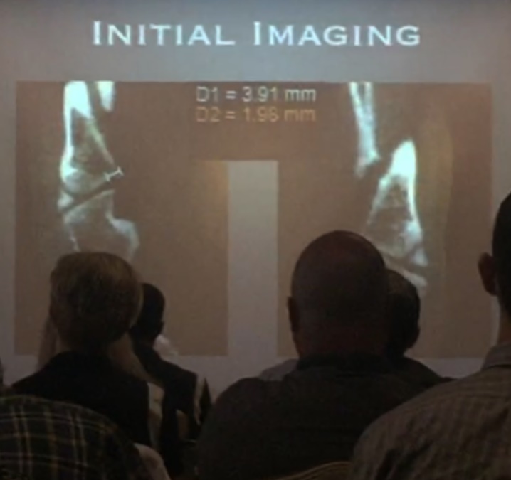
Planning the frontal plane correction of first metatarsal.

“CT scans are very helpful in planning your frontal plane deformity correction of the first metatarsal to get an anatomic position which is really underestimated,” said Dr. Baravarian.

Multi-plane imaging now allows physicians to correct the frontal plane deformity of the metatarsal while simultaneously realigning the sesamoid. The capability is “critical,” said Dr. Baravarian, “for proper outcome with bunion corrections whether you’re doing a LAP or any kind of osteotomy.”

Identifying the cause of hypermobility of the first ray

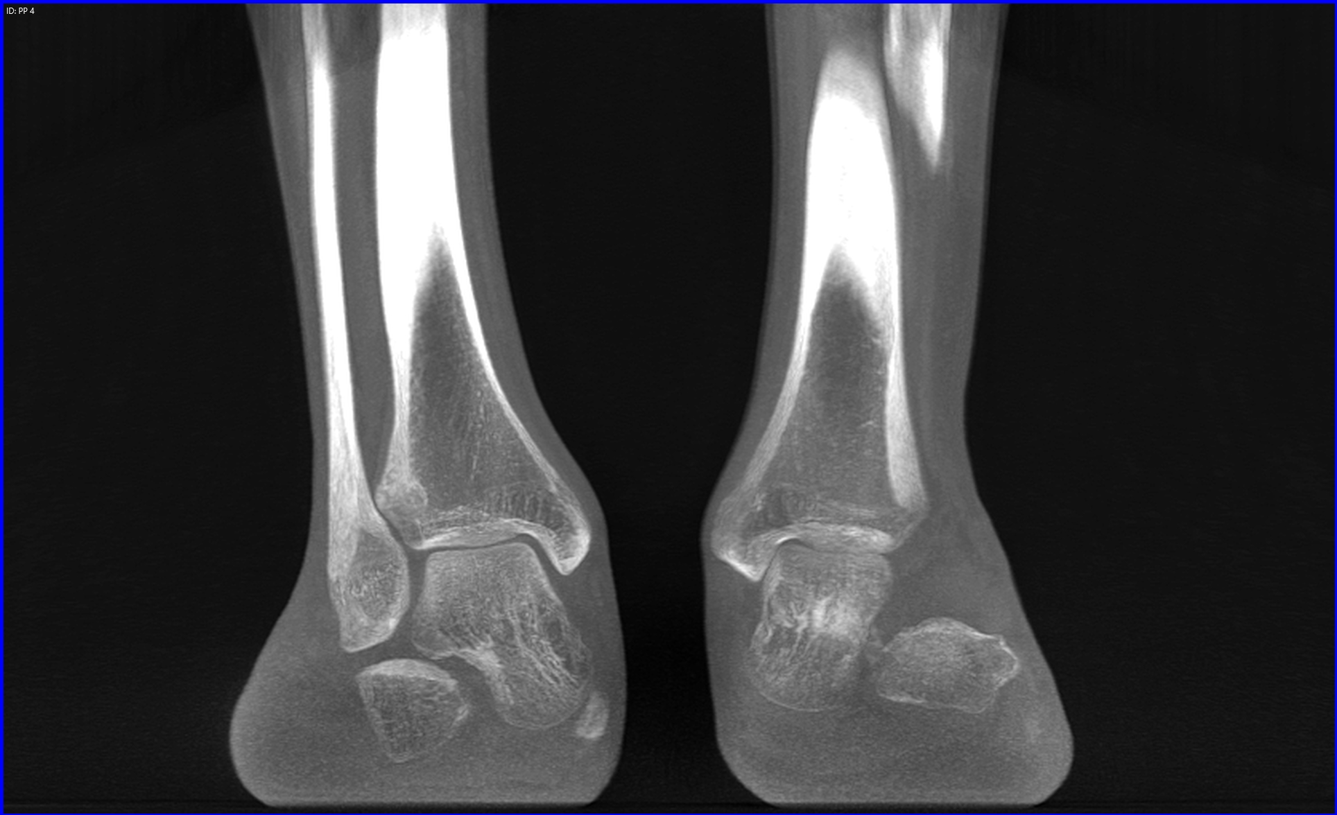
When you look at a patient who has a significant flat foot deformity and a significant bunion deformity with some level of hyper-mobility of the first ray, 3D CT imaging allows you to locate the exact area in need of correction.

“In patients who have PTTD or even a pediatric flat foot case we really need to decide which planes of correction make the best sense,” said Dr. Baravarian.  “If I correct my first metatarsal, is my hind foot going to realign or is there an outstanding deformity that needs correction?”

webinar capture
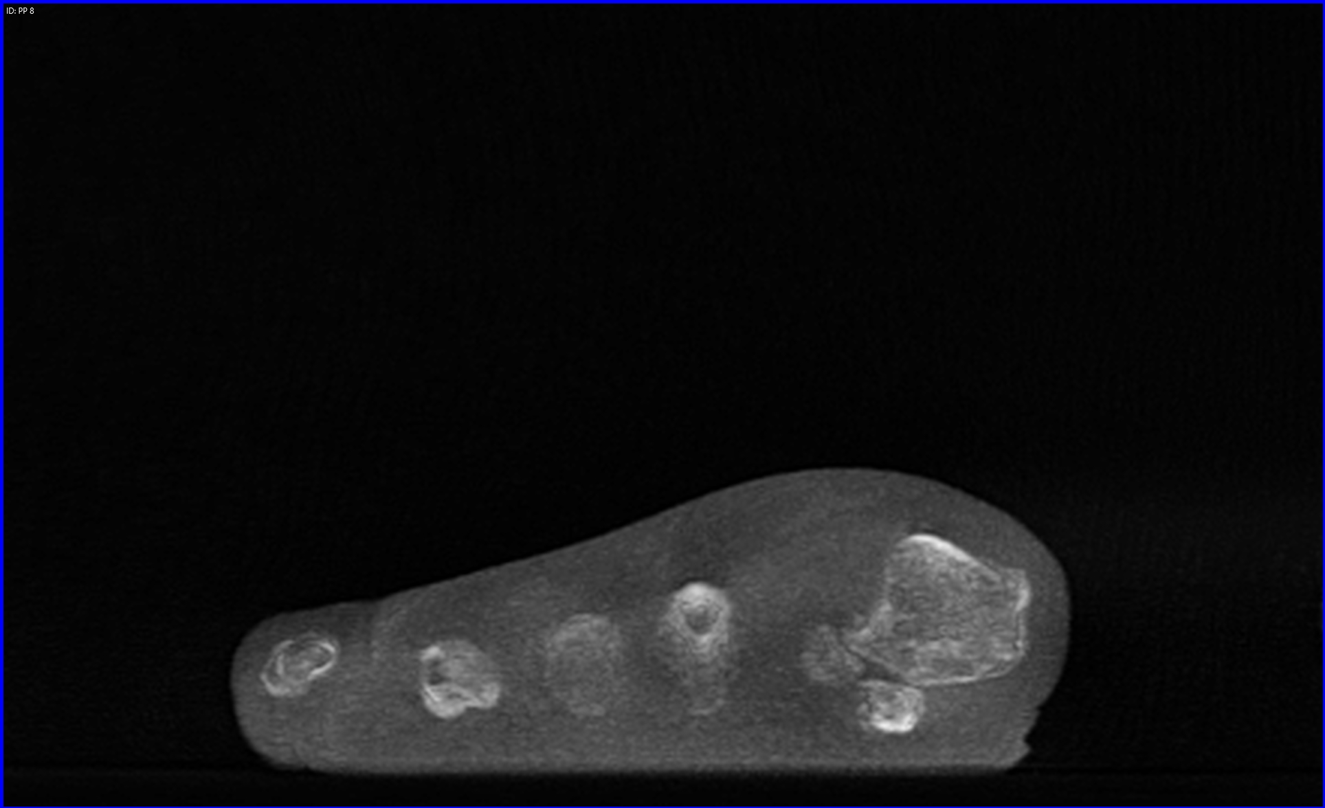
Determining the level of arthritis in hallux rigidus and limitus cases. 

“I’m constantly surprised when I go into surgery and I plan a cheilectomy and I open up a joint and there’s significantly more arthritic changes or some kind of osteochondral legion that I couldn’t really see on the X-ray. Or, I plan to do an osteotomy and I go in and the level of arthritis is not as bad as expected,” said Dr. Baravarian.

It’s very difficult to determine the level of arthritis in a hallux rigidus or limitus case based on a radiograph alone. While MRIs are an excellent option for soft tissue imaging and diagnostic ultrasounds still provide accurate, real-time guidance for proper injections –neither of these methods makes sense for treating anatomic alignment and assessing structural deformities.


“With a 3D CT I’m able to look a little more in-depth into the joint and make a better decision prior to surgery.”

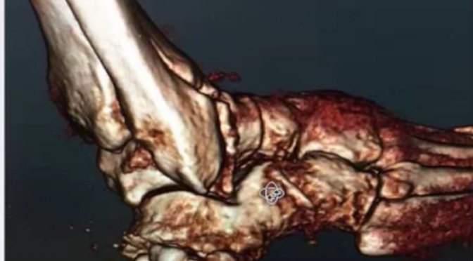
Identifying major deformities in complex fractures

Weight-bearing imaging systems allow you to place a foot in its anatomic position and adjust its deformity to see what level of correction you can get across multiple planes.

It “doesn’t makes sense,” according to Dr. Baravarian, “to plan the correction of a complex fracture without a sense of what’s going on inside the foot and ankle.”

Our traditional method of two-dimensional x-rays doesn’t provide the highest level of certainty that doctors need in order to administer the best care possible to their patients where deformity planning is essential for proper surgical outcomes.

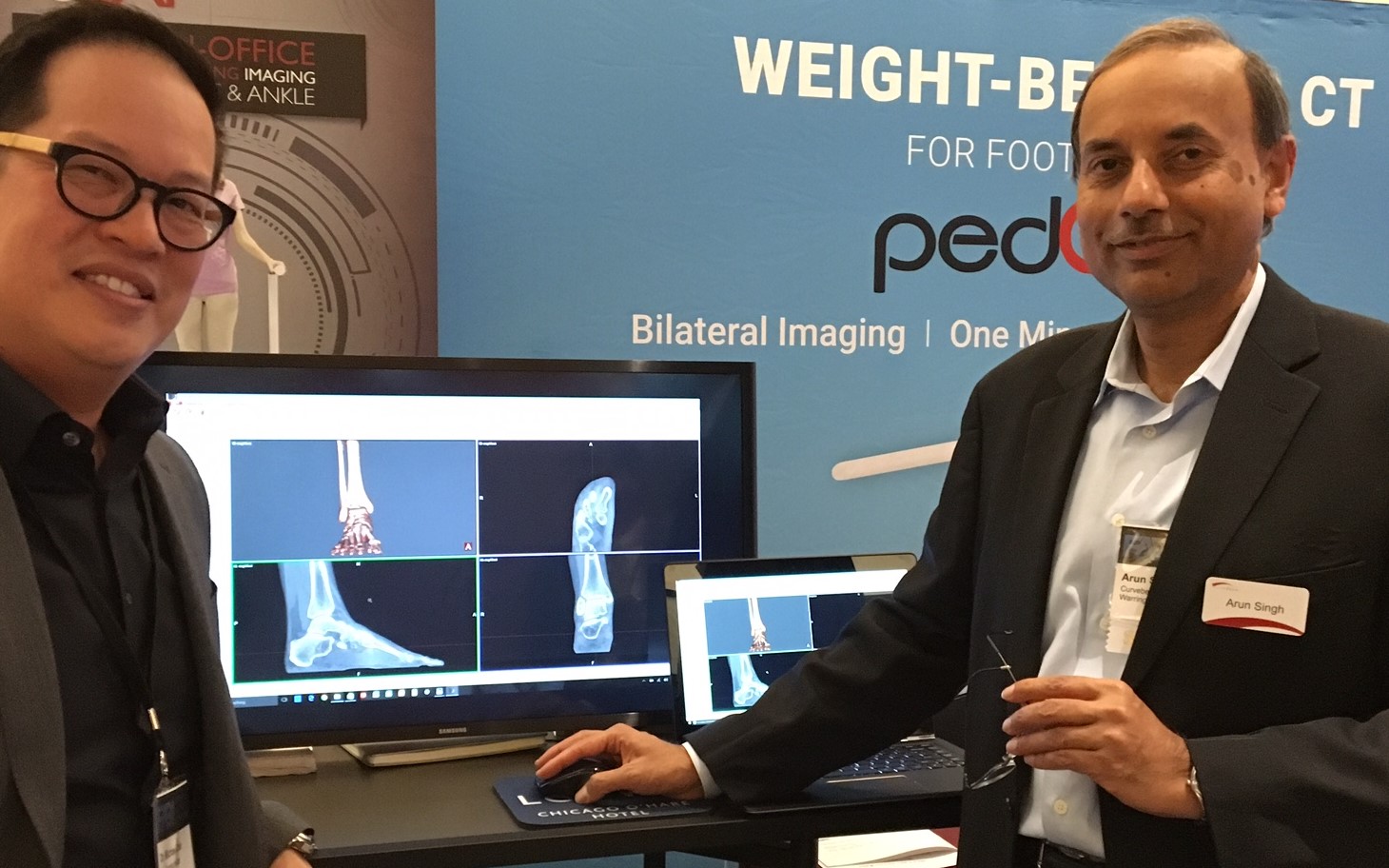
Now, with the help of tools such as the pedCAT, a compact 3D weight-bearing CT imaging system, podiatrists have everything they need to create comprehensive treatment plans and more effective surgeries. Better outcomes. Less risk. And patients back on their feet faster than they ever expected.

You can access the entire webinar here. A FOOT Innovate membership is required to access the content. Membership is complimentary for foot & ankle specialists.

Exclusive Video From The First pedCAT Weight Bearing CT Scientific Users’ Meeting in Berlin

meeting-83519_960_720 (1)The first pedCAT Weight Bearing CT Scientific User’s Meeting recently convened at the 2016 Foot International (EFAS, DAF, I-FAB) in Berlin. The meeting focused on weight bearing cone beam CT (WBCT) technology and the potential applications and challenges it presents.

Fill out this form to access the video.

Look for several interesting items as you watch:

  • Professor Martinus Richter’s work included a time workflow study on the use of X-Ray,  conventional medical CT, and the pedCAT; the results will surprise you. His study also showed how weight-bearing  CT measurements are more accurate than X-Ray measurements.
  • Dr. Arne Burssens’ study on hindfoot alignment answered critical questions: How do we identify varus and valgus, how do we accurately measure them, and how can we be sure that the measurements are clinically useful and reproducible?
  • Dr. Francois Lintz gives us a sneak peek at a new weight-bearing measurement CT tool that will be available exclusively in CubeVue, pedCAT’s software. It will enable users to measure alignment in three dimensions, and distinguish between normal and pathological cases.
  • Dr. Cesar Netto’s study focused on adult acquired flatfoot deformity, comparing  measurements on weight bearing CT vs non-weight bearing CT.
  • Dr. Michael Wachowsky’s study focused on the use of weight bearing CT  with pediatric patients, and asks: With the new weight bearing CT technology, what will be the exact definitions of the measurements?

The challenge created by this new technology is clear: How do we now define what we are measuring, since the current X-Ray methods have just been rendered obsolete?

“We can’t have non-weight bearing CTs and say anything about alignment – that’s over,” concluded Dr. Charles Saltzman. “What do we want to measure, and how do we know what we’re measuring represents reality?”

Fill out this form to access the video.

Panel Recap: Dr. Michael Chin, DPM, Speaks about pedCAT at FABI

Dr. Michael Chin, DPM, and Arun Singh, President & CEO of CurveBeam

His decade old X-Ray system was failing, and feeling like a warrior without his weapon, Dr. Michael Chin, DPM, knew it needed to be replaced fast.

“We could have gotten a DR system, but at the end of the day, I wanted something that was unique, and something that would change the way I practice,” Dr. Chin said.

Dr. Chin participated in a panel discussion about new technology at the Foot and Ankle Business Innovations meeting in Chicago on Jan. 30. Dr. Chin practices at The Running Institute in downtown Chicago.

Dr. Michael Chin, DPM, at FABI

Dr. Chin uses the pedCAT for all of his X-Ray and CT imaging. His X-Ray revenues cover the device’s monthly capital lease payment, and the approximately 20 CTs he and his associate order every month provide his practice with an additional revenue stream.

Dr. Chin said he is able to order a CT scan and his staff can get payer authorization in the same day. This saves his patients from having to come in for a follow-up visit.

“We can fill that slot that we would have used for a follow up with another patient,” Dr. Chin said.

Weight Bearing CT Imaging for Cuboid Subluxations

Dr. Michael Chin, DPM, presented how weight bearing CT imaging has changed how he evaluates cuboid subluxations at the American Academy of Podiatric Sports Medicine meeting held near the West Point Military Academy campus in early September, 2015.

Dr. Chin began using the pedCAT in his office in February of this year.

Not much research is out there on how to use plain radiographs to measure cuboid subluxations, Dr. Chin said in his lecture, titled, “Cuboid Syndrome…The Other Side of Heel Pain.”

Dr. Chin has tested using a bilateral oblique projection to understand the cuboid/ metatarsal relationship, and has been able to observe a slide between the head of the fourth metatarsal and the head of the cuboid.

An MRI could be ordered to see the condition of the peroneal tendon, but the study would be limited because the scan would not be weight bearing, he said.. A traditional CT scan would provide great  visualization of the bone, but would provide no information on anatomic alignment.

The pedCAT weight bearing CT imaging system is excellent for evaluating stress fractures, sesamoids, periosteal changes, or anything medullar, Dr. Chin said. Another benefit is he can measure the exact degree of subluxation between the cuboid and the fourth metatarsal head.

Dr. Chin displayed pedCAT images depicting  pre and post-reduction views of a cuboid subluxation.

chin-lecture
pedCAT scan of a pre-surgical patient with cuboid subluxation. Dr. Chin was able to reduce the subluxation to 2.18 mm.

Dr. Chin practices at The Running Institute in Chicago.

AOFAS Annual Meeting – CurveBeam Symposium Recap

Weight bearing CT is a vital tool for determining the cause of inexplicable pain, and also for avoiding painful surgical complications.

That was the takeaway message from a talk by Dr. Phinit Phisitkul, a clinical associate professor of orthopaedics at the University of Iowa. He shared some of his most interesting cases at a CurveBeam sponsored symposium held during the AOFAS Annual Meeting in Long Beach during the evening session.

We’ve selected three of his cases to share on this blog:

18-year-old male with Noonan Syndrome & severe flat foot: The patient presented with an unusual amount of pain that was difficult to diagnose on plain X-Ray. A weight bearing CT scan revealed he had a severe deformity – a congenital vertical talus. He also had severe impingement.

Vertical Talus - Weight Bearing CT
Vertical Talus – Weight Bearing CT
Impingement - weight bearing CT
Impingement – weight bearing CT

58 year-old male with ankle arthritis: The patient presented with a lot of pain in the ankle joint. A weight bearing CT scan showed a subluxation of the ankle joint and dramatic impingement of the calcaneal fibula. Interestingly, the subtalar joint was in pristine condition. Dr. Phisitkul determined the patient was a good candidate for ankle replacement and hindfoot realignment, and that his subtalar joint could be spared.

Calcaneal-fibular impingement and arthritis - Weight Bearing CT
Calcaneal-fibular impingement and arthritis – Weight Bearing CT

41-year-old female with Hallux Valgus: A weight bearing CT scan revealed a bone spur on the patient’s first metatarsal head. If the doctor had done a normal release, the spur may have ended up pinching the sesamoid. Instead, he performed a lateral release and excised the bone spur.

1st MT Bone Spur - Weight Bearing CT
1st MT Bone Spur – Weight Bearing CT


pedCAT: A Positive User Experience


Baravarian Western

“After using the pedCAT for a year, I am very impressed with how much I need it, and how many different things I use it for,” Dr. Bob Baravarian said at the 2015 Western Foot and Ankle Conference held in Anaheim, Cali. The California Podiatric Medical Association hosted the meeting at the end of June.

Dr. Baravarian cited Hallux Rigidus as one example where pedCAT imaging has been helpful because he is able to properly assess the sesamoids.

Dr. Baravarian confirmed his practice is realizing positive revenue streams as a result of the device.

Dr. Gary Briskin: The pedCAT is the Future

“It’s the future – embrace it. It’s your future – control it.”

Dr. Briskin lectures on weight bearing CT imaging for foot 7 ankle

Dr. Gary Briskin delivered this motivating message at the Foot & Ankle Business Innovations conference in Chicago on Jan. 23.

Dr. Briskin, of University Foot & Ankle Institute in Santa Monica, Cali,  discussed how the pedCAT weight bearing CT system has improved his practice since his group acquired one last year.

His main take-away points were:

– The pedCAT gives you the advantage when it comes to diagnosing pathology.

“You can solve that Lisfranc case no one else can,” Dr. Briskin said. “It opens up the midfoot, which historically has been a problem for us.”

– The pedCAT allows him to clearly determine if bones and joints are completely fused after surgery.

“I do a lot of lapidus procedures,” Dr. Briskin said. “My concern; is this patient fused adequately to start bearing weight?” The exact level of fusion is visible with a pedCAT scan 5 – 6 weeks later, and with minimal scatter.

– Radiologists like the pedCAT images

“I think I’m getting a better CT, because it’s weight bearing and I think the quality is far superior. We get all of our CTs read by a radiologist. We have everything sent digitally. And the feedback we get is they are also impressed with the quality of the images.”

Webinar: Dr. Erik Nilssen – A New Paradigm in Foot & Ankle Diagnosis

FAI webinar

“Patients come to my practice with an expectation of a higher level of care, that they’re getting the best. With the pedCAT, I am able to give them information immediately, and they leave feeling this was well worth the trip.” – Dr. Erik Nilssen

Learn how the pedCAT has changed Dr. Nilssen’s clinical practice in a webinar hosted by Foot & Ankle International and Foot & Ankle Specialist.

We hope you can join us! Sign up here.

CurveBeam Reception at AOFAS: You’re Invited!

If you’re attending the AOFAS/IFFAS Annual Meeting 2014 in Chicago from September 19 – 23, be sure to plan to attend the CurveBeam reception on September 21 at 7:30 p.m.

Sip cocktails and dine on hors d’oeuvres while mingling with colleagues who have integrated the pedCAT into their practices. Meet like-minded forward thinking peers who are considering a pedCAT acquisition at their facilities.

This informal event will feature brief talks by Prof. Dr. med. Martinus Richter, who has published studies on the pedCAT’s efficacy, as well as Dr. Erik Nilssen, MD and Dr. Martin O’Malley, MD.

When: Sept. 21, 7: 30 p.m.

Where: Hyatt Regency Wrigley’s Room (Bronze Level, West Tower)

If you can’t attend the seminar, you can visit us at booth #307 all conference long.