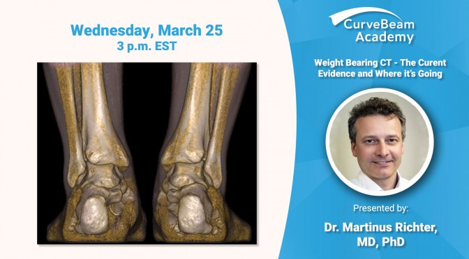
Register today to participate in a virtual lecture delivered by Dr. Martinus Richter, MD, PhD, of Krankenhaus Rummelsberg, who will review the current scientific evidence supporting Weight Bearing CT imaging and future directions for innovation.
The webinar will be held on Wednesday, March 25 at 3 p.m. EST.
Click here to read more about a study conducted by Dr. Richter that found WBCT imaging saved 800+ hours of imaging time a year at his hospital.
View the rest of the sessions in CurveBeam Academy’s Virtual Conference here.
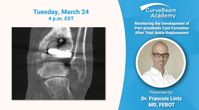
Register today to participate in a virtual lecture delivered by Dr. Francois Lintz, MD, FEBOT, of Clinique de L’Union. Dr. Lintz will discuss utilizing weight bearing CT imaging post-operatively to monitor the progression of cystic formation after total ankle replacement.
The webinar will be held on Tuesday, March 24 at 4 p.m. EST.
Click here to read more about Dr. Lintz’ award winning research on the effect of residual malignment on cyst formation.
View the rest of the sessions in CurveBeam Academy’s Virtual Conference here.
The WBCT Society presented another Open Scientific Meeting on September 13, 2019 at the AOFAS Conference in Chicago. Watch the highlight reel from the meeting above to see some of the key moments or read the summary below!
The WBCT Society is an independent research organization. CurveBeam is a proud sponsor of the Society.
New Leadership:The WBCT Society presented Dr. Martinus Richter, MD, PhD, a plaque for serving as founding president. Dr. Richter’s term ended at the meeting, and incoming president Dr. Alexej Barg, MD, assumed the role.
Semi-Automatic 3D Hindfoot Measurements: Dr. Alessio Bernasconi discussed a study that demonstrated semi-automatic 3D hindfoot alignment measurements were reliable when measuring alignment on pes cavo-varus cases.
New Research Protocols: Dr. Alexandre Godoy, MD, demonstrated how the University of Sao Paulo in Brazil has designed three new research protocols for its CurveBeam LineUP weight bearing CT system. These include:
Cavus varus deformity: perform a Coleman block test clinically and during weight bearing CT to determine if the Coleman block test changes alignment measurements.
Hallux valgus deformity: use weight bearing CT to assess 1st TMT gapping, collapse of medium column, and 1st MT and sesamoid rotation.
Congenital club foot: Investigate residual club foot using WBCT to try to understand relationships between tarsal bones involved in the deformity.
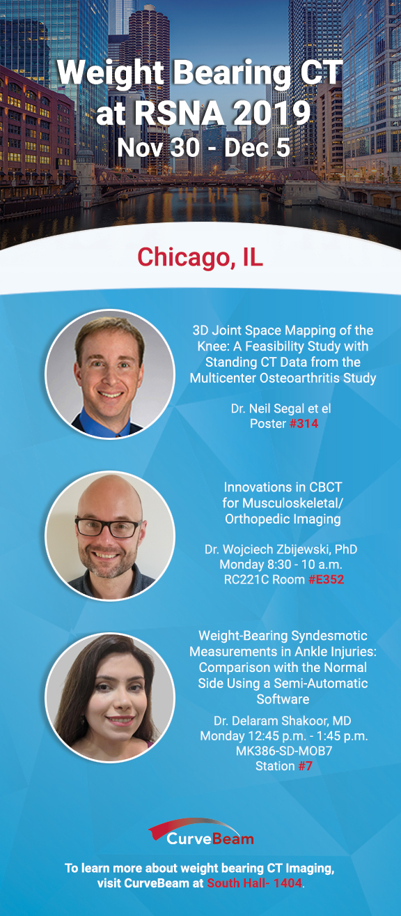
Radiology professionals from around the world will convene in Chicago in December for the RSNA 2019 Annual Meeting. The six-day conference schedule will span a broad range of radiology topics.
If you attend, make sure to visit the posters and presentations above, which detail advancements in weight bearing CT imaging and research.
To learn more about weight bearing CT imaging, visit CurveBeam at South Hall – 1404.
Learn more at the RSNA Annual Meeting at the Innovation Theater on Wednesday, Dec. 4 at 11 a.m. The presentation “Weight Bearing CT: Total Lower Limb Imaging” will be delivered by CurveBeam.
Visit Booth #1404 (South Hall) and be the first to experience CurveBeam’s next generation weight bearing CT system.
This system is investigational only and is not available for sale.
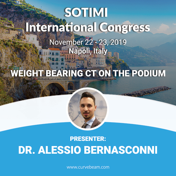
Are you attending the SOTIMI International Congress in Naples, Italy later this month? Be sure to sit in on the following podium presentations on weight bearing CT imaging:
Saturday, Nov. 23
10.00 – 11.00 Comunicazioni: Caviglia E Piede
Misurazioni radiografiche di piede e caviglia nella cone beam weightbearing computer tomography (WBCT) – C. De Franco, V. de Matteo, R. Verrazzo, F. Smeraglia, G. Balati, A. Bernasconi (Naopoli)
Relazione tra instabilita laterale cronica di caviglia e retropiede varo valutato tramite TC cone beam in ortostatismo – A. Bernasconi, F. Lintz, L. Baschet, C. Fernando, N. Mehdi, Weight Bearing CT International Study Group, C. de Cesar Netto (Londra)
Piede cavovaro secondario a Charcot-Marie-Tooth vs piede cavovaro idiopatico: analisi preliminare della morfologia con TC cone beam in ortostatismo – A. Bernasconi, L. Cooper, S. Lyle, S. Patel, D. Singh, N. Cullen, M. Welck (Londra)
Variabiliita’ intra ed inter-osservatore di misurazioni semi-automatiche 3D con TC cone beam in ortostatismo in pacienti affetti da piede cavovaro sintomatico – A. Bernasconi, L. Cooper, S. Lyle, S. Patel, D. Singh, N. Cullen, M. Welck (Londra)
The Weight-Bearing CT Society held its latest Scientific Meeting in Las Vegas this past March during the AAOS 2019 conference. Co-sponsored by CurveBeam, the Scientific Meeting featured an education-packed agenda to discuss the revolutionary transformation weight-bearing technology is having on the imaging industry.
First to present was Dr. Alexej Barg, MD, a University of Utah orthopedic surgeon who also served as the moderator for this event. In his presentation discussing imaging of patients with syndesmosis instability, Dr. Barg explained that conventional non-weight-bearing radiograph imaging cannot predict syndesmotic injuries reliably. And while MRI’s demonstrate sensitivity and specificity of nearly 100%, correlating patient complaints with MRI findings can be difficult. Further, although CT technology surpasses conventional imaging methods when 3D MRI or CT imaging is transferred to 2D, there is a substantial loss of information. However, the torque applied in the natural standing position offered higher contrast and spatial resolution of alignment and degeneration, providing a significant advantage in the accuracy of diagnosing syndesmotic injuries.
Dr. Pablo Wagner, MD, of Clinica Alemana in Chile then took the stage for a presentation titled “WBCT for Assessment of Metatarsal Rotation”. Crediting his brother, Emilio Wagner, MD, for his work with hallux valgus patients, Dr. Wagner explained that metatarsal condyles are visible laterally if pronated, of which 87 percent of hallux valgus cases are. If not corrected, metatarsal pronation will result in worse clinical outcomes and higher deformity relapse rates due to the soft tissue balance lateral to the medical ray axis. However, when compared to challenging, unreliable weight-bearing axial sesamoid views and more useful AP foot weight-bearing views on plan X-Ray, weight-bearing CT scans are the gold standard in quantifying metatarsal rotations.
Following Dr. Wagner, Dr. Cesar de Cesar Netto, MD, Ph.D., discussed “WBCT in Patients with AAFD”. While some symptoms or pain are experienced in patients with Adult-Acquired Flatfoot Deformity, it can be difficult knowing when to be more aggressive in treatment to prevent foot collapse. In a study of 55 male and female patients with stage II AAFD, multiple weight bearing CBCT and MRI variables related to the severity of the deformity were evaluated. Weight-bearing CBCT was found to provide more reliability in predicting patients at high risk for foot collapse.
Dr. Arne Burssens, MD, of the Univesity Hospital of Ghent then deliberated on the “Hidden Aspects of a Medializing Calcaneus Osteotomy Revealed by a WBCT”. Difficult to assess via 2D plain radiographic technology, there is a significant difference when assessing MCO in AAFD using weight-bearing imaging versus non-weight-bearing, offering substantial detail to improve understanding with a higher rate of reliability.
CurveBeam will be on hand at AAOE 2019, exhibiting our innovative imaging solutions for orthopedic specialties and subspecialties in Booth #629.
AAOE provides advocacy, networking and business development for the orthopedic and musculoskeletal healthcare professions. To further promote quality healthcare practice management in the industry, each year the AAOE hosts a conference, gathering orthopedic practice professionals from around the country in one venue to learn new practice management techniques and policies, compare new products and services, discuss changes in healthcare and other issues affecting them each day, and more.
Proud to be a field pioneer, CurveBeam’s design and manufacture of advanced 3D imaging technologies have been setting new standards in orthopedic and podiatric 3D imaging since the company’s founding in 2009. Industry-leading innovation, CurveBeam’s imaging systems utilize progressive Cone Beam CT capabilities to provide cutting-edge imaging at a fraction of the cost of traditional CT equipment.
While at AAOE 2019, stop by Booth #629 and let the CurveBeam team guide you through the benefits of our trailblazing solutions that can positively support the imaging needs of your practice and patients.