Distinguishing Categories of Medial Column Ligamentous Fracture

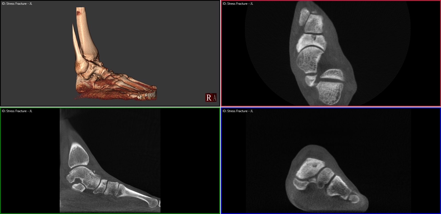
Weight bearing CT scans offer new, three-dimensional perspectives that reveal a better understanding of the biomechanics of flat foot. That was a finding on an academic poster on display at The American Orthotic and Prosthetic Association’s National Assembly in Las Vegas (Sept. 4 – 9).

“IRD, PTTD, Posterior Tib, Flat Foot, Pronation…these are all terms that various professions use to describe some sort of medial column ligamentous breakdown,” according to the poster. “The purpose of this study is to better distinguish some types of pathomechanics of the foot. By categorizing and redefining them, it makes the description of pathology more useful for Orthotic Treatment. This was possible through the use of 3D rendering of the pedCAT machine and CurveBeam software.”

The study was conducted by Ian Engelman, M.S. CPO, and Harold Chamberlin, DPM. They looked at 3D renderings of the bony structures in flat feet, and came up with some new descriptions, as well as their possible ligamentous causes.

For example:

Lisfranc Breakdown
Lisfranc Breakdown – Tarsometatarsal Ligamentous Failure
Lateral Foot Dislocation - Deltoid Ligament Fracture
Lateral Foot Dislocation – Deltoid Ligament Fracture


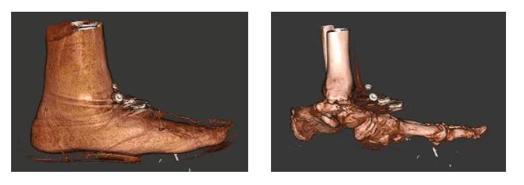
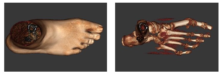
The study concluded that while the exterior contours of the foot suggest certain bony pathologies, the 3D renderings provided by the pedCAT would allow for orthotists to better distinguish the pathomechanics of the foot and ankle.

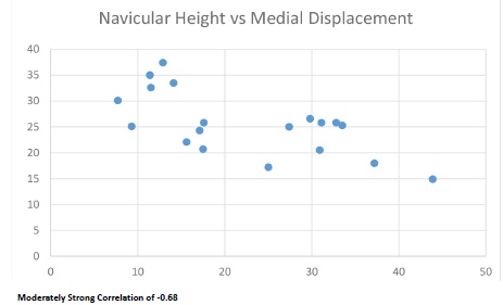
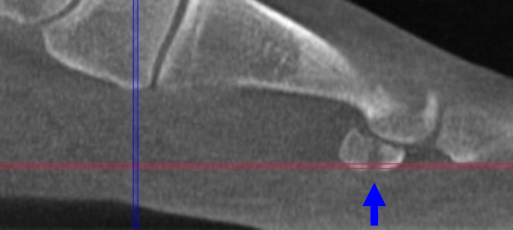
An unexpected result showed a moderately strong correlation (-0.68) between the vertical position of the navicular and medial displacement in a patient population with ranging degrees of medial column breakdown.

Moderately strong correlation of -0.68


pedCAT: Fracture & Fusion Assessments

X-rays of the foot and ankle may not always provide conclusive assessments for post-operative fusions such as of the tarso-metatarsal joint or hind foot joints. Similarly, the physician is often left guessing if a fracture has properly healed.

“The pedCAT takes all the variability out, all the guesswork out of it,” Dr. Martin O’Malley, MD, an Associate Attending Orthopedic Surgeon at the Hospital for Special Surgery in New York, New York, said.

Dr. O’Malley said pedCAT scans allow him to clearly determine if a fusion has healed more than 50 percent, and he decides when to ambulate his patient accordingly.

“I let people walk on it earlier than before, and I keep them off longer than before,” Dr. O’Malley said.

The pedCAT provides a three-dimensional view of fractures that changes the way O’Malley sees this common diagnosis.

“These posterior pieces are often bigger than we thought were based on plain X-ray and they often travel all the way around the medial side as well, which we never thought they did,” Dr. O’Malley said. “You know, we thought it was an infrequent fracture, but now we see it routinely. Now most of my ankle fracture work, I’d say more than half the time, is through a posterior approach. For the first 15 to 18 years of my practice I would do medial/ lateral incisions. Now I’m going to the back of the ankle. And a lot of it is driven by the pedCAT.”


pedCAT Stress Fracture Cross Section


Above is an example of a navicular stress fracture in a collegiate runner that had not healed at all after six weeks of casting. Were it not for the conclusive pedCAT scan, this patient would have been allowed to ambulate.

Watch Dr. O’Malley talk more about the pedCAT here .

To offer your patients state-of-the-art fracture & fusion assessment, consider a pedCAT for your practice.


World Cup Special Edition: Soccer Player with a Sesamoid Injury

The 2014 FIFA World Cup officially begins Thursday, June 12. The world’s top soccer players will all be in Rio, Brazil for one of the biggest sporting events of the decade. The next two weeks will be filled with cheering fans, tears of joy,  and national pride. But inevitably, there will also be injuries. In fact, before the games even begin, a number of players already know they will have to sit them out.

For professional athletes and weekend warriors alike, an accurate diagnosis from the onset can make a huge impact on the speed of recovery.  When it comes to bony injuries of the foot and ankle, the pedCAT is the only tool in the world that can provide a weight bearing, three dimensional image of the entire foot or both feet.

Here’s a case of a 30-year-old soccer player, who was treated by a  California podiatrist. The athlete injured his right great toe and sesamoids while playing soccer. The doctor observed that the patient’s first metatarsophalangeal joint was swollen and ecchymotic, and that he was tender upon palpation of the great toe joint and sesamoid bones.

The doctor took standard non-weight bearing X-Ray images of the patient. The AP and lateral X-Rays showed an obvious fracture with displacement in the medial sesamoid. The lateral appeared to have a fracture, but the doctor could not confirm his suspicion with the X-Ray. The doctor also took an axial X-Ray, but it had no diagnostic benefit since the patient could not extend his great toe.

double sesamoid fracture x-ray

The doctor then took a pedCAT scan of the same patient. The CT images clearly showed a severely displaced right medial sesamoid oblique fracture and a minimally displaced lateral sesamoid fracture. The oblique lateral sesamoid fracture demonstrated 2 mm of plantar displacement with excellent bony contact/ apposition in the dorsal 4 mm of the sesamoid.

pedCAT double sesamoid fracturepedCAT lateral sesamoid fracture displacement

Although the diagnostic information provided by the pedCAT did not alter the treatment plan for the medial sesamoid, it did confirm the injury to the lateral sesamoid. More importantly, the pedCAT was able to provide diagnostic information regarding the geometric nature of the lateral sesamoid fracture.


“I could determine that there was enough bony contact to allow for bone healing with appropriate conservative care management,” the doctor said.


New Study: pedCAT Allows for More Accurate Bone Angle Measurements than Radiographs or CT

A study published in Foot and Ankle Surgery compared three common angle measurements of the foot taken from non-weight bearing CT, weight bearing digital radiographs, and weight bearing pedCAT scans. The study concludes, “The angles differed between radiographs, CT and pedCAT, indicating that only pedCAT is able to detect the correct angles… pedCAT prevents inaccuracies of projection and foot orientation in contrast to radiographs due to the 3D dataset which is principally independent from projection and foot orientation.”

The study also found the average image acquisition time for the pedCAT (average 270 seconds) was 70 percent faster than with radiographs and 35 percent faster than with CT.

The study was authored by Martinus Richter, Bernd Seidl, Zech Stefan, and Sarah Hahn.