Comparison of Three Hindfoot Alignment Measurements: Radiographic Hindfoot Moment Arm, Radiographic Hindfoot Alignment Angle, and TALAS

Dr. Matthew Welck presented findings from a study overseen by Dr. Mark Myerson at the British Orthopedic Foot & Ankle Society (BOFAS) meeting in Bristol, England, in early November.

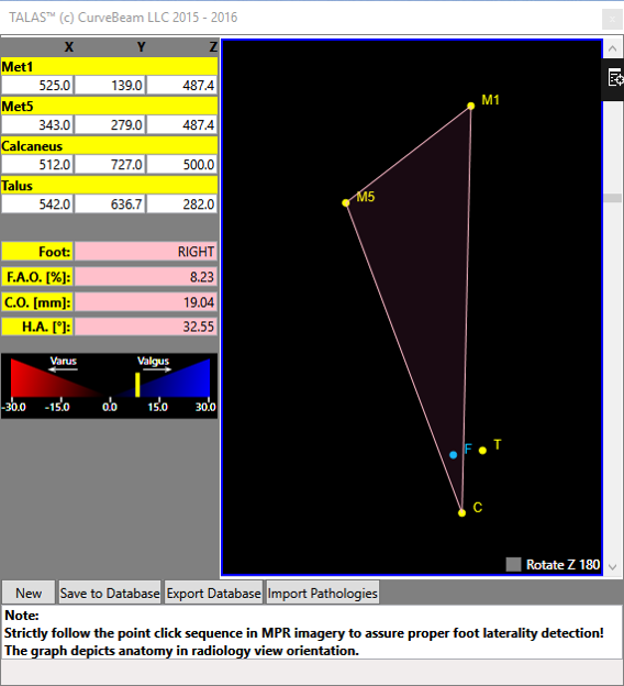
The study compared three hindfoot alignment measurements: radiographic hindfoot moment arm, radiographic hindfoot alignment angle, and TALAS. TALAS is a semi-automatic measurement based on anatomical landmarks on a weight bearing CT scan. TALAS was found to have the best intra-observer reliability.

5th-mt-contact-point
5th MT contact point.

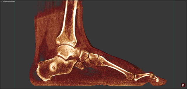
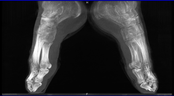
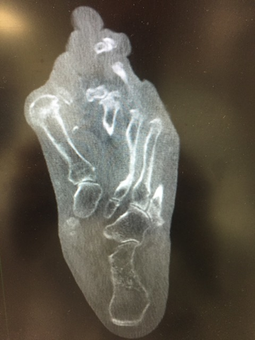
As Dr. Welck explained, the TALAS technique measures offset of midline of whole foot, rather than just calcaneus. He presented an example of a patient with ‘neutral’ alignment to compare hindfoot alignment measurements. The first point was placed on the superior weight bearing portion of the talar dome. The calcaneus contact point was then placed on the lowest point of the calcaneus. The first metatarsal contact point was placed on the lowest point of the first metatarsal head. Similarly, the 5th metatarsal contact point was placed on the lowest point of the 5th metatarsal head. TALAS software provided a mapping of these contact points showing the axis, or the ’true’ hindfoot alignment.Next, Dr. Welck summarized the intra and inter observer reliability among the three measurements. As previously shown in the literature, Dr. Welck explained, there is good intraobserver reliability with the Saltzman hindfoot moment arm technique. Similarly, the hindfoot alignment angle has excellent intraobserver reliability. And there was excellent intra-observer reliability among investigators. Dr. Welck noted that while all three measurement techniques are reliable, the CT Ground Reactive Force Technique was nearly perfect.

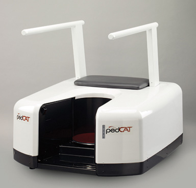
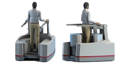
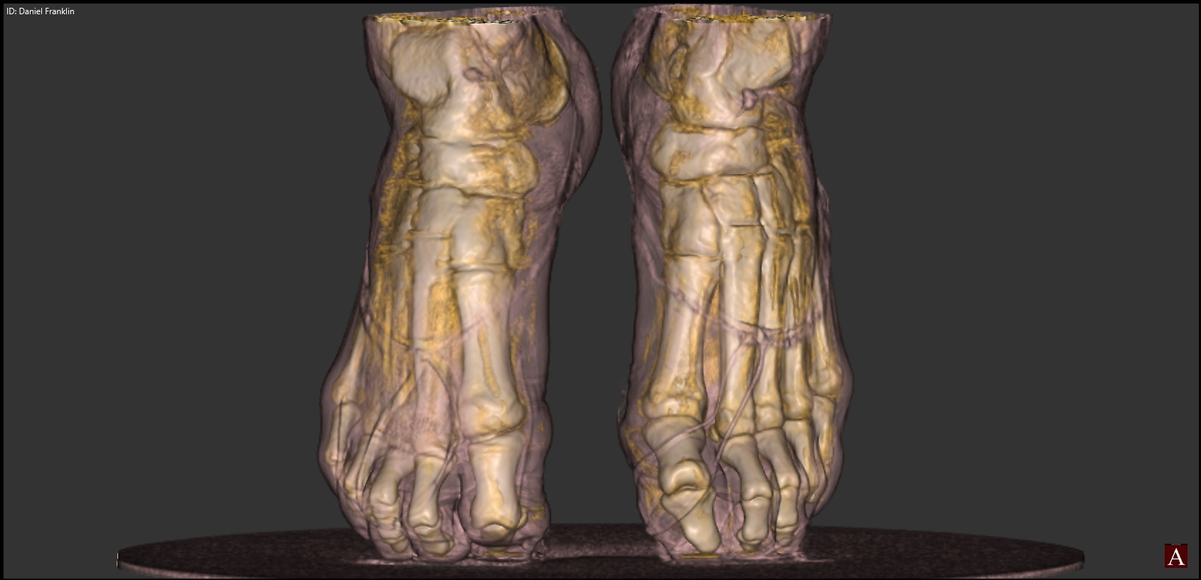
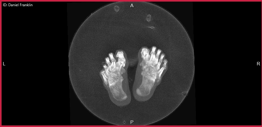
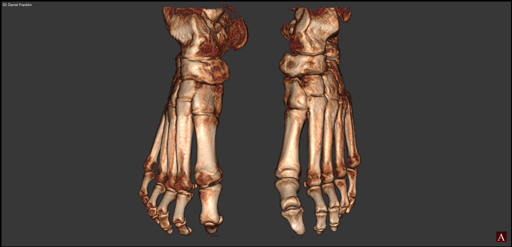
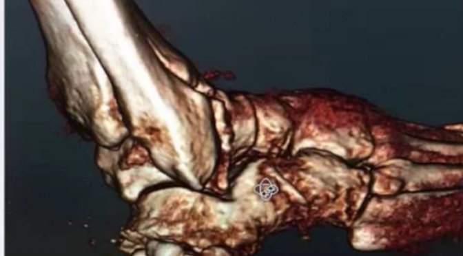
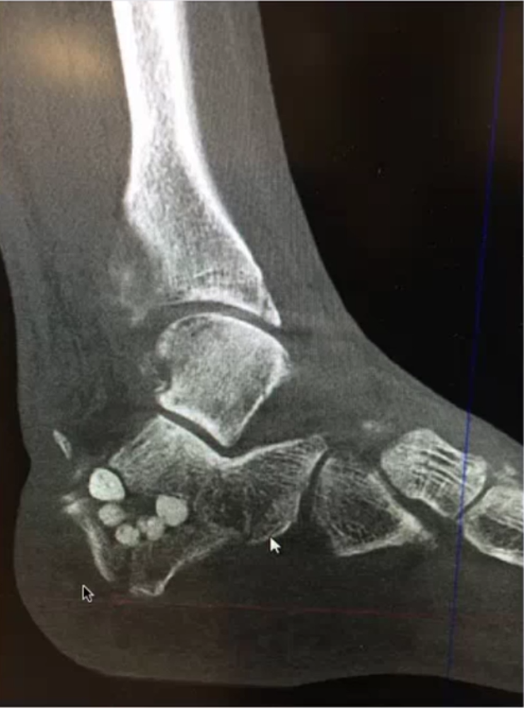
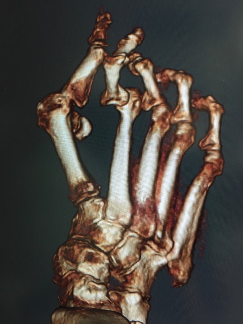
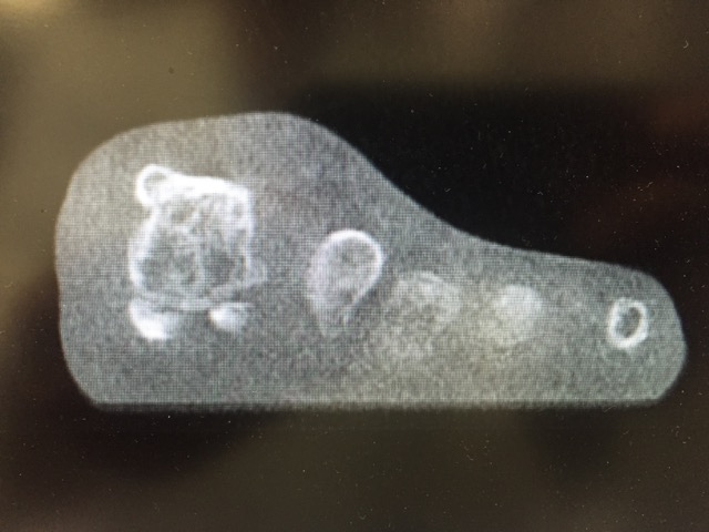
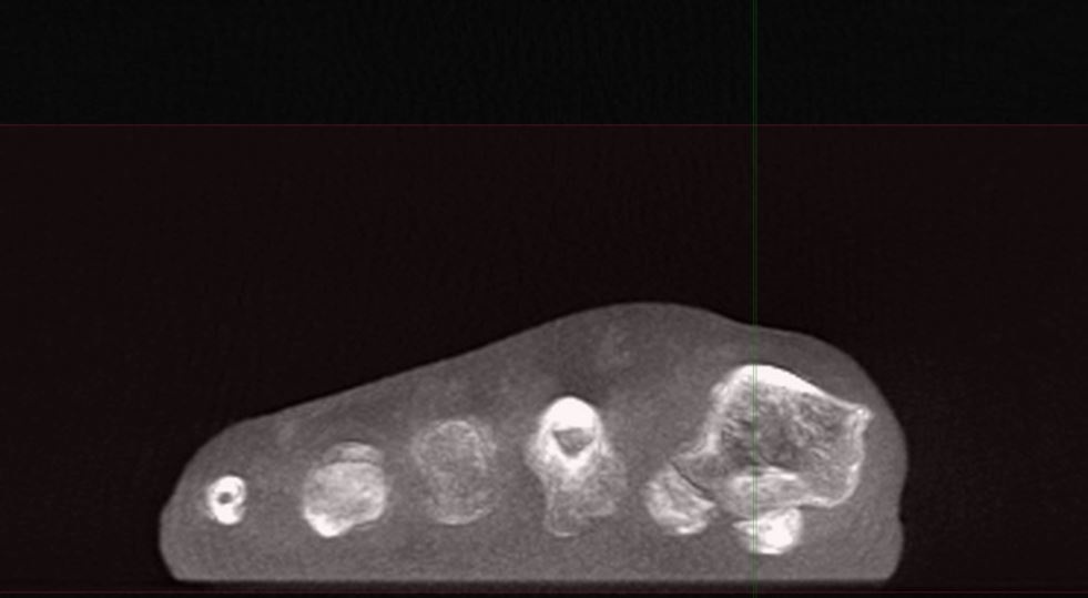
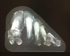
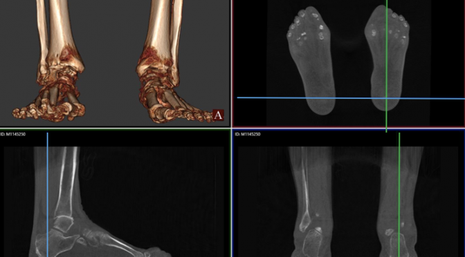
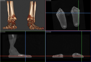
CurveBeam’s pedCAT provides bilateral, weight bearing 3D CT imaging of the foot and ankle, greatly surpassing traditional imaging methods such as X-Ray and CT studies. CurveBeam’s technology also reduces the number of patient trips to the hospital, and exposes patients to less radiation. Because the 3D data generated by pedCAT is far more robust than could be interpreted by available software, CurveBeam engineers are collaborating with leading foot and ankle surgeons to develop a new measurement software – TALAS. TALAS, or Torque Ankle Lever Arm System, is a feature within pedCAT’s visualization application CubeVue.

talas-software
A mapping of the contact points in the TALAS software.

The British Orthopaedic Foot & Ankle Society (BOFAS) is a society of orthopaedic surgeons who have a special interest in surgery of the foot and ankle. The Society works to encourage interest in foot and ankle surgery among orthopaedic surgeons and to encourage both basic science and clinical research in the field.

Value of 3D Reconstructions of CT Scans for Calcaneal Fracture Assessment

Operative fracture treatment of irregularly shaped bones, such as the calcaneus, scapula and scaphoid, demands high-quality images of the area in question for both classification of the fracture and planning of the procedure. However, since plain radiographs do not provide enough pertinent information to this end, computerized tomography (CT) has become the standard for providing the necessary images for treatment of these irregularly shaped bones.

Yet, in a study observing 2D CT scans, only 42% of the evaluators were able to correctly classify the fracture, necessitating the need for some sort of change. Three dimensional CT renderings were proposed to fix the low inter-rater agreement generated by the 2D scans.

The foot in the video above was scanned on a pedCAT weight bearing CT imaging system for the foot and ankle. The calcaneus was segmented using CurveBeam’s CubeVue visualization software. 

To evaluate the effectiveness of 3D CT scans, A standard set of CT secondary reformation scans were presented, followed by a questionnaire describing fracture anatomy and preoperative planning. Subsequently, 3D reconstructions were presented to the evaluates followed by the same questionnaire. After presentation of the 3D images, 49% of the evaluators changed their plan in regard to the approach and 29% in regard to the implants.

Five different data sets (four intra-articular and one extra-articular fractures) were presented to 57 evaluators. All groups, except that of surgeons with more than 20 years of experience, benefited from 3D CTs (Friedman test; P < .01). Inexperienced surgeons benefited more than experienced surgeons and complex fractures more than simple fractures. Specifically, regions of interest such as the middle facet and fractures extending into the calcaneo-cuboid joint were evaluated more precisely.

In regard to 3D CT scans, Böhmer1 described the topographic relationship between the fragments and the surrounding structures as useful for evaluating calcaneal fractures and for preoperative planning because the fractures can be seen from unusual perspectives. Likewise, Choplin2,3 posited 3D scans assist diagnosing foot deformities since the scans improve comprehension of the anatomy, particularly for especially complex fractures. For such complex fractures and anatomy, Pate5 evaluated 202 patients with complex musculoskeletal problems and found 3D CT particularly helpful. Others came to similar conclusions.

When comparing techniques for diagnosing foot deformities, Allon and Mears4 compared plain radiography, 2D CT, and 3D CT of 30 fractured calcanei and concluded that 3D CT improves preoperative planning and the choice of an adequate approach.

Overall, 3D CT scans provide insight previously unavailable through both 2D and plain radiography, which inexperienced surgeons tend to find more helpful in diagnosing and preoperative planning.


References

  1. Bohmer G, Roesgen M, Hierholzer G. Three-dimensional computerized tomography in trauma surgery. A case presentation [in German]. Aktuelle Traumatol. 1992;22(2):47-56.
  2. Choplin RH, Buckwalter KA, Rydberg J, Farber JM. CT with 3D rendering of the tendons of the foot and ankle: technique, normal anatomy, and disease. Radiographics. 2004;24(2):343-356.
  3. Choplin RH, Farber JM, Buckwalter KA, Swan S. Threedimensional volume rendering of the tendons of the ankle and foot. Semin Musculoskelet Radiol. 2004;8(2):175-183
  4. Allon SM, Mears DC. Three dimensional analysis of calcaneal fractures. Foot Ankle. 1991;11(5):254-263
  5. Pate D, Resnick D, Andre M, Sartoris DJ, et al. Perspective: three-dimensional imaging of the musculoskeletal system. AJR Am J Röntgenol. 1986;147(3):545-551.

Weightbearing CT Imaging Methodology with pedCAT by CurveBeam

 
Three-dimensional weight-bearing computed tomography (CT) can be a powerful diagnostic tool, typically used when more information is necessary (e.g. intra-articular fractures, occult fractures and small bone tumors). Unlike conventional CT, which has a fan-shaped X-ray beam, modalities in the pedCAT created by CurveBeam have a cone-shaped X-ray beam. In a Podiatry Today’s article titled “Current Concepts With Weight bearing CT”, Dr. Albert V. Armstrong Jr., dean of the Barry University School of Podiatric Medicine, reviewed three independent studies that examined the efficacy of the technology.

In the first, Yoshioka and colleagues studied 10 patients with posterior tibial tendon dysfunction and 10 control patients, using weight-bearing and non-weight-bearing CT.1 The authors noted that the study clarified part of the clinical condition of the forefoot in flatfoot deformity, saying this may be applicable in basic research of the staging advancement and sub-stage classification of flatfoot.

In the second study, Krähenbühl and coworkers used weightbearing CT to determine the subtalar vertical angle in a study of 40 patients with osteoarthritis and 20 control patients.2 The study authors found that measuring the subtalar vertical angle was a reliable and consistent method to assess the varus/valgus configuration of the posterior facet of the subtalar joint.

In the final study, Geng and colleagues studied weightbearing and non-weightbearing CT scans of 10 patients with hallux valgus and 10 control patients, reconstructing 3D models for the first metatarsal and the medial cuneiform.3 Researchers noted the study furthers an understanding of the physiological and pathological mobility of the first metatarsocuneiform joint.

Weightbearing CT is a safe imaging modality with low radiation exposure that can provide superior images in comparison to conventional CT, as evidenced by the multiple studies. Weightbearing CT can enhance biomechanical evaluation, preoperative planning, postoperative evaluation, wound management, sports medicine, treatment of arthritic conditions (especially degenerative joint disease) and trauma (especially when looking for occult or hairline fractures). It is a promising and up and coming imaging method to replace traditional CT technology.

Cone beam CT allows clinicians to obtain an image of a volume of tissue in one circumferential pass instead of having to take multiple slices with multiple exposures. This leads directly to reduced radiation exposure for patients. Studies indicate, in the example of a bilateral scan of a foot, the pedCAT machine exposes patients to one third the amount of radiation as traditional methods. Another great feature of weight-bearing CT is the ability to perform bilateral scans. One can also view the same patient with the view of the the soft tissue structures removed, leading to increasingly accurate prognosis. In addition, the pedCAT is an excellent tool to illustrate visually to a patient exactly where a bunion, for example, is located underneath the soft tissue. The generated visual displays are much easier to understand for non-trained individuals.

Performing actual weightbearing examinations is possible through pedCAT, a main advantage of the machine. In a specific instance, a podiatrist can view a foot supporting weight, viewing the 3D image and the accompanying 2D images in the sagittal, axial (transverse) and coronal (frontal) planes. This would allow the physician to observe the appendage in its most natural state, allowing a more precise diagnosis of problems.

Read “Current Concepts With Weightbearing CT” by Dr. Albert V. Armstrong Jr. here: http://www.podiatrytoday.com/current-concepts-weightbearing-ct


CurveBeam Announces Development of Extremity CT System for Knee

A new orthopedic CT system promises to improve the standard of care for knee imaging. On the CurveBeam LineUp, patients are scanned while standing upright and fully weight bearing.

CurveBeam LineUP
CurveBeam LineUP provides bilataral weight bearing CT imaging of the knees and lower extremities.

Traditional CT and MR images are acquired in a non-weight bearing position, leading to “missed diagnoses of meniscal damage,” according to Dr. Neil Segal, who has been overseeing research efforts using a LineUp prototype, first at the University of Iowa and currently at the University of Kansas.

Neil Segal, MD
Neil Segal, MD

Although plain radiographs can be acquired while the patient is in a full weight-bearing position, the optimal degree of knee flexion and X-Ray beam tilt to best visualize the joint surface is person specific.

“Difficulty in reproducing the same view of the joint over time impairs ability to detect joint disease, and the 2D nature of radiographs makes these images of overlapping bony anatomy very insensitive for detecting abnormalities until there is advanced joint damage,” Dr. Segal said.

The LineUp was developed by CurveBeam, a Pennsylvania-based company that specializes in extremity cone beam CT systems for orthopedics. CurveBeam anticipates it will submit an application for and receive FDA 510(k) clearance for the LineUp in 2017. The LineUp will be on display at RSNA at Booth #8008 in the North Hall.

CurveBeam introduced the pedCAT, a bilateral weight bearing CT system dedicated to the foot and ankle, in 2012. Since then, the device has been added to the imaging services of hospital foot & ankle sections, orthopedic clinics and podiatry offices.

Like the pedCAT, the LineUp will provide isotropic, three-dimensional volumes of the anatomy with a high resolution output of between 0.2 mm and 0.3 mm slices. The LineUp will be the only cone beam CT system that can provide bilateral, weight bearing scans.

A study led by Dr. Segal focused on osteophytes, one structure

linked to pain in people with knee osteoarthritis. Knees of community-dwelling adults with knee OA were imaged with MRI (reference), fixed-flexion radiographs, and weight bearing CT. The sensitivity and accuracy for detecting osteophytes and subchondral cysts were higher with weight bearing CT imaging in comparison to fixed-flexion radiographs. The study was published in the August 2016 issue of the Journal of Orthopedic Research.

“Clinically, this is a highly meaningful improvement,” Dr. Segal said. “It suggests that weight-bearing CT could replace radiographs as the recommended means of assessing knee OA. This advancement is even more significant given that it was made without significantly increasing the radiation dose (0.01 mSv for SCT vs. 0.005–0.102 mSv for a series of knee radiographs).”

Another research effort led by Dr. Segal indicates weight-bearing CT arthrography studies can provide outstanding delineation of articular cartilage with better differentiation between cartilage and subchondral bone then MRI studies, while also visualizing the cruciate ligaments. In knees with osteoarthritis, meniscal tears not visualized on MRI were detectable on weight-bearing CT.

“Thus, we found that some potential advantages of weight-bearing CT over non-weight-bearing MRI/MRA include 3D measures of meniscal position, detection of pathology not detected in unloaded positions, and ability to bear weight in a functional position, thus better recreating the magnitude of body weight and muscle forces acting around the knee during usual standing,” Dr. Segal said.

CurveBeam CT Imaging Technology is Right On Point

Ballet is an art of extremes. As such, the risk factors are high among dancers for developing chronic injury and weakened tissues throughout the lower half of their bodies. About 60% of ballet injuries affect the legs, hips, ankles or feet. Lateral ankle sprains and Posterior Ankle Impingement Syndrome, or the pinching sensation felt during repeated floor or barre work as the heel bone comes into contact with the talus bone, are chief among them.

By the age of 12, ballet dancers are generally considered ready to begin learning the en pointe technique which utilizes the unnatural convergence of the tibia, talus, and calcaneus to lock the ankles in place.

releve2
pedCAT weight bearing CT scan of a ballerina en pointe.

While we do know this position may facilitate injury, prior to the advent of advanced CT imaging podiatrists had no method of accurately determining the exact anatomical position of either bones or tissue in this position.

CurveBeam, founded in 2009, designs and manufactures Cone Beam CT imaging equipment for the orthopedic and podiatric specialties. In 2012 CurveBeam’s pedCAT system received official clearance from the FDA, and in 2013, CE Mark approval. With the implementation of tools such as the pedCAT and CubeVue, CurveBeam’s custom visualization software, researchers finally have the capability to help ballet teachers better understand the demands of this position before introducing it to young students.

en pointe sag
pedCAT weight bearing CT scan of a ballerina en pointe

Pointe technique, when examined through an advanced imaging system, reveals the posterior portion of the talus resides beyond its articular surface, while the posterior portion of the tibia’s articular surface leaves the articular surface of the dome to rest on the posterior talus. The three bones converge. According to Dr. Jeffrey A. Russell, Ph.D, A.T., FIADMS:

“Attaining the full en pointe position requires contributions from movements between the bones in the foot. Examples of such movements include sliding between the talus and the navicular, the navicular and the medial cuneiform, and the medial cuneiform and the first metatarsal. These small increments of motion combine to provide approximately 30% of the plantar flexion range.”

pedCAT weight bearing CT ballet
pedCAT weight bearing CT scan of a ballet dancer en releve.

“In addition, it is noteworthy that the talus shifts slightly anterior from under the tibia as the ankle-foot complex moves en pointe. This subtlety arises because the converging tibia, talus, and calcaneus form a fulcrum that applies an anterior force to the talus, somewhat like a watermelon seed being squeezed from between one’s thumb and forefinger.”

Recently, Dr. Russell took to an advanced open MR scanner to review the upright and weight bearing position of uninjured ankles in six university-level dancers who had been dancing for an average of 13 years, and dancing en pointe for an average of seven years.

“All exhibited several traits in their ankle MRIs: the posterior portion of the articular surface of the tibia rested on a nonarticular surface of the posterior talus; the synovial sheaths of the flexor and fibularis tendons collected fluid; Kager’s fat pad was impinged by the posterior tibial plafond; and small ganglion cysts were apparent in one or more spots around the ankle and proximal foot.”

pedCAT weight bearing CT ballet
pedCAT weight bearing CT scan of a ballet dancer en releve

It’s an interesting find, and begs the question: do these conditions increase the likelihood of ballet dancers developing ankle osteochondritis or osteoarthritis?

Not only do advanced imaging systems such as the CurveBeam pedCAT – which was not used in this particular study – reveal the bones’ proper anatomic alignment, but they also enable a close investigation of cartilage quality, which isn’t possible with traditional MR imaging. Bilateral, weight bearing three-dimensional views of the foot and ankle are therefore the most cogent means for specialists to create comprehensive treatment regimens and surgeons to better visualize their surgery plans for better operation outcomes.

Most importantly, however, is Dr. Russell’s recent study confirms the use of orthopedic imaging to examine pointe dancers’ ankles in detail will only continue to offer more insight into the demands placed on the ankle by dancing in this way, ultimately leading to safer instruction, more accurate treatment of injuries, and faster recovery times.

Webinar Summary: Advanced CT Imaging in Foot and Ankle Surgical Considerations

We all know that X-rays and MRIs aren’t the be-all, end-all of diagnostic imagery.

Try determining the frontal plane rotation of the sesamoid and first metatarsal with an X-ray. It isn’t possible. Correction of the hallux valgus rotation in bunion surgery depends largely on the repositioning of the sesamoidal apparatus which is impossible to assess without an axial view, and X-rays fall completely short when it comes to assessing these relational details from the vantage point of a single plane.

Such was the topic of discussion during the latest Curvebeam webinar led by Dr. Bob Baravarian, Director of University Foot and Ankle Institute in Southern California. He explained how weight-bearing 3-dimensional CT scans are changing the game of podiatric diagnostics.

Throughout the webinar, titled “Advanced CT Imaging in Foot and Ankle Surgical Considerations,” Dr. Baravarian offered a very straightforward presentation of the often-not-so-straightforward complexities of foot and ankle deformities and how advanced imaging technology can improve both surgical planning and surgical outcomes. He cited the example of hallux valgus among many others as “impossible to imagine treating without 3-D imaging technology” these days – given the results he’s seen with his patients and scope webinar 3of its applications.

Planning the frontal plane correction of first metatarsal.

“CT scans are very helpful in planning your frontal plane deformity correction of the first metatarsal to get an anatomic position which is really underestimated,” said Dr. Baravarian.

Multi-plane imaging now allows physicians to correct the frontal plane deformity of the metatarsal while simultaneously realigning the sesamoid. The capability is “critical,” said Dr. Baravarian, “for proper outcome with bunion corrections whether you’re doing a LAP or any kind of osteotomy.”

Identifying the cause of hypermobility of the first ray

When you look at a patient who has a significant flat foot deformity and a significant bunion deformity with some level of hyper-mobility of the first ray, 3D CT imaging allows you to locate the exact area in need of correction.

“In patients who have PTTD or even a pediatric flat foot case we really need to decide which planes of correction make the best sense,” said Dr. Baravarian.  “If I correct my first metatarsal, is my hind foot going to realign or is there an outstanding deformity that needs correction?”

webinar captureDetermining the level of arthritis in hallux rigidus and limitus cases. 

“I’m constantly surprised when I go into surgery and I plan a cheilectomy and I open up a joint and there’s significantly more arthritic changes or some kind of osteochondral legion that I couldn’t really see on the X-ray. Or, I plan to do an osteotomy and I go in and the level of arthritis is not as bad as expected,” said Dr. Baravarian.

It’s very difficult to determine the level of arthritis in a hallux rigidus or limitus case based on a radiograph alone. While MRIs are an excellent option for soft tissue imaging and diagnostic ultrasounds still provide accurate, real-time guidance for proper injections –neither of these methods makes sense for treating anatomic alignment and assessing structural deformities.


“With a 3D CT I’m able to look a little more in-depth into the joint and make a better decision prior to surgery.”

Identifying major deformities in complex fractures

Weight-bearing imaging systems allow you to place a foot in its anatomic position and adjust its deformity to see what level of correction you can get across multiple planes.

It “doesn’t makes sense,” according to Dr. Baravarian, “to plan the correction of a complex fracture without a sense of what’s going on inside the foot and ankle.”

Our traditional method of two-dimensional x-rays doesn’t provide the highest level of certainty that doctors need in order to administer the best care possible to their patients where deformity planning is essential for proper surgical outcomes.

Now, with the help of tools such as the pedCAT, a compact 3D weight-bearing CT imaging system, podiatrists have everything they need to create comprehensive treatment plans and more effective surgeries. Better outcomes. Less risk. And patients back on their feet faster than they ever expected.

You can access the entire webinar here. A FOOT Innovate membership is required to access the content. Membership is complimentary for foot & ankle specialists.

Weight Bearing CT Advances Planning for Forefoot Reconstructive Surgery

Think of a surgical procedure like a construction project. No contractor would build a building without a full set of plans, cross-sectional diagrams and 3D renderings. Dr. Bob Baravarian, DPM, feels foot surgeons should be just as prepared.

As he explained in Podiatry Today, Dr. Baravarian has always been interested in architecture and real estate, yet was drawn to 159682158975132.Zfw2RIZiU5Lh04gznDby_height640medicine.

Dr. Baravarian sometimes works next to a hip & knee surgeon in the OR. One day, he observed the hip & knee surgeon had a digital tool that laid out an exact surgical plan for the osteotomy he was performing. The plan rendered leg length and acetabular rotation a non-issue.

Soon after, Dr. Baravarian incorporated a pedCAT weight bearing CT imaging system into his podiatric practice. He found he could plan angular corrections to the degree, calculate the exact size of a graft for an Evans procedure, and could even plan the amount of rotation necessary for anatomic alignment of the hindfoot.


Overtime, he began to apply the same methods to forefoot procedures as well.

  • For hallux valgus cases, he scans the patient in the post-corrected position so he can determine if a hindfoot alignment correction is also needed.
  • For hallux rigidus cases, he uses weight bearing CT to evaluate the level of articular damage and the possibility of cartilage graft procedures. “This is impossible to do at such high levels with plain radiographs,” Dr. Baravarian said in the Podiatry Today article.

pt07treatment1 (1)Bilateral, weight bearing three dimensional views of the foot and ankle give specialists the data they need to create optimal treatment plans. The pedCAT, with a scan time of one minute, is a compact, ultra-low dose CT imaging system that is ideal for orthopedic and podiatric clinics. Patients benefit from the convenience of point-of-care advanced diagnostic imaging, and the pedCAT automatically generates all standard X-Ray views in addition to the full CT volume.

As software programs become more advanced, Dr. Baravarian said, surgeons may be able to perform osteotomies and shift the bones in preoperative planning, which will allow even more advanced planning along with far less operative time and less risk to the pt07treatment3 (1)patient.

Dr. Baravarian is the Chief of Podiatric Foot and Ankle Surgery at the Santa Monica UCLA Medical Center and Orthopedic Hospital, and Director of the University Foot and Ankle Institute in Los Angeles.

Read the entire article here.

Need Bunion Surgery? A Weight Bearing CT Scan Could Help You Decide

When a patient appears to have a bunion, a physician typically orders traditional foot X-Rays as part of the clinical evaluation. The X-Rays capture three views of the foot- dorsoplantar, medial oblique, and lateral. But is this enough information to understand complex, three-dimensional object such as the foot? 

A growing number of foot & ankle specialists are advocating that there may be better way to evaluate this common deformity.

“Weight bearing computed tomography scans are beginning to take our understanding to the next level,” Dr. Paul Dayton, DPM, of Des Moines, IA, said in a roundtable discussion on bunions published in the Foot & Ankle Specialist academic journal. “Once you see the connection between coronal rotation and what we have traditionally evaluated on AP radiographs, it opens up a whole new understanding.”

On X-Rays, physicians have to “mentally interpolate” the valgus component of the bunion because they do not adequately capture the frontal plane, said Dr. Robert Weinstein, DPM, FACFAS, of Atlanta, explained in a CurveBeam case study.  

“Since the condition is a tri-plane deformity, we need to understand all of the components and their angular values contributing to the deformity,” Weinstein said. “Better deformity analysis leads to better pre-operative planning, surgical execution, and post-operative results.”

The new data on the coronal position is “exciting” because it explains so many of foot and ankle specialists’ previous questions, Dayton said.  

Weight bearing CT imaging also allows for evaluating deformities that involve external rotation. Rotation of one or more metatarsals, including displacement of the sesamoids, cannot be clearly seen in standard foot X-ray imaging. The lack of the weight-bearing aspect in traditional medical CT imaging means the physician cannot evaluate displacement and rotation under load.

Hallux Valgus Blog Post Image 1
One component of the deformity that is seldom quantified is external rotation, or frontal plane deviation. The presence of rotation of the hallux implies a more complex deformity. A very careful assessment of the first metatarsophalangeal joint architecture, including sesamoid position, their condition, and erosion or flattening of the crista on the inferior first metatarsal head is essential.

It has long been known that in hallux valgus or bunions the relationship between the metatarsal head and the sesamoids is altered,” Andy Goldberg, MD, MBBS, FRCS (TR and Orth), of Stanmore, United Kingdom, told Lower Extremity Review magazine. he said. “The sesamoid bones should sit underneath the metatarsal head, while in hallux valgus the big toe drifts off the sesamoids and the tip of the big toe points outwards. But our research has shown that in many cases the cartilage is worn, which in essence is localized arthritis, and we believe that this could affect the outcome of surgery.”

rotated first met and sesamoids

CubeVue, the pedCAT weight bearing CT imaging system’s custom visualization software, allows physicians to create oblique and frontal-plane images of varying angles and thicknesses from the weight bearing CT data.

Weight bearing CT images could have an impact on the technique a physician selects to repair a bunion, Dr. Dayton said.

“The knowledge that the sesamoids can be in normal position medial and lateral to the crista yet look dislocated on the AP X-ray because of pronation completely changes our mindset about the need for capsular balancing,” Dayton said. “We can see that in those cases supination corrects the deformity.”

Standing CT vs. MRI for Advanced Visualization of Knee Cartilage and Meniscus

At the 2016 OARSI World Congress in Amsterdam, Netherlands, Dr. Neil Segal, MD, MS, and Dr. Ali Guermazi, MD, PhD , presented a poster describing “Advances In Visualization Of Knee Cartilage And Meniscus With Standing Computed Tomography Arthrography”.

Standing CT arthrography (SCTa) has also been shown to have some distinct advantages over MRI, according to the poster presentation. “Potential advantages of SCTa over non-weight-bearing MRI/MRA include: 3D measures of meniscal position and morphology; detection of pathology not detected in unloaded positions; and ability to bear weight bilaterally in a functional position, better recreating the magnitude of muscle and external forces acting about the knee during usual standing….  SCTa can be obtained in multiple knee flexion angles, while an MRI knee coil may permit imaging only with the knee in extension, and SCTa is less expensive than MRI.”

Figure 1: SCTa and corresponding MR arthrography demonstrating outstanding delineation of tibiofemoral and patellofemoral articular cartilage, with better differentiation between the cartilage and subchondral bone on SCTa. Visualization of the boundaries of the menisci was achieved to a similar degree on SCTa and MRA.

As part of the background for their presentation, Drs. Segal and Guermazi stated that “MRI is the standard for non-invasive visualization of cartilage and menisci, and … Absence of weight bearing limits evaluation of the functional position and configuration of these structures…Advances in standing CT (SCT) have allowed 3D imaging of the knees while under physiological loads, similar to fixed-flexed or semi-flexed radiograph protocols.”

Their objective was to evaluate a protocol for SCTa for imaging weight bearing cartilage and menisci and to assess potential advantages over non-weight bearing MRI.

figure 2
Figure 2: Sagittal reformatted SCTa and its corresponding MRA demonstrated outstanding delineation of articular cartilage with better differentiation between the cartilage and subchondral bone on SCTa, while also visualizing the ACL and PCL in the femoral notch.

Although the sample size was small, the results were noteworthy. As shown in the images, SCTa permits evaluation of cartilage and menisci in three dimensions, while the patient is standing and under physiological load. Furthermore, SCTa, “may be useful for assessment of menisci as well as tibiofemoral and patellofemoral cartilage in functional stance,” according to the presentation.

The two participants in the study were a 42-year-old man without osteoarthritis (Figures 1 and 2), and a 67-year-old woman with KL2 knee osteoarthritis (figures 3-5). The participants had a similar contrast agent applied.

Figure 3a: Sagittal SCTa demonstrating minimal cartilage thinning on the left medial tibial plateau Figure 3b: Axial image depicting location of the sagittal slice in Figure 3a
Figure 3a: Sagittal SCTa demonstrating minimal cartilage thinning on the left medial tibial plateau
Figure 3b: Axial image depicting location of the sagittal slice in Figure 3a

The actual imaging techniques used were fixed-flexed (approximately 20°) SCTa (INVESTIGATIONAL ONLY cone beam CT knee imaging system*, CurveBeam, Warrington, PA, USA) and non-weight bearing MRI (Siemens TrioTim, Washington DC, USA).

“Following 2-3 minutes of unloaded knee flexion and extension, a low-dose SCT scan was acquired utilizing cone beam reconstruction. Participants were positioned with the tips of the great toes, patellae, and the anterior superior iliac spines coplanar to each other and the feet 10°externally rotated. Scans were acquired with a 0.3mm isotropic voxel size (20x 35x35cm) with an effective radiation dose of approximately 0.1 mSv. 10 minutes following SCTa, MR arthrography was acquired (NEX=1, ETL=3, Slice thickness=2 mm, Slice spacing 2 mm, Matrix= 240 x 320, FOV=140 mm with axial T1 fat-sat (TR=712 msec, TE=12 msec); coronal T1 fat sat (TR=730 msec, TE=10 msec); and sagittal T1 fat sat (TR=796 msec, TE=10 msec).

Figure 4a: Coronal SCTa demonstrating minimal cartilage thinning on the left medial tibial plateau Figure 4b: Axial image depicting location of coronal slice in Figure 4a
Figure 4a: Coronal SCTa demonstrating minimal cartilage thinning on the left medial tibial plateau
Figure 4b: Axial image depicting location of coronal slice in Figure 4a
Figure 5a: Sagittal SCTa image demonstrating a small tear of the tibial surface of the post. horn of the medial meniscus Figure 5b: Axial image depicting location of sagittal slice in Figure 5a
Figure 5a: Sagittal SCTa image demonstrating a small tear of the tibial surface of the post. horn of the medial meniscus
Figure 5b: Axial image depicting location of sagittal slice in Figure 5a

*The CurveBeam knee imaging system is investigational only and is not available for sale in the US.

pedCAT: Early Diagnosis of Osteomyelitis in the Diabetic Patient

The pedCAT weight bearing CT imaging system could identify bone infection at an early stage, and possibly prevent amputations, researchers at the California School of Podiatric Medicine at Samuel Merritt University determined in a report.

The researchers outlined two cases where “the use of CBCT device enabled us to diagnose and treat osteomyelitis in a timely manner, preventing its spread to adjacent bone and soft tissue, and minimizing the amount of required surgical resection.”

Plain radiograph is the primary imaging modality for the osteomyelitis diagnosis, the report states, but X-Rays may not reveal osteolytic changes for up to 20 days from the onset of infection or until the bone density is reduced by 30 – 50 percent.

Osteomyelitis is one of the most feared complications of diabetic foot ulceration, which often leads to lower extremity amputation and disability. Early diagnosis of osteomyelitis increases the likelihood of successful treatment and preserving ambulatory function. Unfortunately, most of the currently available imaging modalities are of limited use in assessing early stages of bone infection due to their low specificity and sensitivity for early osteolytic changes.

Magnetic resonance imaging (MRI) is more sensitive and specific than X-Ray, and yields greater accuracy in detecting soft tissue abscesses or early osteomyelitis in patients with high clinical suspicion and negative radiographs. In controversial or uncertain cases where MRI is not available, other imaging techniques such as indium-labeled leukocyte imaging combined with radionucleotide bone scan can be used as an alternative. (“Preventive and Therapeutic Strategies for Diabetic Foot Ulcers” – Foot & Ankle International® 2016, Vol. 37(3) 334– 343 – Chris C. Cychosz, BS, Phinit Phisitkul, MD, Daniel A. Belatti, BS, and Dane K. Wukich, MD).

Nuclear Imaging and MRI perform well in detecting early onset of osteomyelitis; but they are expensive to own and operate, are time-consuming in their acquisition of images.

What is desirable for early detection is a method that is accurate, inexpensive, and readily available. Cone beam CT, and the pedCAT in particular, fills this gap nicely.  The device is small enough to fit into most practices, offers high-resolution 3D imaging capabilities, and has a reduced radiation dose compared to traditional CT, according to the report.

Lead author Alexander M. Reyzelman, DPM,  and his associates reported on two diabetic patients who presented with infected neuropathic foot ulcers and were evaluated for potential osteomyelitis using plain film radiographs and the pedCAT CBCT scanner. In both cases, the “pedCAT was instrumental in identifying bone infection. The diagnosis of osteomyelitis was later confirmed by positive findings on bone biopsy. The use of CBCT device enabled us to diagnose and treat osteomyelitis in a timely manner, preventing its spread to adjacent bone and soft tissue, and minimizing the amount of required surgical resection.”

Case 1

A 49 year old diabetic female presented with an infected neuropathic ulcer at the lateral aspect of her fourth digit. The ulcer demonstrated malodor, cellulitis that extended to fourth metatarsophalangeal joint and positive probe-to-bone test. The plain film radiographs and CBCT were utilized in order to rule out osteomyelitis and assess the extent of soft tissue infection. The weight-bearing X-rays of the affected foot revealed subtle lucency at the lateral aspect of the proximal phalanx of the fourth digit, which was contiguous with the ulcer location. However, this finding alone was not sufficient to yield a conclusive diagnosis.  The images obtained using PedCAT clearly demonstrated the break in the cortex and the area of osteolysis involving the proximal phalanx of the fourth digit. The head of the fourth metatarsal and adjacent digits appeared intact. These findings, in conjunction with the clinical appearance of the affected digit, led to a preliminary diagnosis of osteomyelitis. The patient was treated with an arthroplasty of the fourth proximal interphalangeal joint, and has fully recovered. The bone specimens obtained intraoperatively were sent for biopsy, which confirmed our preliminary diagnosis of osteomyelitis.

Case 2

A 53 year old diabetic male presented with an infected neuropathic ulcer at his fifth metatarsal head, which exhibited malodor, edema and erythema extending through tthe plantar lateral aspect of  fifth metatarsal shaft and probed to joint capsule. The X-rays demonstrated no signs of bone involvement, while CBCT revealed distinct areas of cortical lysis and bony fragmentation of the fifth metatarsal head. The proximal two thirds of the shaft of the fifth metatarsal appeared unaffected, with intact cortex, uniform bony density and lack of osseous fragmentation. The patient was treated with partial resection of the fifth metatarsal. The bone biopsy has confirmed our preliminary diagnosis of osteomyelitis.

In the concluding discussion, Dr Reyzelman noted: “Though in our case studies we have not taken advantage of the option allowing to scan the patient in both, a weight-bearing and a non-weight bearing positions, this option could be highly useful for evaluation of complex fractures and dislocations of the foot and ankle.”