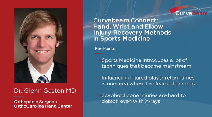
How do professional athletes recover from sports injuries and what are the advancements in sports medicine that are making these recoveries faster, and better? On this episode of the Curvebeam Connect podcast, host Vinti Singh, Director of Marketing at Curvebeam, spoke with Dr. Glenn Gaston, hand surgeon with OrthoCarolina, and hand consultant for the Carolina Panthers and the Charlotte Hornets, about these issues, with a focus on hand and wrist injuries.
As a member of the NFL physician’s society, Dr. Gaston was able to share with Singh, how the NFL’s muscular skeletal committee operates, what it does, how it reviews player injury data, and how it works to find solutions for better player care, and faster injury recovery times.
The focus of the subcommittee’s work is broken into two parts; they look at common metacarpal fractures, hand injuries they see frequently in players, and then they look at injuries such as Scaphoid bone fractures, which are harder to detect, and if left untreated can cause permanent, long-term damage.
“Every single practice, every single game, every single injury to every single player is recorded,” said Dr. Gaston. The committee looks at whether the injury took place on a Thursday or Sunday game, what type of turf the injury happened on, and weather conditions. A lot of considerations go into recognizing patterns and developing the right solutions and methods.
With this research, and the methods used to treat these professional sports athletes, often what gets developed for player injury recovery later becomes the standard used to treat regular injuries.
Learn more at the RSNA Annual Meeting at the Innovation Theater on Wednesday, Dec. 4 at 11 a.m. The presentation “Weight Bearing CT: Total Lower Limb Imaging” will be delivered by CurveBeam.
Visit Booth #1404 (South Hall) and be the first to experience CurveBeam’s next generation weight bearing CT system.
This system is investigational only and is not available for sale.
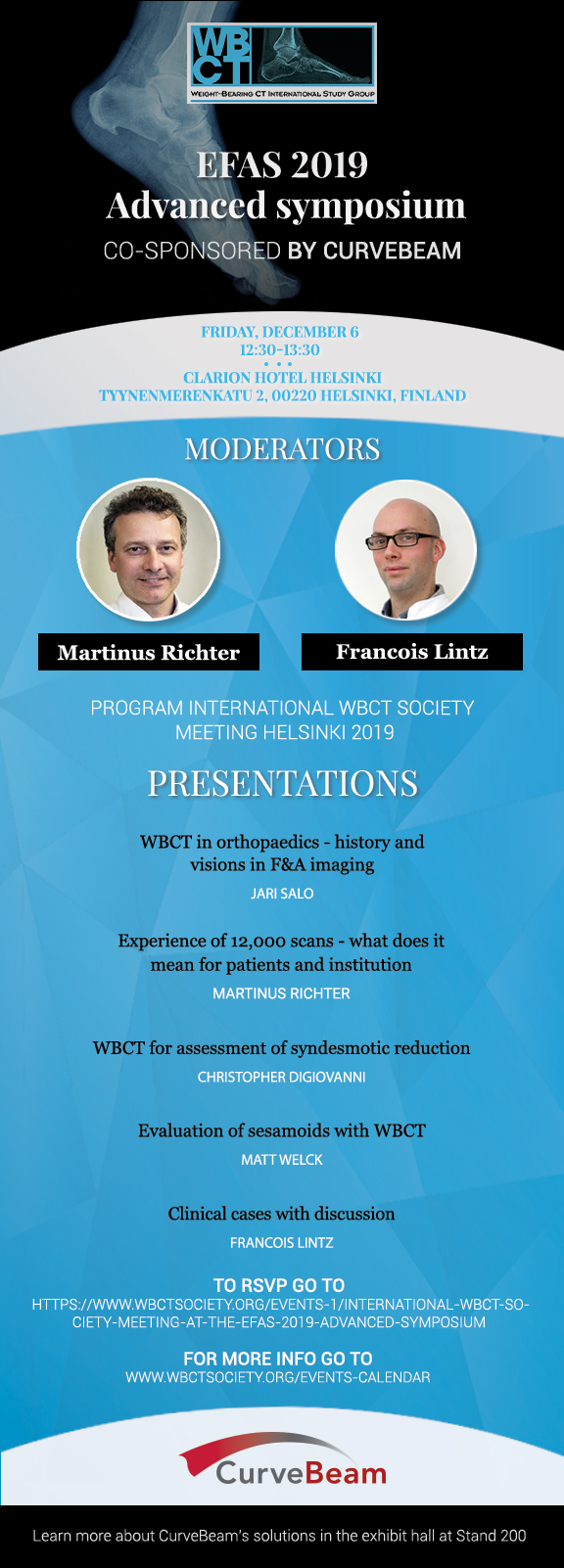
Are you attending the SOTIMI International Congress in Naples, Italy later this month? Be sure to sit in on the following podium presentations on weight bearing CT imaging:
Saturday, Nov. 23
10.00 – 11.00 Comunicazioni: Caviglia E Piede
Misurazioni radiografiche di piede e caviglia nella cone beam weightbearing computer tomography (WBCT) – C. De Franco, V. de Matteo, R. Verrazzo, F. Smeraglia, G. Balati, A. Bernasconi (Naopoli)
Relazione tra instabilita laterale cronica di caviglia e retropiede varo valutato tramite TC cone beam in ortostatismo – A. Bernasconi, F. Lintz, L. Baschet, C. Fernando, N. Mehdi, Weight Bearing CT International Study Group, C. de Cesar Netto (Londra)
Piede cavovaro secondario a Charcot-Marie-Tooth vs piede cavovaro idiopatico: analisi preliminare della morfologia con TC cone beam in ortostatismo – A. Bernasconi, L. Cooper, S. Lyle, S. Patel, D. Singh, N. Cullen, M. Welck (Londra)
Variabiliita’ intra ed inter-osservatore di misurazioni semi-automatiche 3D con TC cone beam in ortostatismo in pacienti affetti da piede cavovaro sintomatico – A. Bernasconi, L. Cooper, S. Lyle, S. Patel, D. Singh, N. Cullen, M. Welck (Londra)
The optimal workflow for in-office Weight Bearing CT (WBCT) imaging is to get insurance authorization, perform the scan, and review scan results all during the initial patient appointment.
Hallux valgus is a tri-plane deformity, and weight bearing allows for a better understanding of coronal plane rotation.
Post-operatively, weight bearing CT can provide a precise view of the rate of fusion healing.
The webcast covered the importance of weight bearing in foot and ankle imaging, the applications of weight bearing CT in common foot and ankle disorders, and how it can be incorporated effortlessly into practices.
According to Dr. Cuttica, “Weight bearing is the functional position of the foot. It allows for us to better determine alignment, to form an assessment of the foot, and to formulate treatment plans. So, weightbearing, obviously as we all know, is very, very important.”
A surgeon at Orthopaedic Foot & Ankle Center (OFAC) in Falls Church, Virginia, Dr. Daniel J. Cuttica, DO, boasts a number of specialties and interests, including foot and ankle surgery, reconstructive surgery, sports-related foot and ankle disorders, cartilage disorders, total ankle replacement, diabetic limb salvage, and dance medicine.
Background
Dr. Cuttica states, “When you evaluate a patient, in addition to clinical exam, you know that imaging is going to be very valuable in diagnosing, treating, and assessing outcomes in foot and ankle.”
However, with weight bearing imaging, you can more reliably identify pathology such as subtle arch collapse, loss of cartilage/joint space, degenerative changes, and impingement.
The Limitations of Conventional CT vs. the Benefits of WBCT
When compared to plain X-Rays, “Computed Tomography (CT) can be very, very beneficial for bone and joint problems, and it does give us a large amount of additional information.” However, Dr. Cuttica explains, “The biggest limitation, at least in foot and ankle, with CT again, is probably your inability to obtain weight bearing images.”
The benefits of CBCTs include:
Easy to operate
Shorter scan time
Patient safety
Optimal patient positioning
Offloads capacity
Flexible siting/easy relocation
There are also many advantages to using WBCT, including:
Ability to obtain weight bearing images
High contrast and spatial resolution
Fast image acquisition time
Decreased radiation (typically 0.01-0.03 mSv vs. 0.07 mSv for Conventional CT)
Relatively small scanner size with portability
Less capitalization cost than Conventional CT
Implementing In-Office WBCT for Foot & Ankle
According to Dr. Cuttica, because of its low radiation dosage and small size, CBCT is ideal for an office setting. For patients, a WBCT scanner in the office is more convenient, can help to avoid unnecessary follow up appointments, and allows for immediate feedback of their diagnosis. For physicians, an in-office WBCT is also more convenient, enabling quicker treatment plan formulation, helping to avoid overbooking, while allowing for more rapid surgery scheduling.
Dr. Cuttica reviewed three office workflow options for in-office WBCT imaging:
Option A – Scan and have patient follow up to go over scans at a later date (not the most efficient)
Option B – Get insurance authorization and perform the scan at the next visit before the patient is seen
Option C – Get insurance authorization, perform the scan, and go over scans at initial appointment (most efficient and most convenient for patients)
Further, WBCT images can be conveniently emailed or uploaded to another doctor or radiologist.
Dr. Cuttica said the most common and beneficial WBCT foot and ankle applications include:
Hallux valgus
Pes planovalgus
Midfoot/Lis Franc injury
Ankle fracture/syndesmosis
Deformity/Charcot
OCD
Bone healing
In Hallux Valgus, WBCT Scans Accentuate Deformities & Guide Treatment
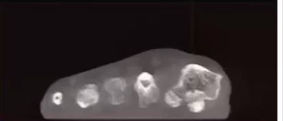
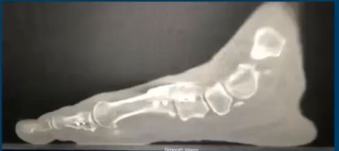
(17:03) In hallux valgus—a triplane deformity—it’s necessary to understand all the components of the malformation. Dr. Cuttica pointed out that WBCT allows for better understanding of coronal plane rotation. As you can see in the image below (17:40), the WBCT imaging clearly shows the first metatarsal joint architecture, the sesamoid position, if there is any flattening/erosion of the crista, as well as a first metatarsal rotation, all of which need to be taken into account when treating bunion deformity.
This screen capture from Dr. Cuttica’s FOOTInnovate webinar displays rotation of the first metatarsal.
(18:16) Dr. Cuttica displayed a typical case of a 47-year-old female with bunion pain who, upon both exam and radiographically, had a hypermobile first ray with some inter-gapping at her first tarsal-metatarsal joint, as well as a moderately sized bunion. (19:43) When Dr. Cuttica performed a WBCT, the rotation of her first metatarsal was visible. Due to the patient’s instability and hypermobility at the joint, Cuttica’s team treated her with the Lapidus procedure—correcting her IM angle and coronal frontal plane rotation—as well as an Akin osteotomy.
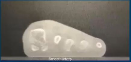
(19:55) At the 6-week post-op exam, the patient’s foot looked fairly healed, and allowed the patient to progress with some activity. (20:19) At the 12-week exam, however, an additional WBCT allowed Dr. Cuttica to better assess the sesamoid position and evaluate the fusion. The sesamoids looked reduced, but it was also revealed that the patient was not fully fused.
This screen capture from Dr. Cuttica’s FOOTInnovate webinar shows the sesamoid position has improved post-operatively.
However, this screen capture from Dr. Cuttica’s webinar revealed the patient’s bones had not fully fused after undergoing a lapidus procedure.
These comprehensive views enabled Dr. Cuttica to better progress with the patient’s treatment—in this case, limiting the patient’s activities as she had a bit more healing to go.
On this CurveBeam Connect Jobcast episode, Director of Regulatory Affairs Ryan Conlon said the most important trait he is looking for is someone with a little bit of creativity. “This role is going to require the associate to read and interpret the regulations or standards, and then use your critical thinking to determine how we’re going to be in compliance with these regulations in a fashion that is minimally impactful to our product performance, company goals, and our timelines and without ever compromising product or patient safety,” he said.
CurveBeam was founded in 2009 by a group of individuals with a proven track record in the advanced and compact CT imaging device domain. We’re an energetic company that is innovating and leading the way in orthopedic CT imaging on a worldwide scale.
Job basics:
Location: Hatfield, Pennsylvania
Days of the week: Monday – Friday
Travel involved: Minimal, if any
Educational requirement: Bachelor’s of Science in Engineering, Science, Regulatory, or a related discipline
If you’re looking for a place where you can work hard and better yourself in an energetic environment, check out Curvebeam’s job openings.
Don’t miss Dr. Albert Armstrong’s forthcoming webinar titled “Podiatric Radiology – Weight Bearing CT Imaging as an Essential Tool for Diagnosis.”
FOOTInnovate will host Dr. Barry’s webinar on Thursday, October 24 at 9 p.m. EST. Click here to register. (A FOOTInnovate account is required to attend.)
Dr. Armstrong is a Professor of Radiology at Barry University. He is a board-certified Diplomat with the American Board of Podiatric Medicine and the American Board of Wound Management. He attended Barry University School of Podiatric Medicine and completed his residency with Mount Sinai Medical Center in Miami, FL.
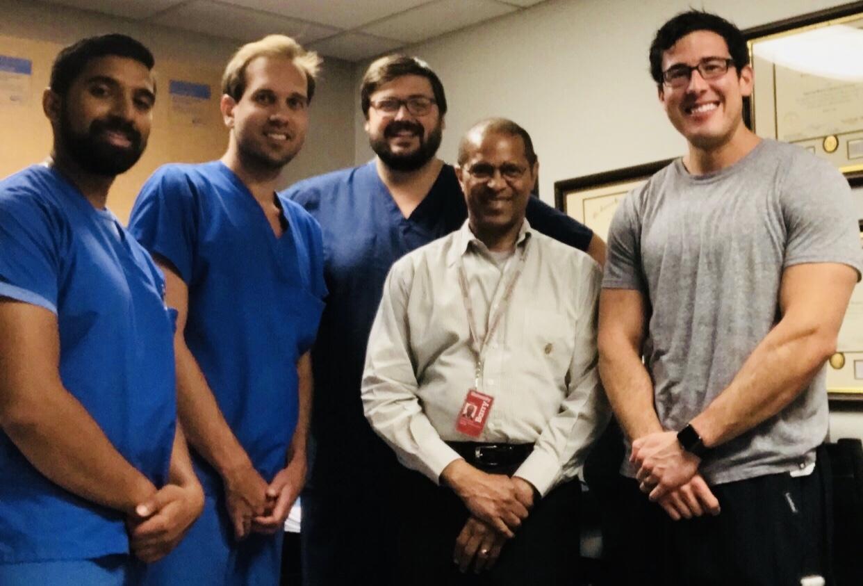
Dr. Albert Armstong, DPM, Professor of Radiology at Barry University School of Podiatric Medicine, in Miami Shores, FL, poses with a group of podiatry residents.
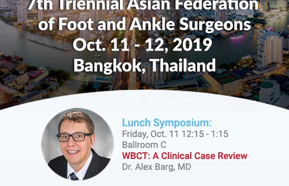
CurveBeam will be exhibiting at the 7th Triennial Asian Federation of Foot and Ankle Surgeons (AFFAS) Scientific Meeting in Bangkok, Thailand. Scheduled for October 11 – 12, 2019 at the Centara Grand and Bangkok Convention Center at CentralWorld in the heart of Bangkok, AFFAS 2019 provides a unique opportunity for orthopedic foot and ankle professionals from around the world to come together to exchange knowledge and innovation in the clinical and research issues currently shaping the specialty.
CurveBeam Lunch Symposium
Join CurveBeam on Thursday, October 11, for a Lunch Symposium in which Dr. Alexej Barg, MD, of University of Utah Orthopedics, will be presenting a Clinical Case Review on WBCT. The Symposium will take place 12:15 p.m. to 1:15 p.m. in Ballroom C.
WBCT Society Symposium
Then, on Friday, October 12 in Ballroom A&B, the International WBCT Society will be hosting a special WBCT Symposium. Dr. Barg’s Presidential address will kick this event off at 11:20am with these highly anticipated presentations to follow:
Time
Presentation
Presenter
11:25am-11:35am
Experience on 11000 Cone Beam WBCT
Martinus Richter, MD, PhD
11:35am-11:45am
Comparative Study of the Alignment of the Hindfoot and Position of the First Metatarsal and Sesamoid of Hallux Valgus with WBCT and WB X-Ray.
Jian-Zhang Zhang, MD, PhD
11:45am-11:55am
Cartilage Imaging with Low Dose WBCT
Oliver Michelsson, MD
11:55am-12:05pm
Comparison Between X-Ray and Weight Bearing Simulation CT on Varus-Type Stage B Osteoarthritis of the Ankle
Kiyonari Tomiwa, MD
12:05pm-12:10pm
Closing Remarks: The Future of WBCT
Francois Lintz, MD, FEBOT
12:10pm-12:20pm
Discussion
Alexej Barg; Francois Lintz,
Please join CurveBeam at our AFFAS 2019 Lunch Symposium on October 11, and be sure to attend the International WBCT Society’s Symposium on October 12. Both events feature renowned orthopedic foot and ankle specialists who will be sharing their expertise on industry-relevant topics.

