Weight Bearing CT Sheds Light on Understanding of Rotational Dynamics in Syndesmosis

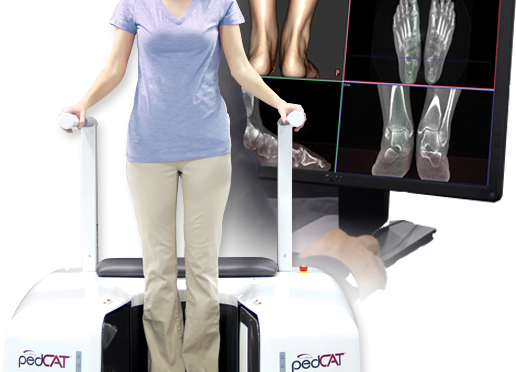
As the orthopaedic and podiatric specialties continue to advance, there is great potential for technology like CurveBeam’s pedCAT system to revolutionize care.

Recent research highlighted the importance of weight bearing scans to the understanding of foot and ankle anatomy, suggesting a role for pedCAT in both a research and clinical setting. “Rotational Dynamics of the Normal Distal Tibiofibular Joint with Weight-Bearing.

Computed Tomography” a 2016 study published in Volume 37 of
syndesmosisFoot & Ankle International sought to determine the normal range of motion for uninjured distal tibiofibular joints. Researchers hoped this reference would be useful as a comparison when assessing injured and repaired ankles.

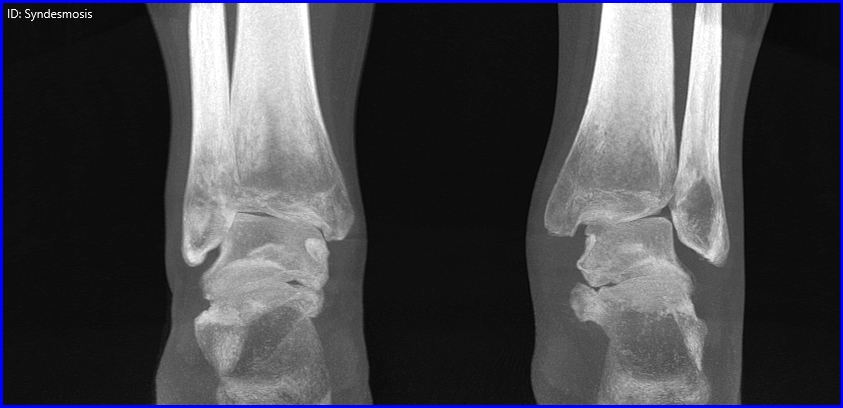
Until recently, all measurements of this motion had been conducted on cadavers or through non-weight bearing scans. In contrast, this study used a weight-bearing CT (WBCT) system to survey the ankles of 32 subjects as they stood on one foot, then the other.

They found a “total movement of 1.5 mm and rotation of 3 degrees” in the syndesmosis as the average across subjects. However, the study also found that intersubject variation was extremely high, meaning different people had vastly different ranges of motion despite similar orthopaedic histories.

These differences were not correlated with sex or age. Intrasubject variation, or the difference in movement between a person’s right and left foot, was significantly smaller and more consistent, less than 1 mm on average.

The study therefore concluded “the contralateral ankle should be used as a reference when dynamic alignment of the distal tibiofibular joint is studied.”

In other words, surveying a person’s uninjured ankle will give a better idea of what is normal movement for that particular individual than comparing the injured ankle to a standardized range, like the one this study endeavored to produce.

CurveBeam’s pedCAT technology could have helped researchers eliminate possible errors in these findings. In the article, researchers admitted “it is possible that we were unable to optimize posture and rotation identically on both sides “ as a result of the limited field of view of the imaging equipment.

The device used in the study could only scan a partial foot in a scan. Test subjects had to stand on one foot, then twist and then switch and twist again. Researchers were unable to measure the force with which the subjects moved on each side.

The pedCAT’s field of view accommodates bilateral imaging, which would have allowed researchers to survey both weight bearing feet at the same time, providing helpful insights. The research indicates the importance of weight bearing measurements of a person’s right and left ankle to determine their normal range of motion.

Being able to accurately assess their syndesmosis on one side will help physicians more accurately assess and repair damages to the other.

CurveBeam’s pedCAT technology is the ideal imaging solution because it allows physicians to scan and survey ankles side-by-side for increased accuracy and ease.

CurveBeam and its technical solutions have the potential to revolutionize orthopaedic and podiatric research and care. Visit us at curvebeam.com to learn how pedCAT and other CurveBeam technologies can make a difference in your practice!

pedCAT Shown to be Most Effective Imaging Option for Mülller-Weiss Disease

The CurveBeam pedCAT imaging system was recently recommended as an incredibly beneficial option for the treatment and analysis of Mülller-Weiss disease. While the disease can be detected in its early stages with magnetic resonance imaging and radiographs, there is a considerable advantage to using weight-bearing computed tomography (WBCT) for diagnosis and evaluation.

Mülller-Weiss involves a painful deformity caused by osteonecrosisKöhler_I and fragmentation of the navicular, most commonly occurring in adults and disproportionately in female patients. The pedCAT, a cone beam CT imaging system, provides an opportunity for early diagnosis and better preoperative analysis, resulting in improved treatment. Standard computed tomography (CT) in itself beneficial for several reasons.

First, it allows for easier examination of the extent of the deformity and for evaluation of any surrounding arthritis. With CT data, surgeons can evaluate and plan for any future attempts at remedying the deformity.

However, because standard CT scans are not weight bearing, they do not provide an accurate view of the relationship between the hindfoot and midfoot deformity. pedCAT WBCT scans allow doctors to go one step further than standard CT scans.

pedCAT 3D datasets can be fully manipulated in order to provide an opportunity for in-depth analysis before proceeding with surgery on a complex deformity. In addition, the pedCAT provides for a better patient experience as the procedure only takes between 19 and 48 seconds, depending on the extent of the deformity.

Weight Bearing CT scan. (Top Left) straight subtalar alignment. (Top right) Anteroposterior view shows navicular bone stock. (Bottom left) Sagittal slice shows features of subtalar varus, superior talonavicular arthritis. (Bottom right) straight subtalar alignment. Source: Foot & Ankle Specialist.

The pedCAT also cuts down on radiation, with a low dose of 1.4 μSv, an amount that is not significantly higher than the 0.7 μSv dosage of the traditional three radiographs of a foot taken to identify Mülller-Weiss. Overall. the pedCAT allows for an accurate and thorough analysis that cannot be achieved through normal X-rays, MRIs nor bone scans.

When creating a treatment plan for Mülller-Weiss disease, the goal is to reestablish a balanced foot, which may require realignment surgery or surgery involving a structural graft.

Due to the complexity inherent in any attempt at treatment, pedCAT imaging is ideal, as it allows doctors to accurately gauge the amount of medial column shortening as well as determine the navicular bone stock and density.

Identifying these key details as accurately as possible is imperative for operations such as calcaneal osteotomy to properly restore alignment or repairing the medial column length.

muller weiss1
Weight bearing CT scan. (Top left) Subtle subtalar varus. (Top right) Anteroposterior view with increased cut thickness allows accurate determination of lateral navicular compression in weight bearing position. (Bottom left) Sagittal slice allows accurate determination of talonavicular and potential subtalar and naviculo-cuneiform arthritis. (Bottom right) Thus demonstrates amount of subtalar varus in weight bearing position. Source: Foot & Ankle Specialist

While a combination of weight bearing X-Ray and unloaded CT scanning are sometimes suggested as an alternative, the degree of the deformity caused by Mülller-Weiss will not be accurately represented.

It is only through WBCT scanning that a true picture of the dynamics within the foot will be understood. pedCAT is a compact and ultra low-dose system, which aims to make such 3D scanning possible for clinics.

Weight Bearing CT Advances Planning for Forefoot Reconstructive Surgery

Think of a surgical procedure like a construction project. No contractor would build a building without a full set of plans, cross-sectional diagrams and 3D renderings. Dr. Bob Baravarian, DPM, feels foot surgeons should be just as prepared.

As he explained in Podiatry Today, Dr. Baravarian has always been interested in architecture and real estate, yet was drawn to 159682158975132.Zfw2RIZiU5Lh04gznDby_height640medicine.

Dr. Baravarian sometimes works next to a hip & knee surgeon in the OR. One day, he observed the hip & knee surgeon had a digital tool that laid out an exact surgical plan for the osteotomy he was performing. The plan rendered leg length and acetabular rotation a non-issue.

Soon after, Dr. Baravarian incorporated a pedCAT weight bearing CT imaging system into his podiatric practice. He found he could plan angular corrections to the degree, calculate the exact size of a graft for an Evans procedure, and could even plan the amount of rotation necessary for anatomic alignment of the hindfoot.


Overtime, he began to apply the same methods to forefoot procedures as well.

  • For hallux valgus cases, he scans the patient in the post-corrected position so he can determine if a hindfoot alignment correction is also needed.
  • For hallux rigidus cases, he uses weight bearing CT to evaluate the level of articular damage and the possibility of cartilage graft procedures. “This is impossible to do at such high levels with plain radiographs,” Dr. Baravarian said in the Podiatry Today article.

pt07treatment1 (1)Bilateral, weight bearing three dimensional views of the foot and ankle give specialists the data they need to create optimal treatment plans. The pedCAT, with a scan time of one minute, is a compact, ultra-low dose CT imaging system that is ideal for orthopedic and podiatric clinics. Patients benefit from the convenience of point-of-care advanced diagnostic imaging, and the pedCAT automatically generates all standard X-Ray views in addition to the full CT volume.

As software programs become more advanced, Dr. Baravarian said, surgeons may be able to perform osteotomies and shift the bones in preoperative planning, which will allow even more advanced planning along with far less operative time and less risk to the pt07treatment3 (1)patient.

Dr. Baravarian is the Chief of Podiatric Foot and Ankle Surgery at the Santa Monica UCLA Medical Center and Orthopedic Hospital, and Director of the University Foot and Ankle Institute in Los Angeles.

Read the entire article here.

Exclusive Video From The First pedCAT Weight Bearing CT Scientific Users’ Meeting in Berlin

meeting-83519_960_720 (1)The first pedCAT Weight Bearing CT Scientific User’s Meeting recently convened at the 2016 Foot International (EFAS, DAF, I-FAB) in Berlin. The meeting focused on weight bearing cone beam CT (WBCT) technology and the potential applications and challenges it presents.

Fill out this form to access the video.

Look for several interesting items as you watch:

  • Professor Martinus Richter’s work included a time workflow study on the use of X-Ray,  conventional medical CT, and the pedCAT; the results will surprise you. His study also showed how weight-bearing  CT measurements are more accurate than X-Ray measurements.
  • Dr. Arne Burssens’ study on hindfoot alignment answered critical questions: How do we identify varus and valgus, how do we accurately measure them, and how can we be sure that the measurements are clinically useful and reproducible?
  • Dr. Francois Lintz gives us a sneak peek at a new weight-bearing measurement CT tool that will be available exclusively in CubeVue, pedCAT’s software. It will enable users to measure alignment in three dimensions, and distinguish between normal and pathological cases.
  • Dr. Cesar Netto’s study focused on adult acquired flatfoot deformity, comparing  measurements on weight bearing CT vs non-weight bearing CT.
  • Dr. Michael Wachowsky’s study focused on the use of weight bearing CT  with pediatric patients, and asks: With the new weight bearing CT technology, what will be the exact definitions of the measurements?

The challenge created by this new technology is clear: How do we now define what we are measuring, since the current X-Ray methods have just been rendered obsolete?

“We can’t have non-weight bearing CTs and say anything about alignment – that’s over,” concluded Dr. Charles Saltzman. “What do we want to measure, and how do we know what we’re measuring represents reality?”

Fill out this form to access the video.

The Face of Technical Support at CurveBeam

When people think of Technical Support when they have a  problem that needs to be solved –  and the sooner, the better. While CurveBeam’s Technical Support can help with on-the-spot issues, it does much more than that. The support comes in many forms: software updates, assistance with calibration, online coaching, and more.

Andrew


All these tasks are headed up by Andrew Kochansz, theTechnical Support Manager at CurveBeam. He joined the CurveBeam family in May of 2013, and has been a long time resident of eastern Pennsylvania.

Andrew has specialized in cone beam CT medical devices since 2006, and previously supported dental/maxillofacial cone beam CT systems. Before that he held positions involving quality assurance, software testing, and computer repairs – all which help make him uniquely qualified to provide support for computer and software dependent imaging systems like the pedCAT. When he is not directly assisting customers, Andrew is also involved in the software testing and verification processes.

When support is required, Andrew is able to remotely log in to most systems and provide assistance within minutes of receiving a call. Sometimes the solution can be as simple as turning the pedCAT off and turning it back on again (“power cycling” it).  If on site required, he coordinates with Customer Relations Director Tami Alexander to arrange for a service technician to travel to the customer site.

Andrew can be reached directly at 267-483-8097 when in his office, from 9:30am – 6:00pm Eastern Time Zone (USA); but he also is available through call forwarding for a couple of hours before and after that. If for some reason he is busy, you can always leave a message with Tami at 267-483-8089, or at the main CurveBeam number 267-483-8081.

For those who need assistance with performing Calibration and QA scans, Andrew would like to remind everyone that video tutorials on how to use CubeVue software, including performing the “manual” QA procedure, can be found on the CurveBeam website.

Andrew can be reached at 267-483-8097 or andrew.kochanasz@curvebeam.com.

The Face of Customer Service at CurveBeam

Satisfied customers are not just the result of well-made products, but the result of continuing support over the lifetime of those products as well. Tami Alexander is the Director of Worldwide Customer Relations at CurveBeam, based out of company headquarters in Warrington, Pennsylvania, United States. She joined the CurveBeam family in October of 2015, moving from the West Coast to take over the position.

Tami Alexander serves as CurveBeam's Director of Worldwide Customer Relations.

Tami has more than 30 years in the medical imaging/radiology world. This includes insurance contracting & credentialing, reimbursement, accreditation and radiation regulations. Prior to joining CurveBeam, she worked at the California and Arizona locations of HealthWest Partners for 13 years, as Director of Operations for five PET/CT imaging centers, and a radiation oncology center.

“I enjoy working with customers in my new role and giving them the support they need long after the sale,” Tami said. “In my previous role,  operations was such an integral part of  the overall experience our customers received  while visiting our centers that I have carried over those same quality standards to meet or exceed our customer’s expectations.”

Tami works closely with Andrew Kochanasz, who is the Technical Support Manager, to ensure that any problems requiring service are quickly addressed. She also works closely with the CurveBeam sales team to coordinate site preparation, installations, and training. If an annual maintenance or any other service is needed at the customer’s site, Tami schedules CurveBeam’s service technicians. If you have warranty questions, or if you would like to purchase additional licenses for CubeVue software, schedule your annual maintenance, or request additional training, Tami is the best person to contact.

Tami would like to let all pedCAT customers know, “Don’t hesitate to give us a call. We are here to help in any way we can. From tech support & customer service, to helping our customers find resources the resources they need for their CBCT systems.”

You can contact Tami at 267-483-8089 or tami.alexander@curvebeam.com.

What Is Cone Beam CT? The Science Behind the Image. Part 1: Overview

Within the last two decades, Cone beam Computed Tomography (CBCT) imaging applications have diversified in both the medical and industrial applications.  Developments in medical diagnostic applications were pioneered in the Dental industry in 1996, when the first commercially produced Dental/Maxillofacial CBCT scanner was introduced.

A number of factors contributed to the commercialization of cone beam CT. First was the introduction of more cost efficient flat panel detectors that were capable of achieving higher resolution. The first dental CBCT systems had image intensifiers coupled with CCD cameras to capture the data used for creating images. These systems were prone to distortions and required frequent calibration. Compared to the image intensifier systems, the new FPDs were less bulky, which in turn allowed the scanners themselves to be designed and manufactured to take up less room. Because the detectors were significantly more sensitive to x-ray photons, the x-ray sources could be made smaller as well.

Faster computer processors using multiple cores, greater working memory capacity, and larger capacity storage drives – all at cheaper prices – made CBCT technology more affordable.

Once CBCT technology was available to the dental specialties, the 3D data was used to create planning and placement tools for dental implants. CBCT technology also revolutionized orthodontics, airway and sinus evaluation, and maxillofacial surgery and reconstruction planning and evaluation.

CBCT applications continue to grow because of the technology’s affordability, equipment design and ease of use.

In the next parts of this series on CBCT technology, we will look into the different components of a CBCT system in more detail.

Need Bunion Surgery? A Weight Bearing CT Scan Could Help You Decide

When a patient appears to have a bunion, a physician typically orders traditional foot X-Rays as part of the clinical evaluation. The X-Rays capture three views of the foot- dorsoplantar, medial oblique, and lateral. But is this enough information to understand complex, three-dimensional object such as the foot? 

A growing number of foot & ankle specialists are advocating that there may be better way to evaluate this common deformity.

“Weight bearing computed tomography scans are beginning to take our understanding to the next level,” Dr. Paul Dayton, DPM, of Des Moines, IA, said in a roundtable discussion on bunions published in the Foot & Ankle Specialist academic journal. “Once you see the connection between coronal rotation and what we have traditionally evaluated on AP radiographs, it opens up a whole new understanding.”

On X-Rays, physicians have to “mentally interpolate” the valgus component of the bunion because they do not adequately capture the frontal plane, said Dr. Robert Weinstein, DPM, FACFAS, of Atlanta, explained in a CurveBeam case study.  

“Since the condition is a tri-plane deformity, we need to understand all of the components and their angular values contributing to the deformity,” Weinstein said. “Better deformity analysis leads to better pre-operative planning, surgical execution, and post-operative results.”

The new data on the coronal position is “exciting” because it explains so many of foot and ankle specialists’ previous questions, Dayton said.  

Weight bearing CT imaging also allows for evaluating deformities that involve external rotation. Rotation of one or more metatarsals, including displacement of the sesamoids, cannot be clearly seen in standard foot X-ray imaging. The lack of the weight-bearing aspect in traditional medical CT imaging means the physician cannot evaluate displacement and rotation under load.

Hallux Valgus Blog Post Image 1
One component of the deformity that is seldom quantified is external rotation, or frontal plane deviation. The presence of rotation of the hallux implies a more complex deformity. A very careful assessment of the first metatarsophalangeal joint architecture, including sesamoid position, their condition, and erosion or flattening of the crista on the inferior first metatarsal head is essential.

It has long been known that in hallux valgus or bunions the relationship between the metatarsal head and the sesamoids is altered,” Andy Goldberg, MD, MBBS, FRCS (TR and Orth), of Stanmore, United Kingdom, told Lower Extremity Review magazine. he said. “The sesamoid bones should sit underneath the metatarsal head, while in hallux valgus the big toe drifts off the sesamoids and the tip of the big toe points outwards. But our research has shown that in many cases the cartilage is worn, which in essence is localized arthritis, and we believe that this could affect the outcome of surgery.”

rotated first met and sesamoids

CubeVue, the pedCAT weight bearing CT imaging system’s custom visualization software, allows physicians to create oblique and frontal-plane images of varying angles and thicknesses from the weight bearing CT data.

Weight bearing CT images could have an impact on the technique a physician selects to repair a bunion, Dr. Dayton said.

“The knowledge that the sesamoids can be in normal position medial and lateral to the crista yet look dislocated on the AP X-ray because of pronation completely changes our mindset about the need for capsular balancing,” Dayton said. “We can see that in those cases supination corrects the deformity.”

Standing CT vs. MRI for Advanced Visualization of Knee Cartilage and Meniscus

At the 2016 OARSI World Congress in Amsterdam, Netherlands, Dr. Neil Segal, MD, MS, and Dr. Ali Guermazi, MD, PhD , presented a poster describing “Advances In Visualization Of Knee Cartilage And Meniscus With Standing Computed Tomography Arthrography”.

Standing CT arthrography (SCTa) has also been shown to have some distinct advantages over MRI, according to the poster presentation. “Potential advantages of SCTa over non-weight-bearing MRI/MRA include: 3D measures of meniscal position and morphology; detection of pathology not detected in unloaded positions; and ability to bear weight bilaterally in a functional position, better recreating the magnitude of muscle and external forces acting about the knee during usual standing….  SCTa can be obtained in multiple knee flexion angles, while an MRI knee coil may permit imaging only with the knee in extension, and SCTa is less expensive than MRI.”

Figure 1: SCTa and corresponding MR arthrography demonstrating outstanding delineation of tibiofemoral and patellofemoral articular cartilage, with better differentiation between the cartilage and subchondral bone on SCTa. Visualization of the boundaries of the menisci was achieved to a similar degree on SCTa and MRA.

As part of the background for their presentation, Drs. Segal and Guermazi stated that “MRI is the standard for non-invasive visualization of cartilage and menisci, and … Absence of weight bearing limits evaluation of the functional position and configuration of these structures…Advances in standing CT (SCT) have allowed 3D imaging of the knees while under physiological loads, similar to fixed-flexed or semi-flexed radiograph protocols.”

Their objective was to evaluate a protocol for SCTa for imaging weight bearing cartilage and menisci and to assess potential advantages over non-weight bearing MRI.

figure 2
Figure 2: Sagittal reformatted SCTa and its corresponding MRA demonstrated outstanding delineation of articular cartilage with better differentiation between the cartilage and subchondral bone on SCTa, while also visualizing the ACL and PCL in the femoral notch.

Although the sample size was small, the results were noteworthy. As shown in the images, SCTa permits evaluation of cartilage and menisci in three dimensions, while the patient is standing and under physiological load. Furthermore, SCTa, “may be useful for assessment of menisci as well as tibiofemoral and patellofemoral cartilage in functional stance,” according to the presentation.

The two participants in the study were a 42-year-old man without osteoarthritis (Figures 1 and 2), and a 67-year-old woman with KL2 knee osteoarthritis (figures 3-5). The participants had a similar contrast agent applied.

Figure 3a: Sagittal SCTa demonstrating minimal cartilage thinning on the left medial tibial plateau Figure 3b: Axial image depicting location of the sagittal slice in Figure 3a
Figure 3a: Sagittal SCTa demonstrating minimal cartilage thinning on the left medial tibial plateau
Figure 3b: Axial image depicting location of the sagittal slice in Figure 3a

The actual imaging techniques used were fixed-flexed (approximately 20°) SCTa (INVESTIGATIONAL ONLY cone beam CT knee imaging system*, CurveBeam, Warrington, PA, USA) and non-weight bearing MRI (Siemens TrioTim, Washington DC, USA).

“Following 2-3 minutes of unloaded knee flexion and extension, a low-dose SCT scan was acquired utilizing cone beam reconstruction. Participants were positioned with the tips of the great toes, patellae, and the anterior superior iliac spines coplanar to each other and the feet 10°externally rotated. Scans were acquired with a 0.3mm isotropic voxel size (20x 35x35cm) with an effective radiation dose of approximately 0.1 mSv. 10 minutes following SCTa, MR arthrography was acquired (NEX=1, ETL=3, Slice thickness=2 mm, Slice spacing 2 mm, Matrix= 240 x 320, FOV=140 mm with axial T1 fat-sat (TR=712 msec, TE=12 msec); coronal T1 fat sat (TR=730 msec, TE=10 msec); and sagittal T1 fat sat (TR=796 msec, TE=10 msec).

Figure 4a: Coronal SCTa demonstrating minimal cartilage thinning on the left medial tibial plateau Figure 4b: Axial image depicting location of coronal slice in Figure 4a
Figure 4a: Coronal SCTa demonstrating minimal cartilage thinning on the left medial tibial plateau
Figure 4b: Axial image depicting location of coronal slice in Figure 4a
Figure 5a: Sagittal SCTa image demonstrating a small tear of the tibial surface of the post. horn of the medial meniscus Figure 5b: Axial image depicting location of sagittal slice in Figure 5a
Figure 5a: Sagittal SCTa image demonstrating a small tear of the tibial surface of the post. horn of the medial meniscus
Figure 5b: Axial image depicting location of sagittal slice in Figure 5a

*The CurveBeam knee imaging system is investigational only and is not available for sale in the US.

pedCAT: Early Diagnosis of Osteomyelitis in the Diabetic Patient

The pedCAT weight bearing CT imaging system could identify bone infection at an early stage, and possibly prevent amputations, researchers at the California School of Podiatric Medicine at Samuel Merritt University determined in a report.

The researchers outlined two cases where “the use of CBCT device enabled us to diagnose and treat osteomyelitis in a timely manner, preventing its spread to adjacent bone and soft tissue, and minimizing the amount of required surgical resection.”

Plain radiograph is the primary imaging modality for the osteomyelitis diagnosis, the report states, but X-Rays may not reveal osteolytic changes for up to 20 days from the onset of infection or until the bone density is reduced by 30 – 50 percent.

Osteomyelitis is one of the most feared complications of diabetic foot ulceration, which often leads to lower extremity amputation and disability. Early diagnosis of osteomyelitis increases the likelihood of successful treatment and preserving ambulatory function. Unfortunately, most of the currently available imaging modalities are of limited use in assessing early stages of bone infection due to their low specificity and sensitivity for early osteolytic changes.

Magnetic resonance imaging (MRI) is more sensitive and specific than X-Ray, and yields greater accuracy in detecting soft tissue abscesses or early osteomyelitis in patients with high clinical suspicion and negative radiographs. In controversial or uncertain cases where MRI is not available, other imaging techniques such as indium-labeled leukocyte imaging combined with radionucleotide bone scan can be used as an alternative. (“Preventive and Therapeutic Strategies for Diabetic Foot Ulcers” – Foot & Ankle International® 2016, Vol. 37(3) 334– 343 – Chris C. Cychosz, BS, Phinit Phisitkul, MD, Daniel A. Belatti, BS, and Dane K. Wukich, MD).

Nuclear Imaging and MRI perform well in detecting early onset of osteomyelitis; but they are expensive to own and operate, are time-consuming in their acquisition of images.

What is desirable for early detection is a method that is accurate, inexpensive, and readily available. Cone beam CT, and the pedCAT in particular, fills this gap nicely.  The device is small enough to fit into most practices, offers high-resolution 3D imaging capabilities, and has a reduced radiation dose compared to traditional CT, according to the report.

Lead author Alexander M. Reyzelman, DPM,  and his associates reported on two diabetic patients who presented with infected neuropathic foot ulcers and were evaluated for potential osteomyelitis using plain film radiographs and the pedCAT CBCT scanner. In both cases, the “pedCAT was instrumental in identifying bone infection. The diagnosis of osteomyelitis was later confirmed by positive findings on bone biopsy. The use of CBCT device enabled us to diagnose and treat osteomyelitis in a timely manner, preventing its spread to adjacent bone and soft tissue, and minimizing the amount of required surgical resection.”

Case 1

A 49 year old diabetic female presented with an infected neuropathic ulcer at the lateral aspect of her fourth digit. The ulcer demonstrated malodor, cellulitis that extended to fourth metatarsophalangeal joint and positive probe-to-bone test. The plain film radiographs and CBCT were utilized in order to rule out osteomyelitis and assess the extent of soft tissue infection. The weight-bearing X-rays of the affected foot revealed subtle lucency at the lateral aspect of the proximal phalanx of the fourth digit, which was contiguous with the ulcer location. However, this finding alone was not sufficient to yield a conclusive diagnosis.  The images obtained using PedCAT clearly demonstrated the break in the cortex and the area of osteolysis involving the proximal phalanx of the fourth digit. The head of the fourth metatarsal and adjacent digits appeared intact. These findings, in conjunction with the clinical appearance of the affected digit, led to a preliminary diagnosis of osteomyelitis. The patient was treated with an arthroplasty of the fourth proximal interphalangeal joint, and has fully recovered. The bone specimens obtained intraoperatively were sent for biopsy, which confirmed our preliminary diagnosis of osteomyelitis.

Case 2

A 53 year old diabetic male presented with an infected neuropathic ulcer at his fifth metatarsal head, which exhibited malodor, edema and erythema extending through tthe plantar lateral aspect of  fifth metatarsal shaft and probed to joint capsule. The X-rays demonstrated no signs of bone involvement, while CBCT revealed distinct areas of cortical lysis and bony fragmentation of the fifth metatarsal head. The proximal two thirds of the shaft of the fifth metatarsal appeared unaffected, with intact cortex, uniform bony density and lack of osseous fragmentation. The patient was treated with partial resection of the fifth metatarsal. The bone biopsy has confirmed our preliminary diagnosis of osteomyelitis.

In the concluding discussion, Dr Reyzelman noted: “Though in our case studies we have not taken advantage of the option allowing to scan the patient in both, a weight-bearing and a non-weight bearing positions, this option could be highly useful for evaluation of complex fractures and dislocations of the foot and ankle.”