Improved CT Foot and Ankle Analysis with True 3D

This video takes a look at EchoPixel’s latest technology, True 3D. A few studies are used as examples of what this powerful visualization platform can allow specialists to see. Using a simple stylus tool, analysts can take full advantage of the program’s 3D rendering capabilities. The program provides intuitive displays for complex tasks, such as making measurements in a 3D space to determine the depth of an implant.

Please enjoy this video as it shows the power of True 3D on a non-weight bearing CT dataset, and imagine the possibilities of pairing EchoPixel technology with bilateral, true weight bearing pedCAT datasets.  Visit echopixeltech.com to learn more.


The Opportunity for Private Office Imaging

Changes in healthcare insurance, regulations, and other factors have shifted the bulk of imaging services to hospitals, but the opportunity for private radiologists is still very promising.

“Hospital outpatient imaging has been migrating from private offices since about 2010,” says Dr. David C. Levin of Thomas Jefferson University, as reported by Aunt Minnie. “But that doesn’t mean that radiologists still can’t make private office ventures work.”

Dr. Levin goes further than just his diagnosis of the medical imaging service industry by prescribing a solution to private radiology offices interested in increase their imaging business.  His business model suggestions include a mix of customer service, marketing, and cost competitive advice that can turn around a struggling radiology office and build a successful and sustainable private practice.

Implement a Cost-Effective Payment Schedule for Patients with High Deductibles

The increasing number of high deductible healthcare plans has created a patient incentive to shop around for low price imaging services. Understanding these plans will help your office develop a fee schedule that is attractive from a cost perspective.

“Patients are going to have higher and higher deductibles, and that’s going to make them shop around for healthcare,” says Dr. Levin. “If you can offer, say, an MRI of the knee for $600, while the local hospital has it at $4,000, you can be sure patients will come to you.”

Marketing – It Really Works

Make prospective patients aware of your services by advertising an elevated quality of care and customer service.

“Hospitals generally don’t market their radiology services, but private practices certainly can,” says Dr. Levin. A well-designed advertising campaign can make your practice stand out and drive new customers to your location.

Don’t Neglect Customer Service

There is so much more that goes into the patient experience besides the treatment itself. Examine the way patients are treated, from the initial contact all the way through follow up, to identify opportunities to improve the experience. Patient frustration can arise at any step in the process. Time saving processes as well as friendly and helpful engagement go a long way to improving the overall patient experience.

Efficiency.  Efficiency.  Efficiency.

Dr. Levin stresses the importance of efficiency throughout the business.

“The more efficient your practice, the lower your costs will be,” he says. Fully utilizing assets and people in a streamlined operation brings down overhead and operating costs, and makes for a lean, cost competitive business with higher margins.

At the end of the day, Dr. Levin emphasizes private radiology practices have an amazing opportunity to improve their imaging services and overall business. Taking the time to evaluate the situation, develop an action plan, and implement constructive changes will certainly pay dividends now and for years to come.

New, Reliable Technology for Measuring Tibiofemoral Joint Space

Recent work by Dr. Neil H. Segal examined the potential of a low-dose standing CT scan for evaluating changes in tibiofemoral joint space of patients suffering from knee osteoarthritis.

The results of Dr. Segal’s study were recently published in the medical journal Skeletal Radiology and reveal that when taken two weeks apart, standing CT images are even more reliable than plain radiographs. The insightful, 3-D, information-packed images suggest that the modified foot/ankle scanner by CurveBeam used to capture the data may soon become a valuable tool for doctors to evaluate and track the disease’s progression, as well as for researchers wanting to study it.

Measurements of the tibiofemoral joint space were obtained from two bilateral fixed-flexed standing CT images taken two weeks apart, utilizing a modified version of a CurveBeam scanner more commonly used to capture images of the foot and ankle. Participants were exposed to an extremely low dose of effective radiation, O.2 mSv – no more than the average person is typically exposed to from the natural environment itself in any two-week period.

The main purpose of the study was to determine whether or not standing CT scans might be a viable alternative to plain radiographs for studying patients’ knee osteoarthritis – and how reliable such scans might be. Thirty people with a range of osteoarthritis features took part, and the Institutional Review Board approved all aspects of the study.

The results suggest the data obtained is extremely reliable. Moreover, the 3D imaging of both the tibiofemoral and patellofemoral joints the standing CT scanner produces offers a wealth of enhanced information for doctors and researchers to draw upon.

All this, the study shows, without a clinically significant increase in either acquisition time or radiation dose.

Dr. Segal’s work comes as a response, in part, to the difficulties and challenges in acquiring meaningful, reliable tibiofemoral joint space measurements using traditional radiographs.

Past studies have shown that the replication of measurements has been poor, with limited information obtained. More aggravating still is the fact that months if not years are usually required between radiographs before any change in the patient’s condition can be detected. An efficient, accurate, time-saving alternative has long been needed.

The modified CurveBeam scanner, by contrast, has the potential to address these challenges. As evidence from the study showed, the standing scanner provided exceptionally reliable images with insignificant doses of radiation emitted.

The next step will be to assess the responsiveness of the standing CT scanner to changes in the tibiofemoral joint over time. If effective, the protocol will no doubt become an invaluable tool for doctors and researchers alike in their work to combat the ravages of knee osteoarthritis and related ailments.

Access the link to the article here.

Preview: Curvebeam at AAHS

In September 2016, we showcased InReach at the ASSH Conference! Take a look above!

Happy New Year from CurveBeam, we’re kicking off 2017 with a bang! This week in Waikoloa, HI, we are showcasing some incredible tech at the American Association for Hand Surgery Conference.

The AAHS was founded on the premise of bringing hand surgeons and hand therapy professionals from the United States, Canada, and other countries around the world who work to assess and manage a variety of common and complex conditions related to the hand and upper limb together to provide an educational forum to expand the professional expertise.

Here are the top reasons our Booth is a can’t miss!

    1. Experience CubeVue

CubeVue is a custom visualization software that can transform your diagnosis ability with 3D technology. It automatically generates all standard X-Ray views, eliminating the need for your plain X-Ray system altogether. Receive a demo at our booth!

      1. Learn how to better practice workflow

It is important to understand how point-of-care advanced diagnostic imaging can benefit your practice workflow. Learn how our imaging solutions can help you!

        1. Receive information about InReach

InReach is a point-of-care extremity CT imaging device that you saw in the video at the beginning of this article! The InReach is an extremity cone beam CT scanner that images the hand, wrist, forearm, elbow and the lower extremities that’s comfortable and low impact on your patients. It’s also FDA 510(K) Pending.

We are proud to surround ourselves with the top of the line medical professionals in the field of hand and upper extremities. Your dedication and hard work in the field is the reason we do what we do, and we are honored to be a part of a community that advances in leaps and bounds each year.

If you’re at the conference this week, don’t be shy, we’d love to meet you!

CurveBeam’s Volunteer Day Warms Community

The holiday season is an excellent time for any company to celebrate and share its values. The team at CurveBeam believes in giving back, just as much as it believes in innovation. To reach out with this spirit, a group of CurveBeam volunteers recently connected with an amazing organization.

Everyone had a simple goal in mind: to make sure kids in the community have the supplies they need for this season and beyond.

Cradles to Crayons serves as a resource for communities in need and a hub of civic engagement in Greater Philadelphia. This organization provides children from birth through age 12 living in homeless or low-income situations with the essential items they need to thrive – at home, at school and at play – free of charge.

Ensuring kids are safe, warm and ready to learn throughout the year

Cradles to Crayons was founded in 2002 to get basic necessities to the kids who need them, including clothing, school supplies, toys, and more. The organization provides all the physical goods that children need to be safe, protected from the elements, and ready to learn.234585951194320-834hbvhp2y7forv0xdvp_height640

By supplying kids with high-quality used or new goods, Cradles to Crayons and its partners make children feel valued.

The organization partners with social service organizations to connect groups that have surplus resources with the communities and families that need them. Cradles to Crayons makes it simple for children, families, and corporations to participate by organizing a clothing collection, hosting a fundraiser, or working in one of its warehouses.

Charity Navigator – an independent group that rates charities on both their organization capacity and efficiency – recently awarded the Cradles to Crayons a four-star rating: their highest ranking. In receiving this designation, Cradles to Crayons was recognized for exceeding industry standards and outperforming most charities in its cause.

In addition, Cradles to Crayons Philadelphia was included in Main Line Today‘s list of local charities to support.

A CurveBeam volunteer party

In lieu of a traditional Christmas party, a group of CurveBeam volunteers spent half a day at Cradles to Crayons, helping prepare items to go out to kids in need in time for the holidays.

The team enjoyed the experience. It was the perfect expression of the company’s ongoing commitment to giving back, and one the team looks forward to repeating next year.

To learn more about CurveBeam’s work and its talented and warm-hearted team, visit CurveBeam.com. Happy New Year!

Curvebeam at RSNA 2016

In November many of the greatest minds and innovators in radiology convened in Chicago for the 2016 RSNA. We had our talented and skilled team representing us and our innovations. While there we unveiled the prototype for the LineUP, a bilateral weight-bearing Cone Beam CT imaging system for the knees and lower extremities. We also showcased  InReach and TALAS. Watch below to recap the show!


CurveBeam’s Top 5 Moments of 2016

Founded in 2009, the news of CurveBeam and its advanced, compact 3D imaging devices spread quickly among orthopedic and podiatric specialists. Just three years later, the CurveBeam pedCAT system cleared the FDA. Without losing any momentum, CurveBeam Europe LTD’s office was opened in London in 2013. Even with all of these notable accomplishments, 2016 shines as the most impressive year for CurveBeam thus far. From our President and CEO, Arun Singh, receiving a lifetime achievement award to our expansion into the Asian market, 2016 has been a year to remember.

Take a look at the top five most significant moments for CurveBeam in 2016.

  1. President and CEO Arun Singh presented the Lifetime Achievement Award

“In recognition and appreciation of [his] visionary contributions to the advancement of Cone Beam Computed Tomography,” the American Association of Dental Maxillofacial Radiographic Technicians presented Arun Singh with the Carl Gugino Lifetime lifetime-achievement-award
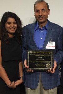
Achievement Award on October 7th. The award was presented to him by Mr. Jerry Peck, who owns dental imaging centers in the San Francisco area. In his speech, Mr. Peck described when Arun installed a Tomax system in one of his offices 27 years ago. He described how Arun worked overnight to create the software code Mr. Peck needed to run the calculations  for the measurements he was looking for.

“This is the type of person, how bright, smart, ingenious, he is. I mean, overnight, all of a sudden, the program is written,” Mr. Peck said.

Mr. Peck went on to describe how Arun cofounded Imaging Sciences and developed both the CommCAT and I-CAT, which were game changers for dental professionals.

“Today, you look at what is the most successful CBCT company in the world, and you built it Arun,” Mr. Peck said.

2. First Weight Bearing Users Meeting is Held

Focusing on the applications of the weight bearing cone beam CT, the first pedCAT Weight Bearing CT Scientific User’s Meeting was held at the 2016 Foot International in Berlin, Germany. “We can’t have non-weight bearing CTs and say anything about alignment – that’s over. What do we want to measure, and how do we know what we’re measuring represents reality?” said Dr. Charles Saltzman as he emphasized the importance of WBCT at the meeting.

3. First Podiatry School is Outfitted with pedCAT System

The Kent State University College of Podiatric Medicine became the first podiatry school to implement the revolutionary pedCAT cone beam CT imaging system. “Kent State University is proud to be the first facility in the Northeast Ohio region to provide this state-of-the-art imaging modality,” said Dr. Allan Boike, Dean and Professor of Foot and Ankle Surgery at KSUCPM. “The CurveBeam pedCAT will allow the college to improve the foot and ankle health of the community while providing the highest quality education for our students and research opportunities for our faculty and residents.”

ben-franklin
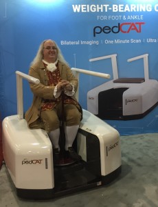
4. Ben Franklin and Susan B. Anthony See the pedCAT System

Philadelphia is a city well known for its rich history, and CurveBeam is proud to be in its next chapter, as the company is now headquartered in this remarkable town. At the APMA “The National” meeting this summer, historical interpreters acting as Ben Franklin and Susan B. Anthony visited the CurveBeam booth to learn more about the revolutionary pedCAT technology.

5.CurveBeam Expands International Presence to Asia

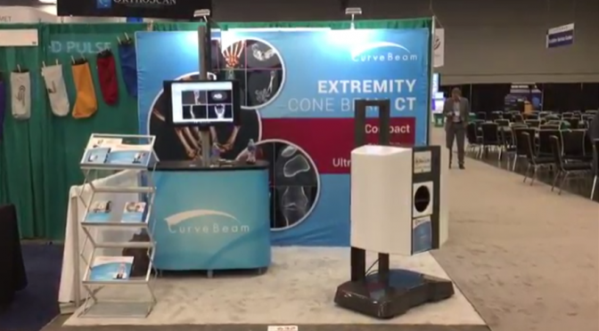
Not only has CurveBeam expanded its domestic presence, the use of the technology worldwide is also growing. The pedCAT system received notable attention at the 2016 National Foot & Ankle Surgery Symposium in Qingdao, China, as CurveBeam’s Chinese partners presented the weight bearing CT technology to the audience of medical professionals.

It’s been quite a ride since our original team pioneered cone beam CT imaging technology for the podiatric specialty. As we gear up to expand into new orthopedic specialties, we could not be more excited for what 2017 will bring!

Comparison of Three Hindfoot Alignment Measurements: Radiographic Hindfoot Moment Arm, Radiographic Hindfoot Alignment Angle, and TALAS

Dr. Matthew Welck presented findings from a study overseen by Dr. Mark Myerson at the British Orthopedic Foot & Ankle Society (BOFAS) meeting in Bristol, England, in early November.

The study compared three hindfoot alignment measurements: radiographic hindfoot moment arm, radiographic hindfoot alignment angle, and TALAS. TALAS is a semi-automatic measurement based on anatomical landmarks on a weight bearing CT scan. TALAS was found to have the best intra-observer reliability.

5th-mt-contact-point
5th MT contact point.

As Dr. Welck explained, the TALAS technique measures offset of midline of whole foot, rather than just calcaneus. He presented an example of a patient with ‘neutral’ alignment to compare hindfoot alignment measurements. The first point was placed on the superior weight bearing portion of the talar dome. The calcaneus contact point was then placed on the lowest point of the calcaneus. The first metatarsal contact point was placed on the lowest point of the first metatarsal head. Similarly, the 5th metatarsal contact point was placed on the lowest point of the 5th metatarsal head. TALAS software provided a mapping of these contact points showing the axis, or the ’true’ hindfoot alignment.Next, Dr. Welck summarized the intra and inter observer reliability among the three measurements. As previously shown in the literature, Dr. Welck explained, there is good intraobserver reliability with the Saltzman hindfoot moment arm technique. Similarly, the hindfoot alignment angle has excellent intraobserver reliability. And there was excellent intra-observer reliability among investigators. Dr. Welck noted that while all three measurement techniques are reliable, the CT Ground Reactive Force Technique was nearly perfect.

CurveBeam’s pedCAT provides bilateral, weight bearing 3D CT imaging of the foot and ankle, greatly surpassing traditional imaging methods such as X-Ray and CT studies. CurveBeam’s technology also reduces the number of patient trips to the hospital, and exposes patients to less radiation. Because the 3D data generated by pedCAT is far more robust than could be interpreted by available software, CurveBeam engineers are collaborating with leading foot and ankle surgeons to develop a new measurement software – TALAS. TALAS, or Torque Ankle Lever Arm System, is a feature within pedCAT’s visualization application CubeVue.

talas-software
A mapping of the contact points in the TALAS software.

The British Orthopaedic Foot & Ankle Society (BOFAS) is a society of orthopaedic surgeons who have a special interest in surgery of the foot and ankle. The Society works to encourage interest in foot and ankle surgery among orthopaedic surgeons and to encourage both basic science and clinical research in the field.

Value of 3D Reconstructions of CT Scans for Calcaneal Fracture Assessment

Operative fracture treatment of irregularly shaped bones, such as the calcaneus, scapula and scaphoid, demands high-quality images of the area in question for both classification of the fracture and planning of the procedure. However, since plain radiographs do not provide enough pertinent information to this end, computerized tomography (CT) has become the standard for providing the necessary images for treatment of these irregularly shaped bones.

Yet, in a study observing 2D CT scans, only 42% of the evaluators were able to correctly classify the fracture, necessitating the need for some sort of change. Three dimensional CT renderings were proposed to fix the low inter-rater agreement generated by the 2D scans.

The foot in the video above was scanned on a pedCAT weight bearing CT imaging system for the foot and ankle. The calcaneus was segmented using CurveBeam’s CubeVue visualization software. 

To evaluate the effectiveness of 3D CT scans, A standard set of CT secondary reformation scans were presented, followed by a questionnaire describing fracture anatomy and preoperative planning. Subsequently, 3D reconstructions were presented to the evaluates followed by the same questionnaire. After presentation of the 3D images, 49% of the evaluators changed their plan in regard to the approach and 29% in regard to the implants.

Five different data sets (four intra-articular and one extra-articular fractures) were presented to 57 evaluators. All groups, except that of surgeons with more than 20 years of experience, benefited from 3D CTs (Friedman test; P < .01). Inexperienced surgeons benefited more than experienced surgeons and complex fractures more than simple fractures. Specifically, regions of interest such as the middle facet and fractures extending into the calcaneo-cuboid joint were evaluated more precisely.

In regard to 3D CT scans, Böhmer1 described the topographic relationship between the fragments and the surrounding structures as useful for evaluating calcaneal fractures and for preoperative planning because the fractures can be seen from unusual perspectives. Likewise, Choplin2,3 posited 3D scans assist diagnosing foot deformities since the scans improve comprehension of the anatomy, particularly for especially complex fractures. For such complex fractures and anatomy, Pate5 evaluated 202 patients with complex musculoskeletal problems and found 3D CT particularly helpful. Others came to similar conclusions.

When comparing techniques for diagnosing foot deformities, Allon and Mears4 compared plain radiography, 2D CT, and 3D CT of 30 fractured calcanei and concluded that 3D CT improves preoperative planning and the choice of an adequate approach.

Overall, 3D CT scans provide insight previously unavailable through both 2D and plain radiography, which inexperienced surgeons tend to find more helpful in diagnosing and preoperative planning.


References

  1. Bohmer G, Roesgen M, Hierholzer G. Three-dimensional computerized tomography in trauma surgery. A case presentation [in German]. Aktuelle Traumatol. 1992;22(2):47-56.
  2. Choplin RH, Buckwalter KA, Rydberg J, Farber JM. CT with 3D rendering of the tendons of the foot and ankle: technique, normal anatomy, and disease. Radiographics. 2004;24(2):343-356.
  3. Choplin RH, Farber JM, Buckwalter KA, Swan S. Threedimensional volume rendering of the tendons of the ankle and foot. Semin Musculoskelet Radiol. 2004;8(2):175-183
  4. Allon SM, Mears DC. Three dimensional analysis of calcaneal fractures. Foot Ankle. 1991;11(5):254-263
  5. Pate D, Resnick D, Andre M, Sartoris DJ, et al. Perspective: three-dimensional imaging of the musculoskeletal system. AJR Am J Röntgenol. 1986;147(3):545-551.

Curvebeam’s Booth Is a Must-See at RSNA 2016!

In just a little over a week, many of the greatest minds and innovators in radiology will convene in Chicago for RSNA 2016, and we at CurveBeam are proud to have our talented and skilled team there representing us. Over the course of the conference, there will be groundbreaking research presented, technological advances displayed and educational classes offered. In the heart of it all, our booth will be featuring some incredible innovations – proving once again that CurveBeam is continuing to find new ways to push the boundaries of our field.

Our core team pioneered Cone Beam CT imaging technology for the dental and ENT specialties. The ability to provide point-of-care imaging revolutionized these industries and allowed not only for improved care, but for the possibility of custom dental implants. Now we are taking that same breakthrough technology and adapting it to the orthopedic field, and we are proud to introduce the results of our efforts at this year’s RSNA. Here are our top three reasons why you can’t afford to miss the CurveBeam booth this year:

  1. See the LineUp

We will  unveil the prototype for the LineUP, a bilateral weight-bearing Cone Beam CT imaging system for the knees and lower extremities. It will not only plugs into a standard wall outlet, but will also be self-shielded and will complete a scan in under a minute. While the technology is still investigational only, it is PACS/ DICOM compliant and is sure to revolutionize the way orthopedic clinics approach radiology.


  1. Experience InReach

While the LineUp is certainly fast, the InReach completes scans of the hand wrist, forearm, elbow and extremities in under 20 seconds flat. We designed InReach technology to bring the idea of point-of-care extremity CT imaging  to upper extremity specialists. While it is also still investigational technology, the progress we’ve made toward this goal is worth coming to see.


  1. Discover TALAS

One of the most crucial elements of orthopedic surgical planning is the precise pre-operative measurement of hindfoot realignment. TALAS is a tool that makes the process not only smoother, but more accurate and precise. It is a semi-automated hindfoot alignment measurement device that has been adapted for weight-bearing CT. Come check it out for yourself, and discover the potential TALAS has to one day be a true game-changer in the field.

“We are tremendously excited to share our recent research and development efforts with the global radiology community,” says CurveBeam President and CEO Arun Singh. RSNA is an opportunity to share and learn together as a community and we are thrilled to be a part of that experience. Feel free to come by and discover our innovative new technology. If you want to learn more before seeing our products in person, visit CurveBeam.com. We hope to see you soon at RSNA 2016!