If CurveBeam was a band, it’d be the Foo Fighters. How’s that for a hook? On this CurveBeam Jobcast, host Tyler Kern got the details on an open software engineer position for Pennsylvania-based CurveBeam, the first company to offer a weight-bearing CT for extremities which has transformed the field of podiatry and orthopedics.
CurveBeam Software Development Manager Dave Rovner explained: “The main guy in that band Dave Grohl was in another band, Nirvana, a pioneer in the music scene. So, how does that relate? Our CEO Arun Singh as well as others at CurveBeam come from another pioneering company in the dental imaging world and CurveBeam really extended that technology to the medical field.”
CurveBeam, a 3D orthopedic imaging leader in the United States, Europe, Australia, and China, is looking for a software development engineer to join its reputed team five days a week in Hatfield, PA. Rovner said the ideal candidate will have a Bachelor of Science in Engineering, Computer Science, or Biomedical Engineering with a software emphasis, as well as two years of experience in programming Python.
Rovner said adaptability within this agile company is essential, as well as good communication skills within small engineering teams.
“Also, we want someone who has the confidence to present new ideas and challenge old ones,” Rovner said. “This is an environment that encourages that.”
At CurveBeam, we pride ourselves on being at the forefront of weight bearing CT (WBCT) technology. The next product in CurveBeam’s family of innovative systems has arrived.
Introducing the HiRise, the next level of weight bearing CT imaging. HiRise is investigational only and is not available for sale.
Vinti Singh, CurveBeam’s own Director of Marketing, demonstrated the HiRise for visitors at RSNA 2019.
What makes HiRise so revolutionary?
The CurveBeam HiRise is the industry’s first solution for capturing bilateral, weight bearing CT imagery of the hip and pelvis.
The HiRise will provide high-resolution, high-contrast imaging of the hip and pelvis that allows for cutting-edge orthopedic care.
Leveraging a rising gantry that surrounds the patient being scanned, the HiRise allows for long-leg views of the entire lower extremity.
The HiRise is also self-shielded, plugs into a standard wall outlet, and offers the largest patient platform in its class at 35 centimeters. The gantry rises high enough to accommodate patients well above 6 feet tall.
Best of all, like CurveBeam’s other solutions, the HiRise is simple to use, offering a fast track to revolutionary orthopedic care.
Providing Complete Care with CurveBeam
The HiRise is yet another addition to CurveBeam’s industry-leading solutions for weight bearing extremity imaging, joining the pedCAT, LineUP and InReach in helping you achieve the weight bearing difference.
With 3D, weight bearing imagery and software that can help you analyze and act upon the insights it provides, you can improve diagnostic information, achieve better patient outcomes and streamline your practice’s workflow and operations.
To learn more about CurveBeam and the HiRise, visit curvebeam.com/.
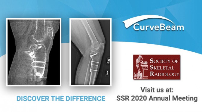
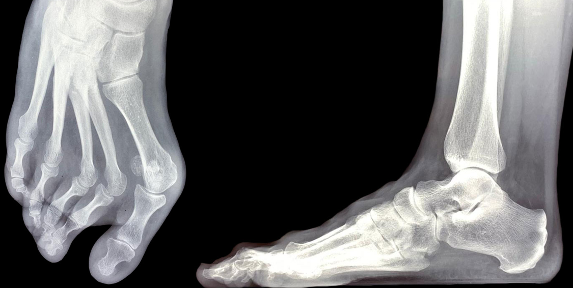
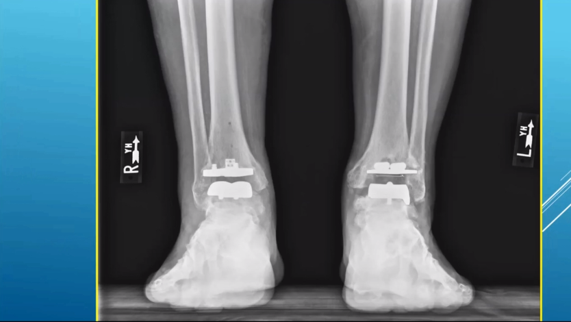
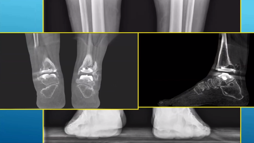
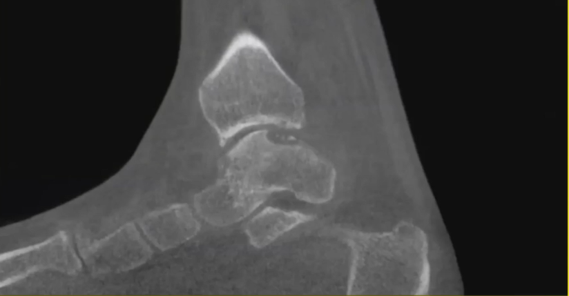
X-rays of knee may not always provide accurate post-operative assessments. For example, in the case below, post-operative X-Rays suggested a posterior shear tibial plateau fracture had been sufficiently healed.
Post-operative X-Rays appeared to show a healed posterior shear tibial plateau fracture.
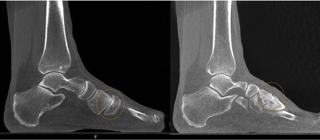
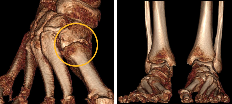
However, a CurveBeam LineUP weight bearing CT exam of the same patient showed a portion of the fracture had not fused as desired.
The weight bearing CT scan revealed a portion of the fracture had not yet healed.
The LineUP provides three-dimensional views of fractures. The treating physician for this case, Dr. Blake Moore, said weight bearing CT has been the most critical addition to his practice since he began it, adding that it “greatly enhances the level of care and sophistication of preoperative planning.”
Are you attending the SSR Annual Meeting? Be sure to visit CurveBeam’s exhibit to learn more about weight bearing CT imaging and how it is revolutionizing orthopedic medicine.
Midfoot arthritis is a challenging problem that causes foot pain and can impede daily activity. Surgery, specifically midfoot arthrodesis, is considered when initial conservative management fails. Arthrodesis should be limited to the symptomatic joints, but it is often difficult to determine which joints are causing the symptoms. Precise anatomic preoperative diagnosis is essential (1).
Cone Beam CT imaging can assist surgeons in understanding complex forefoot deformities and devising the appropriate surgical plan.
For example, in the case below, a patient presented with forefoot pain and was a candidate for surgical revision after X-Ray exams revealed a forefoot deformity.
The patient’s X-Ray displayed a complex midfoot deformity, however the anatomy was too superimposed on the lateral X-Ray to understand the functional position of the tarsometatarsal joints.
Based off of the X-Ray images alone, the treating doctor would have performed a scarf osteotomy and Weil procedures on the 2nd and 3rd metatarsals.
A follow-up weight bearing cone beam CT scan via a CurveBeam pedCAT was ordered.
Sagittal MPR slices revealed 1st tarsometarsal gapping and 2nd tarsometatarsal degeneration.Joint degeneration, as well as instability of the 1st, 2nd and 3rd tarsometatarsal joints, was more apparent on the 3D renderings.
Based on the weight bearing CT scan, the surgical plan was revised to a Lapidus bunionectomy and a 2nd and 3rd tarsometarsal joint arthrodesis.
Will you be attending the AAOS Annual Meeting in Orlando? Visit CurveBeam at Booth #2909 to learn more about weight bearing cone beam CT imaging.
(1) Verhoeven N, Vandeputte G. Midfoot arthritis: diagnosis and treatment. Foot Ankle Surg. 2012;18(4):255–262. doi:10.1016/j.fas.2012.04.004
A loose body is a bone or cartilage fragment that has chipped off inside a joint. If left in place, a loose body can damage a joint surface, cause pain, and restrict movement.
A 2D radiograph is typically the first test performed when looking for a loose body, but overlapping bone may obscure loose bodies. Cone Beam CT imaging provides a 3D image of the foot & ankle at a dose comparable to 2D imaging.
Dr. Albert Armstrong, DPM, MS, BSRS, Professor of Radiology and Medical Director of Advanced Imaging at the Barry University School of Podiatric Medicine, shared such a case in a recent FOOTInnovate lecture.
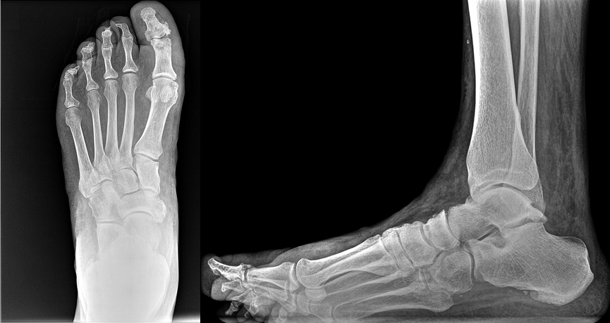
A 38-year-old male patient presented with pain in his left foot, mainly in the great toe. He said his toe felt stiff and would often “lock up.”
A 38-year-old patient presented with pain in his great toe. Due to bony overlap, 2D radiographs were inconclusive as to the cause of pain.
A cone beam CT scan was ordered as a follow-up exam. Because the podiatric clinic at Barry University has a CurveBeam pedCAT on site, the patient was able to get the scan immediately.
The sagittal MPR slices revealed osteophyte formations on the 1st MTP joint, and a resulting loose body.
The cone beam CT scan revealed osteophyte formations on the 1st metatarsophalangeal joint, and a resulting loose body.
Osteochondral lesion of the talus is a broad term that describes injury or abnormality to the talar articular cartilage and adjacent bone. Researchers have shown that radiographs alone miss osteochondral lesions of the talus in up to 50 percent of patients (1) .
Dr. Albert Armstrong, DPM, MS, BSRS, Professor of Radiology and Medical Director of Advanced Imaging at the Barry University School of Podiatric Medicine, shared such a case in a recent FOOTInnovate lecture.
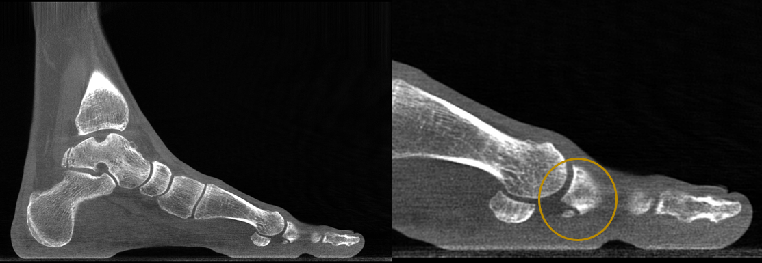
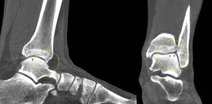
A 40-year-old male patient presented with pain in his left ankle. He was a self-described tough guy who said he has been tolerating the pain for years, but his new job required him to be on his feet all day.
The patient’s X-Rays showed a previous avulsion fracture and some radiolucency in the talar dome, but were otherwise inconclusive:
This 40-year-old male patient’s X-Ray revealed a previous avulsion fracture and some radiolucency around the talus, but was otherwise inconclusive as to why the patient was suffering ankle pain.
A weight bearing CT study was ordered, and it clearly showed two osteochondral lesions in the talar dome, as well as an osteophyte formation on the anterior ankle. Both of these findings pointed to osteoarthritis in the ankle.
The CurveBeam weight bearing CT scan revealed two osteochondral lesions as well as an osteophyte formation on the anterior ankle.
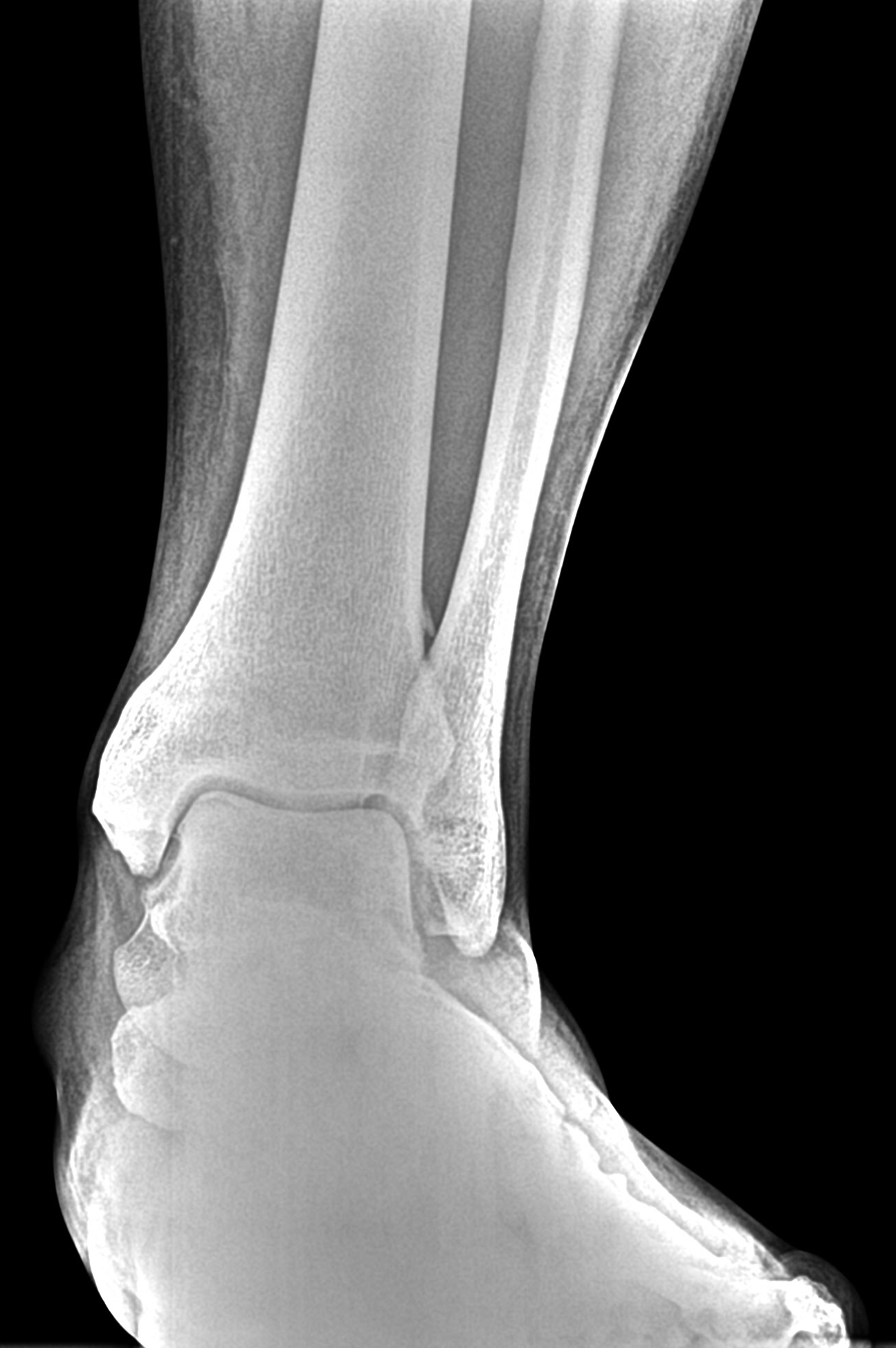
Large lesions and bipolar lesions associated with advanced degenerative joint disease do not respond to simple debridement or cartilage resurfacing techniques. In a case featured in Podiatry Today, a weight bearing CT scan taken after standard X-Rays of a 52-year-old patient with chronic right ankle pain revealed large subchondral cystic changes and bipolar lesions. After reviewing the X-Ray and weight bearing CT. doctors determined the patient to be a good candidate for a total ankle replacement.
Will you be in San Antonio for the ACFAS Scientific Conference Feb. 19 – 22? Visit CurveBeam at exhibit #407 to see several more examples of how weight bearing CT can provide more detailed diagnostic information than X-Ray alone.
(1) Badekas T, Takvorian M, Souras N. Treatment principles for osteochondral lesions in the foot and ankle. Int Orthoped. 2013; 37(9):1697-1706.
Dr. Blake E. Moore, MD, FAAOS, recently delivered a FOOTInnovate webinar titled “Seeing 2020: My First 6 Months with Weight Bearing CT,” diving into specialty views provided by CurveBeam’s LineUp weight bearing CT solution.
Dr. Moore is a board-certified orthopedic surgeon and a member of Atlantic Orthopaedic Specialists in Virginia Beach, VA. Atlantic Orthopaedic Specalists is a private orthopedic group comprised of 21 orthopedic surgeons, including two foot and ankle surgeons, and three hand and upper extremity surgeons.
Leveraging Specialty Views
While the LineUP unit provides 0.3mm multi-planar reconstructions and vivid 3D renderings of patient scans, it’s the machine’s ability to create simulated X-Rays of specialty views with ease that most impresses Dr. Moore.
CubeVue Insta’X tab
“You can get multiple X-Ray views and specialty views that, sometimes, radiation techs that you have in the office may not be necessarily proficient in,” he said. “The image quality is phenomenal there.”
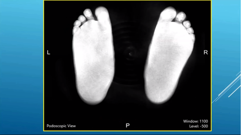
In particular, Dr. Moore has begun utilizing pedoscopic views, which were difficult to obtain prior to the introduction of weight bearing CT.
“I didn’t really utilize (the pedoscopic view), until we had access to weight bearing CT scanning. … You can really see the distribution of weight along the plantar aspect of the foot,” Dr. Moore said. “That can sort of help you guide your treatment in terms of if patients are having medial or lateral column overload.”
Putting Weight Bearing CT into Practice
Dr. Moore presented several case studies that highlight the effectiveness of weight bearing CT and the impact it’s already having on his practice.
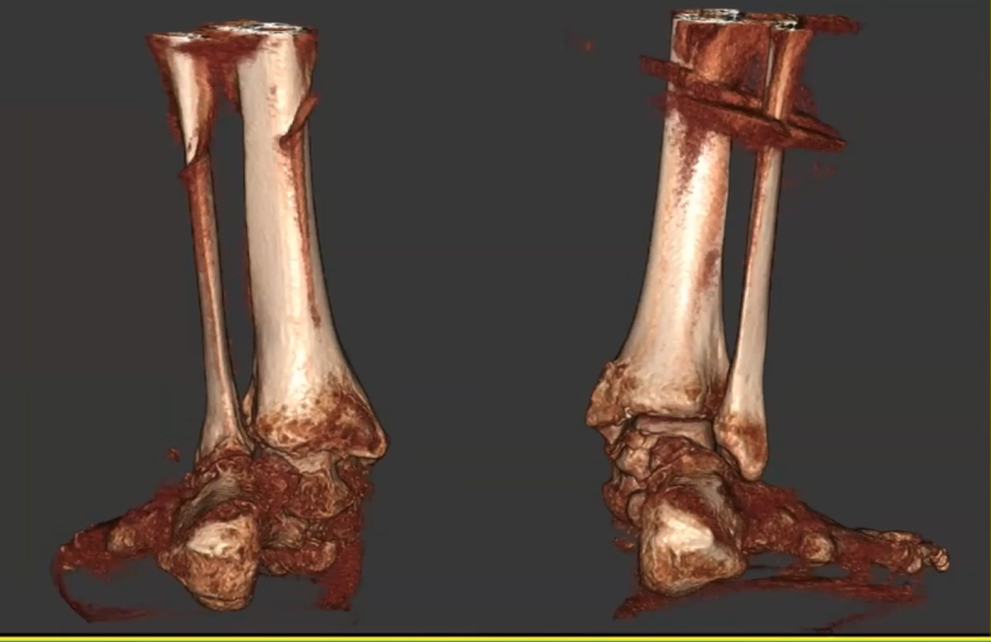
In one case, the bilateral 3D rendering showed how the fibula was impaled into the calcaneus and the lateral aspect of the posterior facet of the subtalar joint, and how the subtalar joint had no congruity at all with the talus.
In this bilateral 3D rendering that Dr. Moore presented on FOOTInnovate, the degree of hindfoot deformity is clearly visible.
“You can really see the difference between the contralateral side and the affected side and as well look at the contribution of physiologic weight bearing to the overall deformity of the foot,” Dr. Moore said. “Seeing the normal side can really help you visiualize where you need to get to to get back to neutral alignment.”
Subtle sub-fibular impingement
Dr. Moore also shared a case in which a subtle subfibular impingement was revealed.
In another case, Dr. Moore reviewed a patient who had undergone total ankle arthroplasty. While some loosening may be observable through traditional scanning, weight bearing CT allowed Dr. Moore to get a more accurate picture of just how significant that loosening was.
Dr. Blake Moore explained that traditional radiographs obtained for post-operative evaluation suggested some early loosening around the tibia on the left side.
However, Dr. Blake Moore explained a weight bearing CT imaging reveled the cystic changes were more significant than the X-Ray suggested, and may need bone grafts.
Finally, Dr. Moore was able to thoroughly evaluate osteochondral lesion of the talus in a patient through CT scanning.
Dr. Blake Moore presented a case with an osteochondral lesion in the talus in his FOOTInnovate webinar.
“I usually get, in addition to an MRI, a CT scan for most of the osteochondral lesions that I treat in order to really get a true idea of the size,” he said. “I think MRI really overestimates the size of these osteochondral lesions and you need to know what the cystic component of that is to pre-operatively plan for these appropriately.”
Preop Templating for TAA
Dr. Moore said weight bearing CT is going to play an enormous role in pre-op templating for total ankle arthroplasty moving forward.
“I think pre-operative templating for total ankle arthroplasty, which is just now starting with the CurveBeam LineUP, is going to be a game-changer for looking at complex deformity and correcting that with patient-specific cut guides. It increases the reliability and reproducibility of the procedure.”
We’re just starting to understand the role that rotation plays when placing these implants, Dr. Moore said.
“When the engineers look at the pre-op templating and make the plans, they really base the rotation off the tibia, and I think that that talar rotation is sometimes difficult to figure out in terms of where that talar implant should be rotated,” he said. “When the templating is currently done, the contralateral affected side is really subtracted immediately. I think that there’s a lot of critical information that can be gained from looking at the normal rotational axis of the unaffected tibiotalar joint.”
Key Financials
Dr. Moore highlighted the immediate financial impact of completing 150 weight bearing scans, which is as follows:
Scans per month: About 35 (90% foot and ankle)
Breakeven: 11 – 12 scans/ month
Reimbursement range: $50 to $750
Average reimbursement: $266
Breaks down to about $39,000 in the first three months
Mobile Weight Bearing CT
Dr. Moore had to decide which location of his practice would house the CurveBeam LineUP machine he utilizes, he said that, were he offered another crack at initial selection, he would have preferred a mobile solution.
CurveBeam offers such a solution, as CurveBeam’s Mobile CT Scanning solutions can bring industry-leading CT capabilities directly to your practice.
“If I could do it all over again, I would have put the unit on a truck so that we could have moved it from office to office,” Dr. Moore said. “But the experience for the patients is first-class.”
Moving Forward
In summary, Dr. Moore said that weight bearing CT has been the most critical addition to his practice since he began it, adding that it “greatly enhances the level of care and sophistication of preoperative planning.”
Custom surgical guides help improve placement and surgical success. Dr. Kristian Buedts, MD, a foot and ankle surgeon at ZNA Middleheim in Antwerp, Belgium recently shared how he uses weight bearing CT scans to create his own surgical guides, and then how he 3D prints those guides at his hospital.
Dr. Buedts highlighted the challenges associated with arthrosis in younger patients, noting that osteo-arthritis typically develops in younger patients as a post-traumatic symptom. More than half the time, the condition presents as asymmetrical wear of the tibio-talar joint (Witteveen 2013), and realignment surgery is often considered to avoid the implications of ankle replacement and joint fusion in younger patients (Krahenbuhl 2017).
These surgeries have been made easier by the accurate 3D representations made possible with weight bearing CT scans, providing professionals like Dr. Buedts with a more consistent pre-operative planning process that included the following steps:
Computer simulation software aids in generation of 3D models
Displacement is calculated by comparing the affected bone to the normal side through superimposition
Virtual osteotomies are conducted
Commercial software helps translate these simulations into custom-made guides
These guides are 3D printed to assist with the actual surgery
In one case, a patient with ankle instability in the ankle joint as a result of ligamentous laxity and a complex cavo-varus deformity with congruent varus deformity in the ankle joint, among other problems, underwent a corrective dome osteotomy.
While cases such as the patient’s are often hard to reproduce and are difficult to operate on, Dr. Buedts and his team utilized weight bearing CT imaging and the 3D printing of pre-operative guides that were used during the procedure to achieve an extremely positive outcome.
To learn more about the possibilities of weight bearing CT imaging, visit curvebeam.com/contact/.
David J. Soomekh, DPM, is a Diplomate of the American Board of Foot and Ankle Surgery and a Fellow of the American College of Foot and Ankle Surgeons.
In this CurveBeam-sponsored FOOTInnovate webinar, Dr. Soomekh delivers a presentation about how exactly weight bearing CT technology has been integrated into his practice.
Click here to watch the FOOTInnovate webinar. A FOOTInnovate account is required to access.
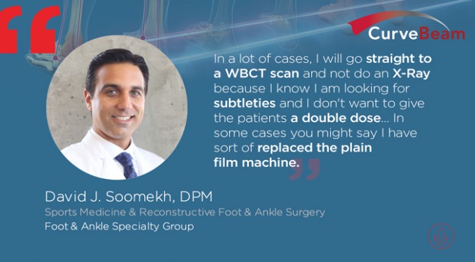
How Dr. Soomekh Leverages the pedCAT
To begin, let’s dive into how Dr. Soomekh uses the CurveBeam pedCAT system in his practice.
“In a lot of cases, I will go straight to a WBCT scan and not do an X-Ray because I know I am looking for subtleties and I don’t want to give the patient a double dose, ” Dr. Soomekh said. “In some cases you might say I have sort of replaced the plain film machine.”
Dr. Soomekh said that the pedCAT is instrumental in his ability to:
diagnose fractures that are difficult to appreciate on plain film due to bone overlap;
detect stress fractures;
appreciate joint relationships in dislocations more easily;
and get an accurate 3D view of joint changes and subchondral lesions associated with arthritis.
In addition, the pedCAT assists Dr. Soomekh with pre-operative planning and an in early detection of osteomyelitis.
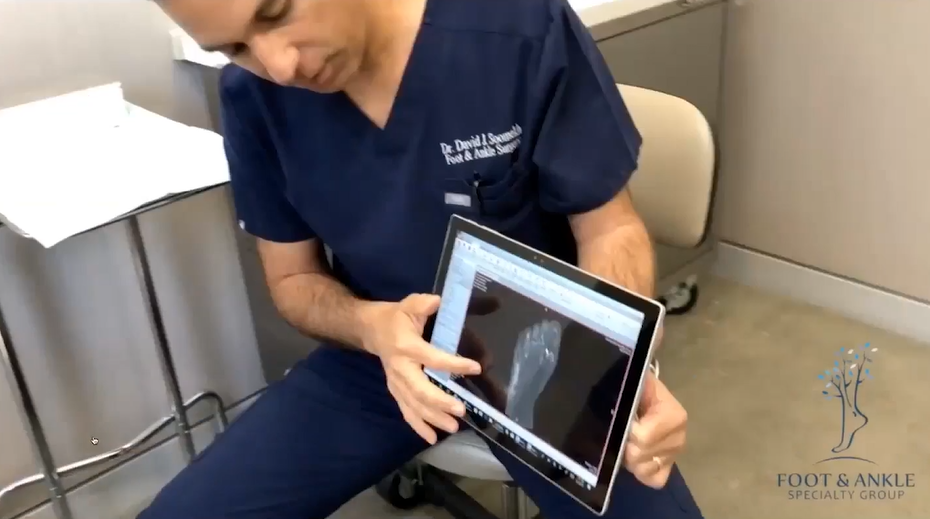
Dr. David Soomekh, DPM, shares patients’ pedCAT scans with them via a tablet. He said the datasets wirelessly upload to the viewer in less than 10 minutes.”
“I [view images on] a mobile device. … It’s wireless, and the image still comes up in about six to eight minutes after it’s processed,” Dr. Soomekh said. “I’ll bring it into the room and show the patient the views and the pathology.”
pedCAT Reimbursements and Authorization
Though the rates presented by Dr. Soomekh are specific to Southern California, they provide valuable insight into the operational benefits of a pedCAT system.
For Medicare, Dr. Soomekh said his reimbursement rate is $130 for a scan. With PPO insurance, the rate can range from $180 to $280, and as high as $350. Dr. Soomekh’s cash-pay rate is $200.
While Medicare doesn’t require authorization, Dr. Soomekh’s practice has implemented some best practices for securing authorization for PPO insurance.
His staff will make a pre-authorization call while the patient is in-office, and billing is held until authorization is received. Occasionally, a peer-to-peer call is required. The patient signs a consent form he or she will be responsible for the cost of the CT if it is not covered by insurance.
“In most cases, I don’t wait until I get the authorization before I go ahead and take the scan,” Dr. Soomekh said. “Some patients ask me to wait until they get the authorization, and we’ll do that. … Otherwise, the staff will be doing an authorization call while we’re doing the scan. If we get authorization right then, we’ll let the patient know.”
Results of Weight Bearing CT in Action
Though a thorough watching of Dr. Soomekh’s webinar will reveal many cases in which weight bearing CT is beneficial, let’s take a look at how the pedCAT helps Dr. Soomekh assess Hallux Valgus.
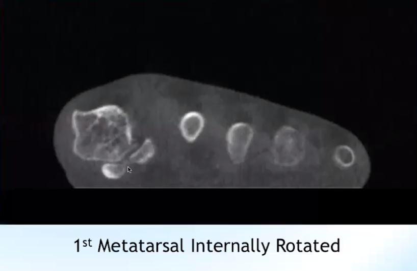
The system helps Dr. Soomekh in pre-operative planning as he assesses the degree of frontal plate rotation of the first metatarsal and its association with the sesamoids. He can also appreciate the degree of transverse plane angulation at the first metatarsal cuneiform joint, among other benefits.
The image below, for example, provided Dr. Soomekh with a look at the internal rotation of the first metatarsal in a younger patient.
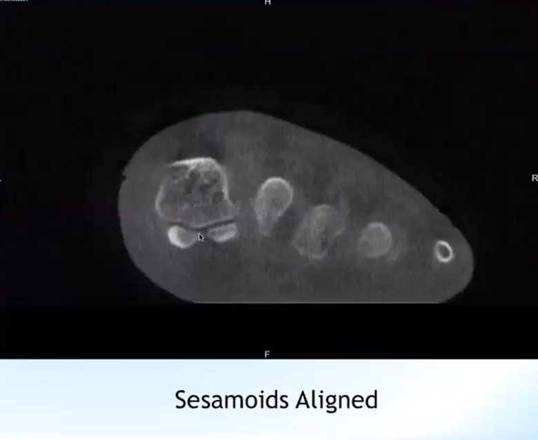
The image below shows the result of corrective surgery for the same patient.
“The sesamoids are underneath, now, in the appropriate position, and the angulation is closed,” Dr. Soomekh said.
pedCAT Benefits
To conclude the webinar, Dr. Soomekh outlined the major benefits of adopting the use of a pedCAT machine at his practice.
These include:
Excellent image quality
Instant results
Immediate and focused treatment for the patient
Patients benefit from “one-stop shop” experience
Increased practice revenue
Images can be sent to radiologist for an official report
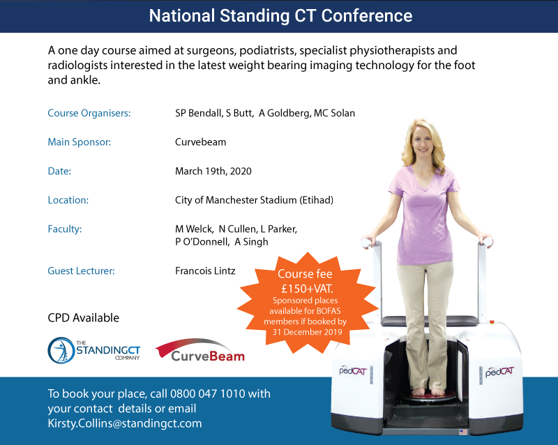
UPDATE: The Standing CT Conference has been re-scheduled to Thursday, June 11, 2020. Please contact kirsty.collins@standingct.com.
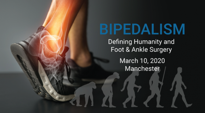
On March 19, CurveBeam will again proudly serve as the primary sponsor for “Bipedalism: Defining Humanity and Foot & Ankle Surgery,” the National Standing CT Conference.
The event, which provides a one-day course aimed at surgeons, podiatrists, specialist physiotherapists and radiologists interested in the latest weight bearing imaging technology for the foot and ankle, is set to take place in Manchester, England’s City of Manchester Stadium, also known as the Etihad Stadium.
The conference is organized by Standing CT Company, which provides weight bearing CT services via mobile imaging centers to hospitals and orthopedic practices in the United Kingdom and Europe. Standing CT Company exclusively utilizes CurveBeam’s weight bearing CT systems.
The conference will feature a variety of speakers and courses for attendees.
Course organizers include Stephen P. Bendall, MBBS, FRCS, FRSC (Orth), an orthopedic foot and ankle surgeon at Princess Royal Hospital in Haywards Heath, Sajid Butt, MB BS, FCPS, FCPR, a radiologist at Royal National Orthopedic Hospitals in London and Stanmore, Andrew Goldberg, MD, and Matthew Solan, BSc, MB, BS, FRCS (Tr & Orth), Medical Director, Chair of MAB and consultant orthopedic surgeon.
Other faculty include CurveBeam President & CEO Arun Singh, Matthew Welck, MBChB. BSc (hons). MSc. FRCS (Orth), Nicholas Cullen, MBBS, BSc(Hons), FRCS, FRCS(Tr&Orth), Lee Parker, BM MRCS FRCS (Tr. & Orth.) Paul O’Donnell, MBBS, MRCP, FRCR, and Francois Lintz, MD .
Question-and-answer sessions will follow each of the four main discussions.
To book your place at this critical conference, call 0800-047-1010 and provide your contact details or email Kirsty.Collins@standingct.com.


