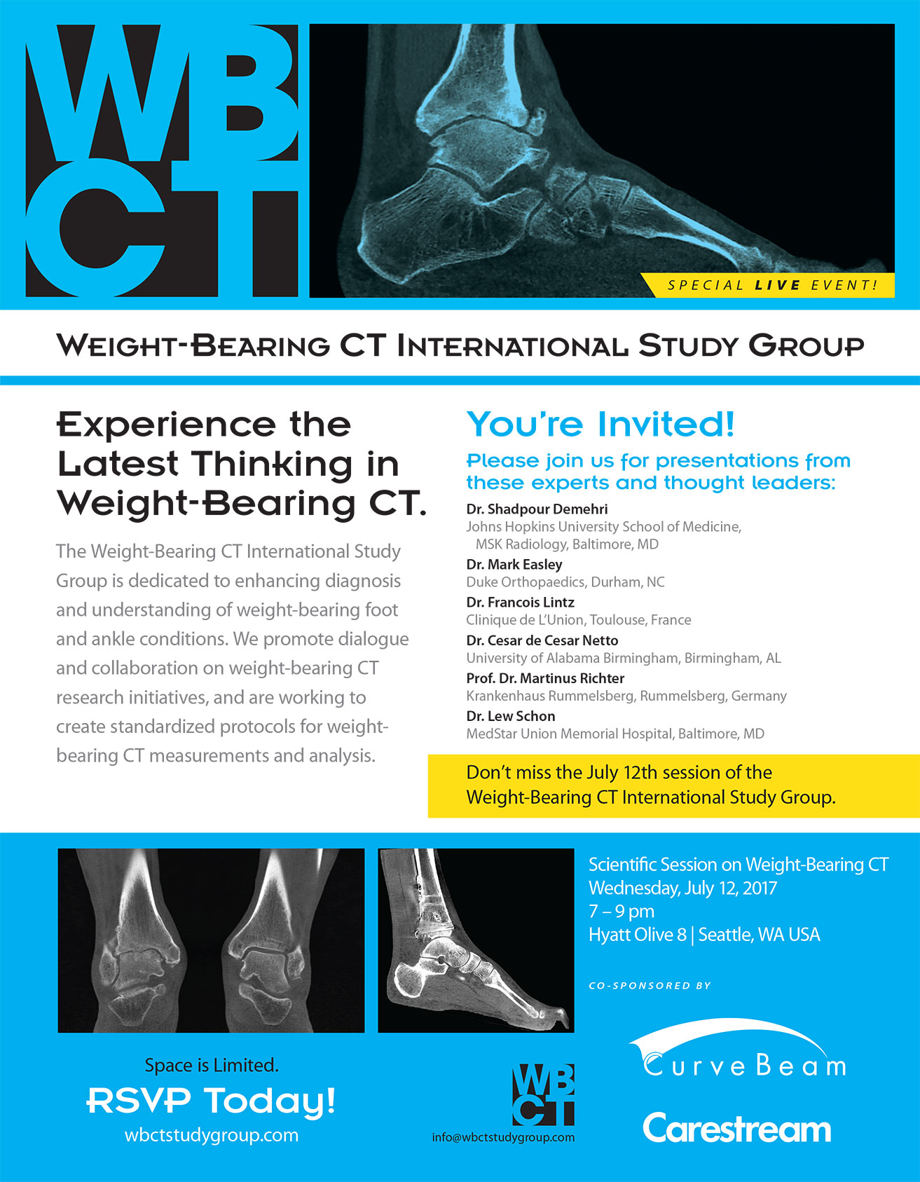
Be Our Guest At A Special Scientific Event!

The Weight-Bearing CT International Study Group invites guests to join us on Wednesday, July 12, 2017 from 7pm – 9pm at the Hyatt Olive 8, Seattle for an informative scientific session on the latest in weight-bearing foot and ankle research. Curvebeam, along with Carestream, is proud to co-sponsor the special live event with presentations from expert orthopedic physicians and renowned thought leaders from around the world. The discussion is an opportunity to promote dialogue and collaboration on weight-bearing CT research initiatives, while advancing the group’s mission of enhancing diagnosis and understanding of weight-bearing foot and ankle conditions, as well as creating standardized protocols for weightbearing CT measurements and analysis.

Space is limited so reserve your spot at wbctstudygroup.com. We look forward to seeing you at the event!

Be-Our-Guest-At-A-Special-Scientific-Event-full

Webinar: Pitfalls In Hindfoot and Ankle deformities tackled by weightbearing CT

Arne_BurssensYou are invited to join FOOTinnovateTM and Dr. Arne Burrssens for the webinar “Pitfalls In Hindfoot and Ankle deformities tackled by weightbearing CT” sponsored by Curvebeam.
Click here to register.

Dr. Burrssens is an orthopedic surgery resident at Ghent University in Flanders, Belgium. During his one-hour presentation on Monday, June 5, 2017 9:00 PM – 10:00 PM (W. Europe Standard Time), he will discuss his personal experience with weight bearing CT and reveal how it has improved his patient care. He’ll also share a few specific cases to provide a deeper look into his work.

Dr. Burrssens is co-author of the article “Weightbearing CT in normal hindfoot alignment — Presence of a constitutional valgus?” published in February 2017 by the journal Foot and Ankle Surgery. The purpose of the study was to assess hindfoot alignment using weightbearing CT.

The research concluded that weightbearing CT provides an objective assessment of hindfoot alignment with reproducible results, which can aid a surgeon during a calcaneal osteotomy.

PLEASE NOTE: You must register for this session at least 15 minutes prior to the start time. Click here to create a free account.

CurveBeam and Carestream Health Announce Collaboration to Promote Weight-Bearing CT Awareness and Research

Warrington, Penn. – June 6, 2017 – Medical device companies CurveBeam and Carestream Health announced a collaboration to support and facilitate education and research for weight-bearing computed tomography imaging.

The ability to capture weight-bearing CT images allows foot and ankle specialists to study foot biomechanics in three dimensions. Significant clinical research has already demonstrated weight-bearing CT has the potential to be the new standard of care for an initial diagnosis. However, much work remains to be done to increase awareness among orthopaedic specialists,” said Vinti Singh, marketing manager for CurveBeam. “Through collaboration, CurveBeam and Carestream can best support these efforts.

Before the introduction of weight-bearing CT imaging in 2012, patients could get either a weight-bearing radiographic 2D X-ray or a non-weight bearing medical CT scan. X-rays can at times obscure and distort anatomical details, while non-weight bearing CT scans may inaccurately represent joint space and alignment. The American Orthopaedic Foot & Ankle Society recommends weight-bearing imaging when possible to get the most accurate assessment.

The image quality and detail captured with 3D weight-bearing CT exams allow the surgeon to view and measure anatomical abnormalities of lower extremities under natural load conditions,” said Helen Titus, Carestream’s Worldwide Marketing Director for Ultrasound & CT. “The goal of this collaboration is to spread the word about this new imaging modality among orthopaedic specialists.”

The companies will work together to support the Weight-Bearing CT International Study Group, which is comprised of orthopaedic researchers from leading institutions in the U.S. and Europe. The group is working to create standardized protocols for weight-bearing CT measurements and analysis.

The companies’ first joint initiative is to co-sponsor an evening scientific session during the American Orthopaedic Foot & Ankle Society (AOFAS) annual meeting in Seattle, WA. Speakers at this session include Dr. Mark Easley of Duke Orthopaedics in Durham, NC, and Dr. Lew Schon of MedStar Union Memorial Hospital in Baltimore, MD. For more information about the Study Group, and how to RSVP for the session, please visit www.wbctstudygroup.com.
Both companies offer weight-bearing solutions for extremity CT imaging. The devices employ cone beam CT technology and are designed for point-of-care settings, as well as hospitals and imaging centers.

To view Carestream’s latest news announcements, please visit www.carestream.com/news.

CARESTREAM is a trademark of Carestream Health.

Keep Your Orthopedic Practice Alive Through Diversification

Medical providers are always looking for ways to create optimal patient outcomes while lowering costs. Dwindling reimbursement can dissuade people from pursuing medical careers—or their chosen specialty. Medical Student Education published a study finding 15 percent of fourth-year medical school students reported payment as the primary factor driving their career choices.

These concerns pose unique challenges for orthopedic surgeons. If we fail to improve revenues for orthopedic practices, then many of these practices will fail. If that happens—at the same time baby-boomers are reaching the age of needing more musculoskeletal care—our nation will experience a true healthcare crisis.

Not long ago, developing a financially successful practice hinged on a physician’s clinical expertise. But expertise alone will no longer serve to attract patients and generate revenue. Our long-term goal should be to generate revenue from multiple sources, diversifying practices to ensure practice viability.

The American Academy of Orthopedic Surgeons, AAOS, created a 45-page guide, Enhancing Your Practice’s Revenues: Pearls and Pitfalls (A Primer for Orthopedic Surgeons), recommending orthopedic practices offer in-office imaging.

Imaging has become an integral part of many orthopaedic offices,” the report says. “In-house imaging provides an ancillary revenue stream, improves practice efficiency, and may play a role in overall patient satisfaction. The types of imaging services offered vary depending on a variety of factors, which include but are not limited to practice size, geographical location, political considerations, and orthopaedic specialties represented in the office.

Revenues from imaging services can add significantly to your bottom line. A reasonably busy MRI, for example, may generate more revenue and profit for the practice than an orthopaedic surgeon, while X-ray often generates an additional $50,000-$100,000 or more of operating profit per orthopaedic surgeon.”

For orthopaedists, the history and physical has long been key to the successful diagnosis of patients with musculoskeletal patients. Traditional film X-rays provided them an additional diagnosis tool. Imaging technology continues to evolve, allowing orthopaedists to diagnose diseases, disorders and conditions more quickly and with even greater accuracy. The result is a win-win situation for patients and doctors alike.

The CurveBeam pedCAT is designed to fit into your practice and workflow, with a footprint of about 4’ x 5’. The unit plugs into a standard wall outlet and gives you the bilateral, weight-bearing 3-dimentional views of the foot and ankle you need to create a comprehensive treatment plan.

The pedCAT scans are reimbursed through CPT code 73700 – CT lower extremity without contrast.

The pedCAT is a compact, ultra-low dose CT imaging system that is ideal for orthopedic and podiatric clinics. Patients benefit from the convenience of point-of-care advanced diagnostic imaging, scan time is one minute, and the pedCAT automatically generates all standard X-ray views in addition to the full CT volume. All of this contributes both to improved patient care and the bottom line.

To learn more about how the pedCAT can enhance your practice, visit CurveBeam.com today!

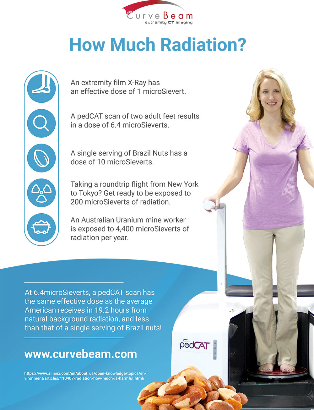
A pedCAT Scan: Less Radiation Than A Serving of Brazil Nuts

Most people may not realize it, but humans are exposed to natural background radiation all the time. The source is naturally occurring radioactive materials and cosmic radiation from outer space. Geography plays a huge factor in how much radiation a person is exposed to, but the average  American adult receives an effective dose of about 3000 microSieverts a year.

So how does the Curvebeam pedCAT scan compare to radiation from other activities? See below!

A-pedCAT-Scan-Less-Radiation-Than-A-Serving-of-Brazil-Nuts-infographic

The pedCAT Weight-Bearing CT Scan Put CurveBeam a Step Ahead of the Competition

It’s hard to forget about your feet—socks and shoes, not to mention pain at the end of a long workday. As the foundation of your mobility, foot health is vital to overall health. But foot health involves more than just keeping them clean and dry. It is vital to wear properly fitted shoes—custom insoles (avoid flimsy over-the-counter insoles) and supportive sandals are also a good idea. In fact, feet are so important to us that they even get their own specialists.

An expert in the field of podiatry, Dr. Kyle Kinmon of Boca Raton, FL recently shared some advice on WPTV for maintaining optimal foot health. Kinmon used part of his television segment to introduce viewers to the latest pedCAT technology from CurveBeam: a weight-bearing CT scan for diagnosing foot and ankle issues.

In fact, Kinmon’s office is the first in South Florida to offer a 3D foot scanner, the pedCAT technology from CurveBeam. Introducing the tool to TV viewers on WPTV, Kinmon said, “This is called the pedCAT. It’s the newest technology in the world of foot and ankle. It’s a weight-bearing CT scanner that, in 19 seconds, will provide us with every possible image you could want to see in the foot and ankle.”

To learn more about Dr. Kinmon’s practice, please visit www.certifiedfoot.com.

Dr. Kinmon, a fellow of the American College of Foot and Ankle Surgeons, has contributed to research in the field of podiatry with many peer-reviewed publications, including an award-winning article in the Journal of the American Podiatric Medical Association and an article on pediatric flat foot for New Balance shoe company’s health line. Kinmon is also the Founding Director of Residency Training for Bethesda Memorial Hospital’s three-year Podiatric Surgical Residency Program, further supporting residents in surgery at three area residency training programs.

While Kinmon is highly trained in foot and ankle surgery, he always considers surgery the last resort. In the vast majority of cases, surgery is not required, and the condition is resolved using conservative treatment alone. Still, even for someone as skilled as Dr. Kinmon, diagnosing foot and ankle issues can be challenging.

The pedCAT’s bilateral, weight-bearing 3D views of the foot and ankle give specialists the information they need to create comprehensive treatment plans. Its compact (4’ x 5’), ultra-low dose (2-5 micro Sieverts per scan) CT imagine system plugs into the wall and is ideal for orthopedic and podiatric clinics. Scan time is one minute, and the pedCAT automatically generates all standard X-ray views in addition to the full CT volume. The pedCAT is PACS/DICOM compatible and supports DICOM modality worklist (HIS/RIS) and PACS integration.

One can definitely understand why Kinmon was excited to share the news about pedCAT with television viewers. With the pedCAT from CurveBeam, his job is now a lot easier and his diagnoses more accurate than ever.

To learn more about the exciting new pedCat from CurveBeam, visit http://curvebeam.com/products/pedcat today!

CurveBeam Announces FDA 510(k) Clearance for InReach Cone Beam CT Imaging System for the Upper Extremities

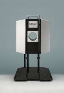
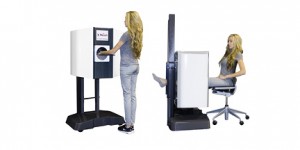
InReachMay 8, 2017 – Warrington, Penn. – CurveBeam announced it has received FDA 510(k) clearance for the InReach, a Cone Beam CT imaging system primarily designed for the hand, wrist &elbow; & lower extremities in non-weight bearing position.

The InReach is an ultra-compact CT scanner that provides high-contrast 3D datasets of bony anatomy, which could potentially replace radiographs as a first line of diagnosis.                                                                                              

The InReach is ideal for the point-of-care because of its small footprint, its self-shielded design, and standard power requirements. Point-of-care 3D imaging allows for faster diagnosis and more accurate treatment plans.

curvebeaminreach

“The InReach will revolutionize the speed and accuracy of assessment of upper extremity conditions that specialists have traditionally found challenging to diagnose with plain X-Ray, such as scaphoid fractures,” said CurveBeam President & CEO, Arun Singh. “The InReach continues the company’s mission to elevate advanced diagnostic imaging capabilities to enhance orthopedic care.”

The InReach is designed with patient comfort in mind. Patients’ hand, wrist or elbow is positioned in a height-adjustable bore while in standing or sitting position. The unit can also accommodate non-weight bearing, lower limb imaging. Scan times are less than 30 seconds.

The InReach device is supplemented by CubeVue, CurveBeam’s custom visualization software.  CubeVue gives orthopedic specialists unprecedented access to multi-planar slices and vivid 3D renderings of the anatomy previously not easily accessible to specialists. CubeVue’s Insta-X feature provides Digitally Reconstructed Radiographs, potentially eliminating the need for radiographic exams altogether. 

The InReach is the second extremity CT imaging system CurveBeam has introduced to the market.

CurveBeam is the leader in Weight-Bearing extremity CT imaging, starting with the introduction of its pioneer product, the pedCAT, in 2012. The pedCAT is the only CT system that allows for bilateral, true weight bearing imaging of the lower extremities. Since 2012, the pedCAT has been integrated into leading foot & ankle orthopedic and podiatric practices around the world.

CurveBeam is currently developing its next generation multi-extremity device, the LineUP, which will provide bilateral Weight-Bearing images of the knees in addition to feet, as well as hand, wrist & elbow. CurveBeam anticipates the LineUP will be submitted for FDA review by July, 2017.

Discussion Looks to Provide Blueprint for Foot and Ankle Deformity Correction

Foot and Ankle Specialist (FAS), a bi-monthly journal for orthopedic surgeons and podiatrists, recently published a roundtable discussion focused on providing insight into the difficult process of deformity correction. For surgeons dealing with the lower extremities, even the central principles of understanding and treating deformities can be extremely complex. Noman Siddiqui, Guido Laporta and John Herzenberg, all of whom carry numerous distinctions in the field of podiatry, all weighed in on the discussion.

Deformity management in cases involving the lower limb can involve high levels of pain. Because of this, while analysis is fairly similar to deformity analysis in other regions of the body, treatment is as much an art as a science. Much of the discussion involves presentation of cases and remarks by the specialists as to how they would care for the patient.

The roundtable ranges from topics on improving education in the field to proper care of patients who are treated with external fixation. Of particular interest is the repeated insistence by the panel that the most essential way to improve the field is to increase the amount of information available to a surgeon before an operation occurs. They remark that, even though patients receive a thorough physical as well as a biomechanical exam, gait analysis, and functional testing, more extensive testing and imaging is needed.

All three of the medical experts point out the need for a computed tomography (CT) scan for the more difficult cases. These scans help to erase any gray areas in the diagnosis and help to evaluate the “mal-union” or “non-union” present in malformed feet and ankles. Dr. Herzenberg even recommended 3D CT technology in order to get a complete picture of the situation, and both a weight and non-weight bearing evaluation.

“[Foot] deformity is complex and may require second-level radiographic studies such as a CT scan,” remarked Herzenberg. “Recently, we have added a standing 3D CT machine in our office, which offers the same advantage from a CT standpoint that we get from plain films taken weight bearing versus non–weight bearing.”

These orthopedic specialists are not alone in their desire for more comprehensive scanning technology when treating deformities. More and more clinics are taking similar steps to those used by Herzenberg’s clinic, The Pediatric Orthopedics Department at Sinai Hospital in Baltimore, which now uses the CurveBeam pedCAT. It is quick, efficient and thorough, and was designed with patients in mind, limiting their discomfort. This is especially important since, as mentioned earlier in the roundtable, pain can be significant when dealing with foot and ankle deformities.

The discussion presents a comprehensive look at the current practices in a complex corner of healthcare, and presents insights into the best way to manage orthopedic deformities in the lower extremities. The specialists involved even go so far as to give advice for those preparing to deal with these cases, and examine current technology that allows them to do so to the best of their ability. To read the roundtable discussion in its entirety, click here.


Weightbearing CT Revolutionizing Hindfoot Alignment Measurements

A new study, led by famous Belgian M.D. A. Burssens, emerged in the February issue of Foot and Ankle Surgery. The study examines the use of traditional hindfoot angle measurements versus weight bearing CT and shows how weight bearing CT can help contribute to the pre-operative planning by providing further insights into a physiological hindfoot alignment.

Hindfoot malalignment is a widely-accepted cause of foot and ankle disability and problems. For preoperative planning and clinical follow-up, reliable radiographic assessment of hindfoot alignment is of utmost importance and can affect future health. The long axial radiographic view and the hindfoot alignment view are commonly used for this purpose. The main goal of this study is to obtain measurements from a population with clinical and radiological absence of hindfoot pathology. These values were then compared to hindfoot measurements obtained from the long axial view based on the anatomical axis of the tibia and the calcaneal axis, to point out possible differences attributed to that measurement method.

The study has many key findings related to traditional hindfoot predictive measurements. One major finding questions the commonly held belief that non-symptomatic feet have a slight valgus. When traditional hindfoot angle measurements were applied, the study showed that non-symptomatic feet have a neutral alignment. These results show a more neutral alignment of the hindfoot as opposed to the generally accepted constitutional valgus.

Another major finding was that the weight bearing CT clearly showed the talus. The talus is usually superimposed in plain x-rays and hard to see and can affect the mathematical calculations of the predicted hindfoot angle. So, unlike the Saltzman view, these calculations included the talus.

The ultimate goal of all orthopedic surgeons is to influence the correct alignment of bones. Even the slightest miscalculation in measurement can have lasting consequences on post-surgery recovery and future mobility. As the study notes, “Although surgical hindfoot corrections are frequently performed either extra-articular by osteotomies or intra-articular by arthrodesis, still numerous debate exists on the amount of correction and the ideal foot position after arthrodesis.” This finding could have repercussions on hindfoot position during fusion or in quantifying the correction of a malalignment. Due to the use of weight bearing CT, the inferior calcaneus point can be used during pre-operative planning of a hindfoot correction as an anatomical landmark due to its shown influence on load transfer.

A disadvantage of the study was that it was not truly spatial, as only the coronal plane was used in measurements. In the future, 3D segmentation models will allow for better spatial calculations. Until 3D weight bearing CT is an available option, it is recommended foot & ankle specialists exercise caution when planning from 2D radiographs. This study will contribute to the pre-operative planning by providing further insights into a physiological hindfoot alignment. For more detail, access the article here.

Weight Bearing CT International Study Group Presenters Share Important Findings

The American Academy of Orthopaedic Surgeons (AAOS) held its annual meeting at the San Diego Convention Center from March 14-18, 2017. The Weight Bearing CT International Study Group Meeting on March 18 was an important part of this event. The group promotes dialogue and collaboration on weight bearing CT research initiatives, and is working to create standardized protocols for measurement and analysis.

At the meeting, three presenters shared their findings:

“Using Weight Bearing CT to Understand the Adult Acquired Flatfoot,” Scott Ellis, MD, Hospital for Special Surgery

Dr. Ellis presented three studies that considered the role of weight-bearing CT in Adult Acquired Flatfoot (AAFD). Researchers sought to understand the nature of pain in AAFD, and to determine the cause of and the nature of deformity for AAFD.

The first project assessed the cohort of Stage II patients with lateral hindfoot pain compared to control patients with just medial pain. The study found that subtalar arthrosis was significantly more common in the group with hindfoot pain. Arthrosis is typically undetected on a plain radiograph, but is obvious on a 3D weight-bearing CT scan. The researchers found an important role for weight bearing CT in management of AAFD and in determining the many factors contributing to this condition.

A second study by Bruce Sangeorzan used simulated WB to determine the possible etiology of AAFD. Researchers correlated the angles of the coronal subtalar joint position in patients with AAFD versus a control group. They found that AAFD patients exhibited significantly more valgua in the angle than in the control patients and that the inftal-suptal correlated with a number of measurements related to flatfoot.

A third study at the Hospital for Special Surgery compared peritalar subluxation between AAFD patients and controls using weight-bearing multiplanar imaging. The study found that weight-bearing CT better identified the source of pain in AAFD, and showed where the deformity occurs with respect to talus. According to the study’s results, weight-bearing CT overcomes the limitations of both standard CT and plain radiography.

“Weight-bearing CT of the Foot & Ankle: Literature Review and Indications for Use in the Clinic,” Alexej Barg, MD, University of Utah, Department of Orthopaedics

This study examined different imaging modalities, including conventional radiographs, CT-Arthography, weight-bearing CT, MRI, and ultrasonography. Researchers found that a weight-bearing CT scan offers a tremendous amount of additional information over conventional methods, allows rapid evaluation of complex foot and ankle anatomy with thin-section imaging, improves diagnostic accuracy compared to radiographs, and helps minimize the radiation dose to the patient. An additional benefit is that a cone beam CT scanner is relatively small and requires less capitalization cost than a conventional CT unit.

Dr. Barg also offered a comprehensive literature review of relevant weight-bearing CT studies. He concluded his presentation by sharing cases of weight-bearing CT scanning in clinical use. He found the weight-bearing CT scan is a significant improvement over conventional X-rays, especially in the ability to compare results of different tests and patients through standardized measurements with reliable identifications of anatomic landmarks.

“Rotation of the First Metatarsal in Hallux Valgus Deformity,” Phinit Phisitkul, MD, Department of Orthopaedic Surgery and Rehabilitation, University of Iowa Hospitals and Clinics

Hallux valgus is a complex triplane deformity that may result from intrinsic factors, such as genetics or anatomy, as well as extrinsic factors, such as wearing high heels with a narrow box toe. Previous studies generally did not rely on weight-bearing CT scans. This study reviewed 85 patients who underwent weight-bearing CT using the CurveBeam pedCAT between January 2014 and May 2016.

The study concluded that weight-bearing CT of the foot is helpful in preoperative planning for complex hallux valgus deformities. Researchers also found that pronation of the first metatarsal is more highly correlated with foot structure, for example pes planus, than hallux valgus deformity.

CurveBeam is an innovator setting the standard of care in orthopedic imaging. CurveBeam’s automatic measurement tools lead to better patient outcomes, streamlined practice workflow, and increased revenue. Check out a replay of the 2017 Weight Bearing CT International Study Group Meeting at https://www.youtube.com/watch?v=LHIAF-ExOsY&t=11s .