One Step at a Time? Knee Replacements Are Advancing by Leaps and Bounds!

An increasing number of U.S. residents over the age of 65 has expanded the need for longer-lasting knee implants. Orthopedic companies have responded by developing new technologies that have resulted in more durable implants. This, according to ODT, has in turn led to “an increase in the number of younger patients receiving them.” At the same time, older knee implants will continue to deteriorate, and therefore need to be replaced. This means growing opportunities for these same orthopedic companies.

In other words, these are not your parents’—or grandparents’—knee implants. Past implants were primarily made of metal or metal/polyethylene, but newer artificial joints use water-resistant ceramic or plastic in at least one component, and are designed to address metal sensitivities. And with computerized navigation and electromagnetic probes, surgeons can replace knee joints with increasing accuracy. Often the whole knee no longer has to be removed and replaced; rather the bone can have a small part shaved off and resurfaced. These advancements in surgical materials and techniques have resulted in a tripling of total knee replacements in people ages 45 to 64 from 2000 to 2010.

Rather than a total knee replacement, a patient often only needs a unicompartmental, or partial, knee replacement. This usually involves replacing only the inside (medial side) of the knee, as this is often the first part to wear out. The outside (lateral portion) of the knee can be done in a similar fashion, with little pain, a quick recovery, and only a small scar in each case. For those with knee arthritis, unispacers are now used—especially in younger patients—to separate the knee surfaces and keep them from grinding together.

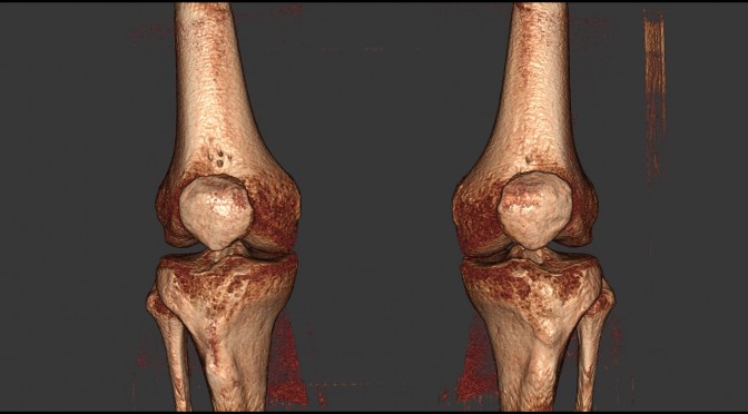
(Read about how weight bearing CT imaging has been used in osteoarthritis research here: http://www.curvebeam.com/products/lineup-investigational-only/studies/)

Add to this the fact that these less intrusive methods only require local anesthesia, and newer pain medications and pain management methods are now available. Modern knee surgeries also mean faster recovery time—and an earlier return to work and other daily activities.

There’s little question that increased innovations are less invasive, making knee surgeries more common. New materials, and even biomaterials developed using stem cell research to create individualized natural cartilage, are on the horizon, meaning this well-established, mature market is set for some serious changes.

CurveBeam was founded in 2009 by a group of individuals with a proven track record in the advances and compact 3D imaging device domain. The company designs and manufacturers Cone Beam CT imaging equipment for the orthopedic and podiatric specialties.

To learn more about how CurveBeam technology can help you get the best images for your patient and their needs, visit us at http://CurveBeam.com today!

In-Office CT Imaging Equipment Helps Urgent Care Centers Better Address Orthopedic Care

According to a 2015 survey by the Urgent Care Association of America, there are more than 7,000 urgent-care centers in the United States. The industry is expected to grow 4 to 6 percent per year in the near future, mainly due to a shortage of primary-care providers, overfilled emergency rooms, a growing aging population, and an increase in the number of insured patients. There has been a similar increase in orthopedic urgent care, driven chiefly by patient’s demands for decreased wait times and lower costs for orthopedic care. In order to better address these needs, urgent care facilities are increasingly adding in-office CT imaging equipment.

As healthcare changes and evolves, doctors need to pay attention to trends as well as patient expectations in order to grow their practices. In a recent article in the Orlando Sentinel, Dr. Alejandro Badia relates why he started an orthopedic urgent-care center. “Every patient that came to see me had already been somewhere, and that somewhere didn’t do much for them,” he stated. “I said to myself, ‘Why can’t somebody reach me more directly?‘” Badia, a practicing hand surgeon, established OrthoNOW orthopedic urgent care in 2010, and began franchising the concept in 2014. “Our mission is to change the way expert orthopedic health care is delivered,” said Badia.

OrthoNOW and its franchisees are capitalizing on the rapid growth in urgent care. As related in a recent article in Becker’s ASC Review, representatives of Ambulatory Surgery Centers (ASC) have witnessed a similar trend. Sandy Fragale, administrator at Orthopedic & Sports Medicine Specialists of Green Bay, WI, said, “It’s a lower cost alternative to urgent or emergency care. It’s less expensive than the ER or urgent care. Additionally, the patients get to see a physician, which is a big bonus.” Josh Siegel, MD, Northeast Surgery Center in Newington, NH added, “Our walk-in clinic allows us to be available when our patients need us and with no appointment necessary. In many cases, our patients are able to come to the walk-in clinic and see an orthopedic specialist the same day.” Dr. Siegel also highlights the tremendous cost savings to patients who might pay the ASC $200 instead of the $2,000 typically charged by a hospital ER.

CurveBeam designs and manufactures Cone Beam CT imaging equipment specifically designed for the orthopedic and podiatric specialties. CurveBeam wants to help orthopedic care centers serve patients better and more efficiently. The company was founded in 2009 by a group of individuals with a proven track record in the advanced and compact 3D imaging device domain, and is still privately owned and operated. CurveBeam’s systems have been installed across the United States, Europe, Australia, and China. The pedCAT, a compact, ultra-low dose CT imaging system, works in conjunction with CubeVue, CurveBeam’s custom visualization software. Cone Beam CT scans are virtually the standard of care for advanced orthodontics and oral surgery treatment planning. CurveBeam hopes to set the standard in orthopedic imaging worldwide.

To learn more, visit CurveBeam.com today.

3 Leading Orthopedic Doctors to Present at the Weight-Bearing CT International Study Group’s Scientific Session in Lisbon, Portugal Next Month

The Weight Bearing CT International Study Group promotes dialogue and collaboration on weight bearing CT research initiatives. The group is dedicated to enhancing diagnosis and understanding of weight bearing foot and ankle conditions, and is working to create standardized protocols for weight bearing CT measurements and analysis. Its next event is a Scientific Session on Weight Bearing CT on September 30, 2017 in Lisbon, Portugal. We hope you will join us there!

The WBCT International Study Group Mission will be presented By Dr. Arne Burssens of the Ghent University Department of Physiotherapy and Orthopedics. Dr. Burssens is the lead author of several studies on weight bearing CT, including “Weightbearing CT in normal hindfoot alignment — Presence of a constitutional valgus?” and “Measuring hindfoot alignment in weight bearing CT: A novel clinical relevant measurement method.”

WBCT& its Clinical Advantages will be presented by Dr. Martinus Richter of Krankenhaus Rummelsberg in Germany. Dr. Richter is the inventor of the Intraoperative Pedography (IOP) system and the International Federation of Foot & Ankle Society’s Vice President/Program Chair. He recently discussed his experiences using the pedCAT, a compact, ultra-low dose CT imagining system.

Finally, the WBCT Global Database Project will be presented by Dr. Francois Lintz, an orthopedic foot and ankle surgeon at Clinique de l’Union in Toulouse, France. Dr. Lintz recently presented the CurveBeam webinar entitled “TALAS Software for Quick and Efficient 3DWBCT Hindfoot Alignment Measure: A Revolution in Foot and Ankle Diagnosis.

A promising innovation in foot and ankle radiographic imaging, the potential of 3D Weight Bearing Computed Tomography (WBCT) based on Cone Beam technology cannot be overstated. Researchers realized that different approaches and methods toward weight-bearing CT have been used and need to be discussed to arrive at the optimal approach for further research.

The International WBCT Study Group is comprised of active and passive members from relevant international surgical and radiology foot and ankle societies interested in conducting and publishing research projects together. This group is independent from industry but cooperates with the different manufacturers of WBCT devices, including CurveBeam, the maker of pedCAT.

Planning to join us? RSVP at the bottom of this page: wbctstudygroup.com.

We look forward to seeing you there!

New Findings on the Impact of Weight on Arthroscopic Osteochondral Talar Reconstruction; MRI Over CT for Detecting Edema

A recent study published by the American Orthopaedic Foot and Ankle Society on the impact of weight on arthroscopic osteochondral talar reconstruction suggests that the AT-AMIC procedure can safely and reliably be performed—regardless of a patient’s weight—resulting in a significant improvement in quality of life.

The goal of the study was to evaluate healing as well as the functional outcomes after an AT-AMIC for two categories of patients: those with a BMI score greater than or equal to 25, and those with a score of less than 25. Using both CT and MRI, the study’s authors— Federico Giuseppe Usuelli, MD, Camilla Maccario, MD, Chiara Ursino, MD, Nicola Serra, PhD, and Riccardo D’Ambrosi, MD—analyzed 37 arthroscopic osteochondral reconstructions undertaken between January 2012 and June 2014 at a site renowned for foot and ankle procedures. The 37 patients could be divided nearly evenly into two groups: a healthy weight group and an overweight group, according to standard BMI recommendations.

As the study unfolded, the authors found that “being overweight leads to an overuse of the ankle joint and increases the stress on the soft tissues and joints.” The most interesting discovery, however, was “the significant difference between the measurements obtained with the CT and those with MRI at each time point,” especially in the overweight group.

With the healthy weight group the differences were minimal and nearly undetectable, except for measurements taken at T0 and at T1. With the overweight group, however, lesions became more visible when measured with MRI. It was these observations that led the authors to hypothesize that “weight plays a key role in the edema surrounding the osteochondral lesion,” and that such lesions are better identified and measured with MRI.

Conversely, the strength of CT scan is its ability to focus on the condition of the subchondral bone plate,” the study said. “The integrity of the subchondral bone plate is crucial for the vitality of the articular cartilage.”

In addition, “we hypothesized that in overweight patients a load focused on the talus could lead to edema, with an over-estimation of the lesion measured with MRI.”

All of the study’s patients demonstrated substantial improvement at the study’s final follow-up. As a result, in the authors’ view, a patient’s weight should not be used as a “negative predictor” for how that person will ultimately respond to an AT-AMIC procedure.

Advanced CT Technology Increases Presence in Sports Medicine with Decreasing Footprint

Football season is about to begin and many teams are already in full practice mode. The fast pace and high impact nature of football leads to numerous injuries during practice and certainly on game day. A handful of players from each team could find themselves in the care of consulting radiologists and doctors who use ultrasound, plain films, CT, MRI, and other procedures such as arthrography to diagnose trauma.

Whether for professional athletes or weekend warriors, a fast and accurate diagnosis is critical for a quick recovery. Because of this, there has been a substantial growth in the use of Computed Tomography (CT) in the diagnosis of athletic injuries, particularly in the sport of football.

Every football team is concerned with the health and safety of their players. Given the high-speed collisions and powerful hits that are part of the game, keeping players healthy is a difficult task.

Advancements in medical technology, and radiology specifically, are being applied to sports medicine to diagnose the severity levels of the seemingly inevitable athletic injuries. In particular, cone beam CT has gained favor for is compact footprint and incredibly high accuracy imaging performance.

The simplicity and convenience of cone beam CT make it ideal for high school and college sports as well. Being able to image and diagnose potential football injuries quickly will instill confidence in parents that their players are receiving the best treatment possible.

While cone beam CT technology can be utilized effectively for any body part, it is predominantly used for orthopedics. Cone beam CT is a tremendous asset, especially when applied to diagnosing complex musculoskeletal injuries such as complicated wrist and ankle fractures. In fact, the pedCAT system is the only cone beam CT tool in the world that can provide weight bearing, three dimensional images of one or both feet. When it comes to football injuries, this is a huge advantage!

CT technology has truly found a place in serving the athletic community for its ability to expedite diagnoses of injuries and get players back on the field as quickly as possible. Cone beam CT has taken this convenience and accuracy to the next level with a small footprint and built-in shielding to enable high accuracy imaging to be performed right in the stadium or training room.

Founded in 2009, CurveBeam has earned a reputation for advancing compact 3D imaging with high-tech cone beam CT solutions. Designing and manufacturing the world’s most innovate cone beam CT systems, CurveBeam continues to set the standard in the medical industry.

Learn more about the our state-of-the-art CT technology today!

New, Improved Tool for Measuring Hindfoot Alignment Made Possible Using 3D Biometric TALAS Software

A groundbreaking study that outlines a new semi-automatic tool that can be used to quantify hindfoot misalignment was recently published in Foot and Ankle International. Titled “3D Biometrics for Hindfoot Alignment Using Weightbearing CT” , the study outlines a new 3D metric tool called Foot and Angle Offset (FAO).Use of this new tool eliminates many of the operator and anatomical biases that can negatively impact the accuracy of the 2D radiograph-based measurements of hindfoot alignment (HFA) that are currently in widespread use.

One of the researchers’ achievements was finding a plausible way to use the forefoot instead of the tibia to measure HFA in clinical practice. Recent investigation has shown that use of the forefoot rather than the tibia as a reference in HFA measurements is a step forward in reducing anatomical bias related to measuring HFA projection and rotation. However, up until now use of the forefoot to measure HFA was too complicated for application in clinical practice using older, 2D methods. This study demonstrates that 3D processing of weightbearing CT data makes using the forefoot to measure HFA much more practical, representing one breakthrough in HFA measurement which could result in better outcomes for patients if further studies can validate FAO for clinical use.

An even more impressive achievement was that researchers established use of an entirely original, 3D analytical tool as an effective way to use data from weight-bearing CT to measure HFA. FAO is a measurement of the offset between the talus projection on the ground plane and the hind-foot-to-forefoot midline of the foot. Essentially, this is a 3D measurement of the severity of varus and valgus, which corresponds to the torque generated in the ankle through the combined actions of body weight and ground reaction force. The researchers set out to prove that the FAO calculated using weight-bearing CT data from clinically normal, varus, and valgus cases would be significantly different.

To test their hypothesis, the researchers performed a retrospective cohort study in which they processed existing weightbearing CT data, collected through the CurveBeam pedCAT system, through the new CurveBeam Torque Ankle Lever Arm System (TALAS), an automatic measurement tool capable of calculating FAO. The researchers’ hypotheses were proven correct which establishes FAO as an efficient tool for measuring the severity of hindfoot malalignment using weight-bearing CT. The authors claim “the originality of this study lies in the description of an entirely new concept (3D biometrics) for measuring HFA that avoided projection, rotation, and operator errors related to traditional 2D methods.

The researchers did note several limitations to the study. First, there is no “gold standard” or “true” measurement of HFA, and typically HFA measurement reliability is found by assessing intra and inter-observer reliability. They also state that they did not compare their measurements with a traditional HFA measurement method such as the Saltzman view. Despite the study’s limitations, researchers ultimately concluded that using 3D biometric software to asses FAO “may represent the way forward to make the best of [weight-bearing] CT.”

CurveBeam Celebrates the History of Independence Day

Independence Day is one of CurveBeam’s favorite holidays. While every American loves celebrating the Fourth of July, CurveBeam’s location in Warrington, Pennsylvania, just outside Philadelphia, where the Declaration of Independence was written and ratified, gives the holiday additional significance for us.

CurveBeam is proud to be an American company, and we are proud that all our products are made right here in the U.S.A. So this year, we’d like to take a minute to look at the history of this celebration and the big role Philadelphia played in America’s founding.

The Revolutionary War had been raging for over a year when, on June 11, 1776, the Second Continental Congress met in Philadelphia. A committee was formed—including Thomas Jefferson, Benjamin Franklin, and John Adams—to draft a Declaration of Independence from Great Britain. Jefferson, considered the most eloquent writer, drafted a copy that, after much debate and many changes, was adopted by the Continental Congress on July 4, 1776.

The next day, copies were distributed. On July 6, The Pennsylvania Evening Post was the first newspaper to print the Declaration of Independence, and on July 8, 1776, the first public reading was held in Philadelphia’s Independence Square.

The first celebration of Independence Day was held July 4, 1777. Congress was adjourned in Philadelphia to the ringing of bells and the bursts of fireworks. No doubt John Adams was pleased to see the fireworks, since he had envisioned their use to celebrate American Independence when he wrote to his wife on July 3, 1776, about what they were preparing to do in Philadelphia.

As important as this day has been, it was only in 1870 that Congress established Independence Day as an official holiday, and then making it a paid holiday for federal employees in 1938.

CurveBeam is proud that our pedCAT is manufactured entirely in the United States. Our factory headquarters are located in Warrington, Pennsylvania, with the factory floor housed in the same building as our administrative, sales, and research and development offices. We are equally committed to creating opportunities in our community, with most pedCAT components sourced from local manufacturers.

We would like to extend an open invitation to all foot and ankle specialists to visit us. We’d be more than happy to give you a tour and show you how the pedCAT’s made. Learn more and reach out to us at CurveBeam.com today!

Weightbearing CT Helps Orthopedists More Effectively Treat Hindfoot and Ankle Deformities

Dr. Arne Burssens, orthopedic resident at Ghent University Hospital in Flanders, Belgium, held a webinar on June 5, 2017 titled “Pitfalls in Hindfoot and Ankle Deformities Tackled by Weightbearing CT.” Dr. Burssens discussed his clinical experience with CurveBeam pedCAT weightbearing CT and how it has improved patient care. He also shared a few specific case studies to provide a deeper look into his work, and concluded by answering questions from attendees.

Dr. Burssens covered two main topics. The first, “Quantification of Hindfoot Alignment,” touched on deformities, normal alignment, and post-operative correction. The second, “Quantification of Syndesmotic Lesions,” discussed syndesmotic ankle sprains and Maisonneuve fractures.

In his hindfoot studies, Dr. Burssens used a combination of the Cobey method and the Saltzman inferior point to measure the hindfoot angle using weightbearing CT. This method examines the intersection of the anatomical tibia axis and the talocalcaneal axis as the patient’s foot is aligned to the 2nd ray in the axial plane. The method creates a standardized foot position to avoid rotational errors and eliminates the need to take additional measurements.

The goal of the first study was to obtain useful and reproducible clinical measurement methods to determine hindfoot alignment. Using the CurveBeam pedCAT, 30 valgus and 30 varus malalignment cases were studied. The results showed a high correlation with both clinical reconstruction and talar shift.

The second study looked at patients with minor ankle trauma who still complained of pain after some weeks. The patients had normal hindfoot alignment, so density analysis of the inferior calcaneus point was studied to see if additional weightbearing was causing deformation. The results found a more neutral alignment in these patients than the generally accepted measurement. This condition would not typically be detected using non-weightbearing CT or X-Rays.

Dr. Burssens also wanted to see if weightbearing CT results could be improved through the use of 3D alignment, rather than just through the axial plane method. Engineers at Materialise conducted a 3D modeling study that incorporated the tibia anatomical axis, talocalcaneal axis, the calcaneal inferior point, and the talar point representing the center of gravity. The 3D hindfoot angle was defined as the interaction of both the tibia anatomical axis and the talocalcaneal axis. This 3D method demonstrated superior reliability when using computer generated landmarks.

Another study investigated the influence of solitary calcaneal medial osteotomy (CMO) on hindfoot alignment. Twelve valgus misalignments were studied pre- and post-op after fixation with double screw or step plate. The results showed a significant improvement in hindfoot post-operative alignment, and weightbearing CT showed good correlation with TALAS software.

Dr. Burssens concluded from these studies that weightbearing CT gives an easily interpreted view of hindfoot configuration that helps overcome superimposition and rotational errors, and shows a clear visualization of the subtalar joint. The improved hindfoot measurement methods presented help show deformities, neutral alignment, and post-deformity correction. They also complement TALAS software. 3D measurement shows promise to provide even more accurate trackable data for numerical modeling, leading to more evidence-based surgical corrections.

In the second topic, Dr. Burssens presented two case studies of syndesmotic ankle sprain patients. He used weightbearing CT to determine the extent of the ankle lesion. In order to create an objective mode, three points were calculated: the most lateral, superior, and anterior tip of the fibula. The lesion was then quantified by its deviation from a normal fibula.

Dr. Burssens concluded that weightbearing CT allows surgeons to detect and quantify subtle lesions in a syndesmotic ankle sprain. In Maisonneuve fractures, the data can serve as essential tool for pre-operative planning. More research is needed and a control group is currently being measured to provide a reference.

To learn more about the CurveBeam pedCAT weightbearing CT, visit CurveBeam.com.

CT Scans: Customer Experience and Perception Drive Value

While doctors and radiologists understand the value generated from a CT scan, the same may not be true of the patient. Thus, it is important for clinicians to think from the perspective of the individual being treated to best deliver the most positive experience possible. What patients want is high-quality medical care from actively engaged providers who can also aid in decision-making when it comes to assessing options and risks.

To feel more comfortable with something so unfamiliar, a detailed explanation of what to expect goes a long way to providing well-being for those on the receiving end of a CT scan. Clinicians must realize that truly successful medical care involves affording their patients with a pleasurable experience, and not just a favorable outcome.

Dr. H. Benjamin Harvey, director of radiology quality improvement at Massachusetts General Hospital spoke at the International Society for Computed Tomography (ISCT) annual meeting, saying, “It’s the patient experience that matters — how good a job the patient thinks you did.” When it comes to CT imaging, “how the patient feels their imaging experience went is as important as how it actually went.” From the patient’s perspective, perception truly matters.

Quality care goes beyond process compliance. The manner in which the care was received, as well as the eventual outcome, bears great significance. Discovering the wants of the patient will best guide clinicians in delivering an optimal experience. These desires are usually quite simple: ease of registration, comfort of exam, timeliness in communication, and accuracy of reporting and billing. Having to undergo any type of medical treatment can be inconvenient and the cause of trepidation, so removing hassles makes for greater satisfaction.

Most patients do not understand what makes a good radiology report. However, they will understand and enjoy quick paperwork, a speedy intake, a friendly-face at the front desk, even easy-to-find parking. “We think it’s silly, but when you spend 30 minutes driving around looking for a parking space for your CT exam, it shapes and reflects on how you think about that experience,” says Dr. Harvey.

CurveBeam Cone Beam CT systems are compact and designed for the point-of-care. Patients can undergo a CT exam at their specialists’ office and avoid a trip to the imaging center or hospital altogether. Because scans are DICOM compliant, the scans can easily be shared with an off-site radiologist.

Finally, most patients prefer that their radiologist communicates with both them and their referring physician, who is the point person when it comes to their medical care. This way, everyone is on the same page which will facilitate the most effective treatment possible. For physicians and health care providers, a good customer experience which is comfortable and convenient enables patients to form a more positive perception of the procedure, and create the highest value.