Pedography Plus PedCAT Clarifies the Relationship Between COG and FC

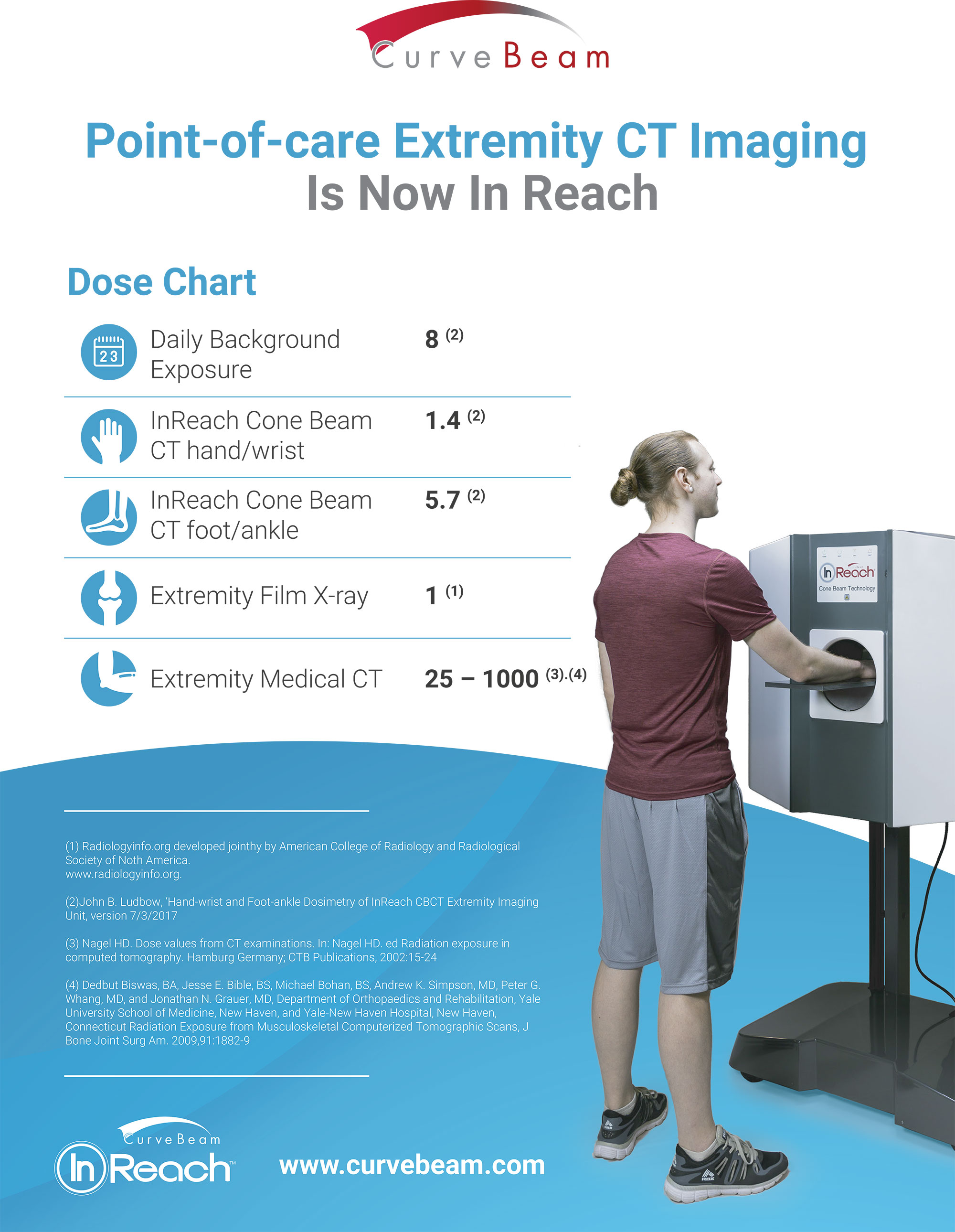
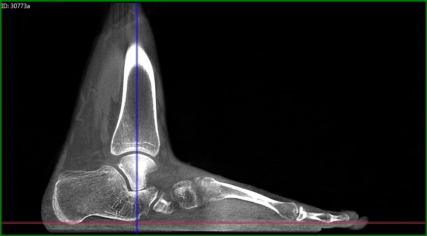
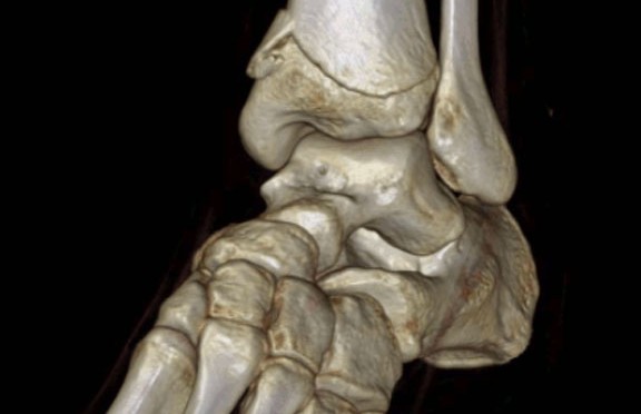
For those needing to analyze the relationship between morphology-based foot center (FC) and force/pressure-based center of gravity (COG), a study by Martinus Richter, M.D., Ph.D., Francois Lintz, M.D., Stefan Zech, M.D., and Stefan Meissner titled Combination of PedCAT with Pedography Shows Relationship of Morphology (Bone) Based Foot Center (FC) and Force/Pressure Based Center of Gravity (COG) showed that combining pedCAT with pedography fulfills those needs. The pedCAT gives you exact, bilateral, weight-bearing 3-dimentional views of the foot and ankle. By inserting a customized pedography sensor into the PedCAT, a new range of analyses was made possible.

In this study 36 patients/72 feet of patients 18 years of age and older were analyzed using pedCAT/pedography. Feet were scanned in a standing position with full weight-bearing. “The morphology based definition of the FC was performed with the pedCAT data following the TALAS algorithm. This algorithm takes different bony landmarks (Posterior calcaneal process, center of talar dome/tibial plafond, metatarsal heads) into consideration and calculates the FC. The force/pressure based COG was defined with the pedography data using a software based algorithm.”

While there is no relevant movement in the COG during the combined pedCAT/pedography scan, there was a 22.6 mm average distance between COG and FC, and FC was found to be distal to COG in all feet and lateral in 49 feet (68%), with no difference found between right and left feet. In other words, as expected, a difference between FC and COG was discovered using this method. Since there is a standard/typical shift between COG and FC, additional pedography may not be needed to predict COG based on FC.

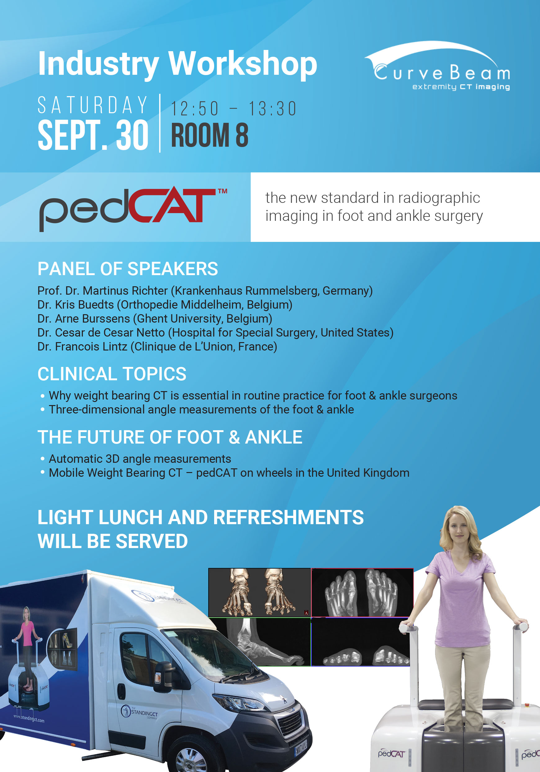
Curvebeam, founded in 2009 by a group of individuals with a proven track record in the advances and compact 3D imaging device domain, designs and manufacturers imaging equipment for the orthopedic and podiatric specialties. The pedCAT from CurveBeam may be small (4’ x 5’), but it is increasingly being shown through research to make the podiatrist’s job a lot easier and their diagnoses more accurate than ever.

To learn more about the exciting new pedCat from CurveBeam, visit http://curvebeam.com/products/pedcat today!

Webinar: Clinical Insights from Dr. Cesar de Cesar Netto

curvebeam webinar

Current research in orthopedics and podiatry have revealed many advantages of weight-bearing CT procedures over traditional ones.

To offer greater insight into Weight Bearing CT benefits, Dr. Cesar de Cesar Netto, of the Hospital for Special Surgery, is offering a lecture via Webcast on Tuesday, November 14. His lecture is titled, “Weight Bearing CT as an Essential Diagnostic Tool in Clinical Practice.”

Dr. Netto explained, “I will be demonstrating cases that would not normally be indicated for a medical CT exam. I will review how those cases presented on X-Ray, and then compare those to weight bearing CT scans of the same patient. I will show how weight bearing CT can often illuminate a pathology or deformity in ways that radiograph can often miss altogether.”

Prior to 2012, doctors and patients had fewer choices for imaging. X-rays can obscure bony detail, and non-weight bearing CTs lack accuracy with regard to join space and alignment. Today’s weight bearing CT scans improve upon the weaknesses of prior imaging options, and can result in a higher level of care for orthopedic and podiatry patients.

To register for this lecture, click here. To watch more webinars, click here.

CT Has Numerous Advantages over X-Ray in the Taylor Spatial Frame Treatment

Winner of the Medical Device Excellence Award, the Taylor Spatial Frame (TSF), is regularly employed to treat complex fractures, bone deformities, and nonunion (http://www.smith-nephew.com/). Once TSF is implemented in a patient, a surgeon inputs information about the initial deformity into computer software that analyzes it and creates a “virtual hinge” of the deformity, the first and most important step in the creation of a corrective daily plan.

The TSF’s six-axis movement allows for precise corrections, but treatment can be imperfect without the correct input parameters in deformity, frame, mounting etc. CT is just as accurate as x-ray when scanning these parameters, but it is far more efficient—reducing errors and delays in treatment.

Following the initial correction, up to one-third of patients will experience persisting deformities known as residual deformities. These deformities can result from incomplete or insufficient corrections, which can be resolved by inputting new information about the deformity into the software that creates prescriptive correction plans. With that said, inaccuracies in the mounting parameters will nearly always result in unexpected translation-angulation deformities.

According to the study, “Calculating the Mounting Parameters for Taylor Spatial Frame Correction Using Computed Tomography,” by Kucukkaya et al. (2011), the exacting calculations of mounting parameters are integral to rapidly and effectively resolving residual deformities, and computed tomography (CT) techniques are uniquely precise in these calculations. Intraoperative fluoroscopy and postoperative x-ray techniques require that the effects of magnification be calculated.

If mounting parameters change during treatment, which they often do, recalculation is necessary. These issues make exact calculations nearly impossible. In contrast, Kucukkaya et al. (2011) found that CT calculations account for such changes and complexities, reducing residual deformities as well as treatment delay. CT’s advantages are particularly obvious in deformities with rotational elements, which are especially prone to deformities.

The Taylor Spatial Frame relies on the input of accurate data. The precision and flexibility of CT sets it apart as the best method for calculating the necessary parameters. The one drawback of CT is the relative increase in radiation exposure, though these effects are not uniform throughout the body and are negligible in knee and ankle cases. This minimal risk can be reduced even further by utilizing Cone Beam CT instead of the conventional narrow beam.

By using a wide beam in a cone shape, CurveBeam systems reduce the number of scan rotations required around the region of interest and thus the exposure to radiation. Similarly, the systems dramatically lower mA settings and the need for close positioning to the region undergoing imaging. This nearly eliminates CT’s radiation, its sole drawback in TSF. The Taylor Spatial Frame, when paired with CurveBeam cone beams, increases the effectiveness of deformity treatment while reducing the occurrence and persistence of residual deformities. This is accomplished with risk of radiation nearly eliminated.

To learn more about the advantages of cone beam CT in Taylor Spatial Frame treatment just visit Curvebeam online today!

Exposure to Low-Level Radiation is Less of a Risk than Obesity, Smoking, and Air Pollution

While the risks to health from medium- and high-level radiation have been studied for years and are relatively well-understood, the risks of exposure to low-level radiation are less clear. A new study published in the Proceedings of the Royal Society B1 found that low-level radiation exposure poses less risk to health than other issues, such as obesity, smoking, and air pollution.

The study, funded by the Oxford Martin School at the University of Oxford, was published to give the layperson a place to start when discussing public policy. “Our target audience is a civil servant, activist, journalist, worker or student who is new to the field and needs to get to grips with the terminology, see an overview of what is currently known, and understand where (and why) there is still uncertainty,” Angela McLean, a professor of mathematical biology at the University of Oxford and the lead author of the study, told HCB News.2

The paper examined a full range of radiation exposures, ranging from low-dose all the way up to acute high-dose. Researchers studied the effect of radiation on workers in the nuclear, medical, and mining industries, as well as background environmental exposure from substances such as radon. The authors also reviewed studies of real-life incidents of radiation exposure such as the Japanese Life Span study of atomic bombing survivors, as well as those of emergency workers in the Chernobyl Nuclear Power Plant and the Fukushima Daiichi Nuclear Power Plant accidents.

The results? Dr. McLean says, “We know a great deal about the health risks from radiation thanks to exceptionally careful studies of groups of people exposed to different levels from nuclear bombs or accidents, medical exposure of patients, naturally occurring sources (such as radon), and workers in the nuclear industry and medicine. From these studies it is clear that moderate and high doses of radiation increase the risk of developing some types of cancer.”3 But perhaps more importantly, the study showed that obesity, tobacco smoking, and exposure to ambient particulate air pollution pose a greater risk in shortening the years in a person’s life span than the effects of low-level ionizing radiation.

CurveBeam designs and manufactures Cone Beam CT imaging equipment specifically designed for the orthopedic and podiatric specialties, including the pedCAT, a compact, ultra-low dose CT imaging system. This technology allows doctors to make a better diagnosis the first time, eliminating the need for additional scans and, therefore, reducing low-level radiology exposure to patients. Best of all, the practice has access to the results right away.

To learn more, visit Curvebeam.com today.


1 http://rspb.royalsocietypublishing.org/content/284/1862/20171070
2 https://www.dotmed.com/news/story/39096?utm_campaign=2017-09-14&utm_source=DOTmed+Non Registered+News&utm_medium=email
3 https://www.sciencedaily.com/releases/2017/09/170913104428.htm

A Sure Fit for the Mountains

Anyone who has been skiing knows that the difference between a fantastic day of carving and a night of aches and pains can come down to the fit of your ski boots. New boots are the easiest way to improve both the comfort and quality of your skiing experience, and the only way to achieve a perfect fit is with a custom boot.

One company, Surefoot, has created a design unique to the industry that offers customers the perfect pair of made-to-measure ski boots.

Poorly fitted ski boots waste a great deal of movement, causing fatigue and lack of control, and can even result in injury. Ski boots consist of a hard shell, with a separate liner and insole. The boots attach to the skis via mechanical bindings, creating a rigid connection between the skier and the ski. In order to prevent injury and loss of control, this connection must be as direct and immobile as possible.

To turn a ski, you apply pressure to the edges of the skis, which bite into the surface of the snow and pull you in that direction. Today’s skis feature advanced designs and materials, making them highly responsive to the skier’s input. However, most people have irregular feet, with high or low arches, or directional biases, which can cause unintentional inputs. For example, many people have pronated feet, which cause the skis to naturally turn towards each other. Your body compensates for these normal biases by adjusting the leg muscles; however, this means that you are constantly fighting your own body, wasting valuable energy, and giving up control.

That’s where made-to-measure boots come in. They place your feet into a neutral position, compensating for any natural biases of the foot, allowing you to carve comfortably with minimum fatigue and maximum control.

Surefoot supplies its customers with the perfectly fitted boot, thanks to its unique measurement system. Their proprietary foot scanner measures the customer’s foot in 538 individual places, creating a topographical map. This map is used to mill the insole, which is then added to the ability-specific shell, chosen based on the customer’s particular skiing habits. Finally, a custom liner is injection molded into the boot.

This process removes the hotspots and pressure points common in many stock boots, increasing comfort and vital blood-flow. It also puts the skier into a neutral, balanced stance—and all with virtually no break-in period!

As a leader in orthotics weight bearing CT imaging, CurveBeam cares deeply about innovations in the orthopedic and podiatric industries. Surefoot’s unique method of fitting boots is a brilliant use of cutting-edge industry technology. In order to ski comfortably and maintain peak control, skiers need boots that work with their individual bodies. Surefoot’s proprietary system allows them to measure and manufacture the most precise fit possible.

Just like Surefoot, CurveBeam allows orthopedic doctors to quickly and accurately serve their patients’ individual needs with the latest and most effective imaging technology. CurveBeam’s unique Cone Beam CT technology employs a wide, cone-shaped beam. One rotation captures the region of interest. CurveBeam systems have a fixed tube current (mA) of 5, which is well below ultra-low-dose settings of traditional medical CT. Its image quality of high contrast, hard tissue features is equivalent to conventional CT. The flat panel detector is positioned closer to the anatomy being imaged, which means less X-Ray dose is required to capture similar signal strength, as compared to conventional CT.pedCAT system allows foot & ankle specialists to obtain a true weight bearing, bilateral scan of the feet & ankles in 48 seconds. The pedCAT is compact and plugs into a standard wall outlet, so can easily be placed in a podiatric or orthopedic clinic. Scans are high resolution and ultra-low dose.

To learn more, visit CurveBeam online today! To learn more about the pedCAT, click here.

Measuring Flexible Adult Acquired Flatfoot Deformity with Weight-Bearing High-Resolution 3D Cone-Beam CT Scanners

Adult acquired flatfoot deformity is a common, complex disorder associated with the failure of various soft-tissue structures involving and affecting the arch of the foot. It can differ in both severity and location, which contributes to the complexity of analyzing the problem among patients. Unfortunately, it is often difficult to characterize this deformity with standard radiography. Evidence strongly suggests, though, that weight-bearing (WB) imaging is vastly superior to non-weight-bearing (NWB) imaging.

Weight bearing CT image acquired via CurveBeam pedCAT. The pedCAT has the largest field of view in its class, allowing for an entire adult foot or feet to be captured in a single scan.

Using high-resolution cone beam CT, orthopedic surgeons can obtain measurements analogous to traditional radiographic parameters of adult acquired flatfoot deformity, according to a recent study in The Journal of Bone & Joint Surgery.

The study also found weight bearing images better demonstrated the severity of osseous derangement. WB scans show increased deformities across almost all parameters compared to NWB conditions, including a 78 percent decrease in forefoot arch angle in WB scans compared to NWB scans. Additionally:

  • On the axial view,
    • the talus-first metatarsal angle and talonavicular coverage angle increased by 57% and 43% respectively;
  • In the coronal plane, 25% to 39% decreases in the navicular-to-skin and navicular-to-floor distances;
    • 22% and 38% decreases in the medial cuneiform-to-skin and medial cuneiform-to-floor distances, respectively;
    • 16% difference in the calcaneofibular distance;
    • 9%, 19% and 36% increases in the subtalar horizontal angle at the posterior, middle and anterior positions, respectively;

Although a relatively small sample size was used (12 men and 8 women), the differences discovered were so significant as to be adequate to support the researcher’s conclusions.

Another limitation of the study was researchers used a cone beam CT extremity scanner that could not visualize the whole length of the first metatarsal, especially the distal aspect, due to the limited field of view. This impediment greatly contributed to the low reliability of all measurements that involved the assessment of the first metatarsal axis. The CurveBeam pedCAT has the largest field-of-view in its class, and a single scan can capture both entire feet.

Weight-bearing (WB) radiographs have been the typical tools used to determine the degree of deformity in flexible adult acquired flatfoot deformity, but the use of WB computed tomography (CT) is on the rise because of its ability to create 3D images better able to show doctors the full complexity of the structural failures. WB 3D extremity cone-beam CT has recently been shown to outperform even multidetector CT when evaluating foot & ankle bone anatomy—and with less radiation exposure.

High-resolution 3D cone-beam CT imaging allows you to reliably assess flexible adult acquired flatfoot deformity during physiological upright WB. Its results are at least analogous to traditional radiographic measurements, and WB is shown to be considerably more accurate than NWB. More clinical work is needed to support these conclusions, but with more widespread adoption of high-resolution 3D cone-beam CT imaging, we expect to see these results reproduced.

Weight Bearing CT Technology Touted at Chinese National Foot & Ankle Conference

The CurveBeam pedCAT was on display at the 20th National Conference on Foot and Ankle Surgery in Beijing in early September. We had the privilege of meeting with new and current clients, sharing advancements in the industry, and networking with key industry professionals. Dr. Francois Lintz, a pedCAT user and orthopedic surgeon at Clinique L’Union in France, gave a keynote speech on current trends in orthopedic surgery.

Professor Shang, host of the conference, is the director of the Tongren Hospital Foot & Ankle Center, which is the first hospital in China to utilize the pedCAT. During Professor Shang’s education session, he shared his first-hand experience with pedCAT in the Tongren hospital. He also discussed the value in using the pedCAT in a research capacity.

CurveBeam’s distributing partners in China note that BeBeijing is an exciting place for orthopedic treatment. pedCAT’s ability for diagnosis and international research potential is considered especially valuable throughout China as physicians can use it as a tool to prove, through research, that their abilities and skills are on par with the world’s academic elites.

The PedCAT allows Chinese doctors to see point-of-care advanced diagnostic imaging and provide patients with comprehensive treatment plans. The single scan protocol captures both feet and ankles in one volume, a feature great for physicians and patients.

The conference was a great experience to advance the CurveBeam brand throughout Asia. From conversation with researchers to strategy sessions with potential clients, we are already looking forward to next year’s conference.

CurveBeam designs and manufactures Cone Beam CT imaging equipment for the orthopedic and podiatric specialties. To learn more about pedCAT’s value as well as our other innovative products, please visit the website at CurveBeam.com.

Skeletomuscular Imaging Use Increased Dramatically, 1994-2013

Use of radiography, ultrasound, MR, and CT—the four most common imaging tests used for orthopedic extremities—on Medicare patients experienced a jump from 1994 to 2013, according to a study published in the latest issue of The American Journal of Roentgenology by the Harvey L. Neiman Health Policy Institute,

CT grew by 754 percent. Although experiencing the smallest jump in growth at only 43 percent, radiography remained the most used billing specialty. The use of ultrasound grew by 528 percent, while MR grew by 619 percent. Hospitals should expect to see the use of this equipment continue to rise.

Knowing patterns of imaging technology use and growth is important for hospital to plan for future equipment. For example, the relatively small increase seen in radiography masks the fact that, because radiography is by far the most used form of imaging technology, the number of uses has risen sharply, indicating hospitals may need to invest in more radiography equipment to meet the increased demand.

At the same time, such dramatic growth in other forms of imaging means an increased need for more machines as well. After all, even if your CT scanner only had 10 patients in 1994, at 754% growth, that same scanner would be used on 7,540 patients in 2013. That’s over 20 patients a day with it up and running all 365 days of the year.

An increased interest in skeletomuscular care is likely one of the main drivers of this increase. After all, increasing the use of advanced imaging can increase information accuracy and thus improve patient care. The upcoming Appropriate Use Criteria (AUC) in 2019 will also make it more likely that the use of these machines will become more fine-tuned to ensure the right scans are being used at the right time for the right needs.

To learn more about growth and advancement in imaging processes at Curvebeam, visit us online today!