A Mobile Solution for Weight-bearing CT in the UK

Industry-Workshop-featured-image

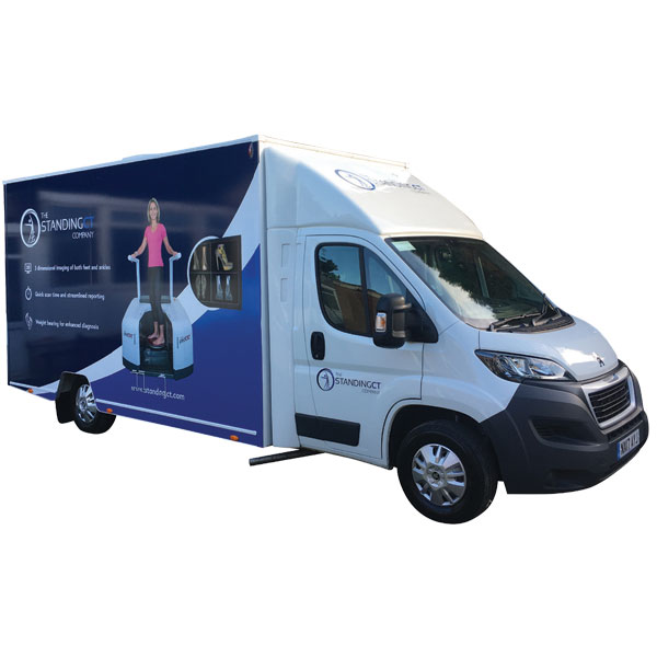
Britain’s national healthcare system can make getting a CT scan difficult, and weight-bearing scanners are almost unheard of. However, an inexpensive mobile option which will allow more doctors to perform the scans they need is now available. Weight-bearing CT scanners are available to visit in three hospitals around the UK and growing thanks to The Standing CT Company. Their goal is to be able to provide every doctor in the country with access to these scans.

Standing CT uses the CurveBeam pedCAT, the only weight-bearing scanner that scans both ankles and feet in a single pass. The pedCAT is used by leading surgeons and hospitals around the world, providing easier access for patients and superior results for clinicians. The pedCAT has everything a radiographer is looking for. It’s a compact, ultra-low dose CT imaging system that provides a bilateral, weight-bearing, 3-Dimensional view of the foot and ankle. With a less than one-minute scan time the pedCAT generates not only a CT image but digitally reconstructed X-ray views as well.

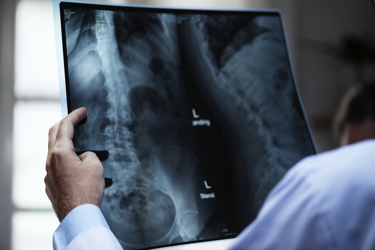
Unless a patient is standing, it is difficult to capture the proper relationships among the bones, muscles, and ligaments. For too long hospitals did not have the proper technology to create optimally useful images of feet and ankles

The Standing CT Company was founded in 2016 to solve this problem, bringing doctors and experienced entrepreneurs together to develop a solution. They created mobile units so hospitals could provide high-quality foot and ankle scanning. Standing CT Company gained regulatory approvals in May, and received their first patient in June 2017. While the first pedCAT unit in the UK was integrated four years ago at Royal National Orthopaedic, Standing CT Company is the only company offering a mobile option. By working with surgeons to learn what and where their greatest needs lie, Standing CT Company will continue to expand their reach throughout the UK over the next 3-5 years.

The mobile scanning unit is brought directly to the doctors and patients in a van containing a mobile pedCAT visiting one hospital on specific days of the week. A radiographer and healthcare assistant work together to transport the van and assist with patients. The van can run on generator power for 10 hours. This new technology offers specialists a standard model, serving hospitals and communities that would otherwise not have the capital to purchase the technology on their own.

Hospitals throughout the UK need weight-bearing scanners, and this mobile technology allows surgeons to perform the scans they need without the hospital having to purchase the expensive technology.

Learn more about Standing CT today at www.standingct.com. To learn more about the CurveBeam pedCAT, visit https://www.curvebeam.com/products/pedcat.

Study Finds Low Levels of Radiation May Actually Help Fight Off Cancer

anatomy-3003099_1280It has long been accepted that there is no such thing as a safe dose of radiation, and to suggest that radiation could actually have positive health benefits would be laughable. The standard belief was that our bodies had natural cancer barriers and that doses of radiation would significantly diminish these barriers, leading to a higher risk of cancer. Recently, however, two articles published in the Journal of American Physicians and Surgeons included findings that show low-dose radiation to be safe and even has the potential to be beneficial to patients.

In the article “Small Radiation Doses Enhance Natural Barriers to Cancer,” Dr. Bobby R. Scott outlines the current literature available on the topic. Through his analysis of studies concerning the effects of x- and gamma rays on tissue interactions, natural immunity and cancer-facilitating inflammation, Scott found that low doses are actually far safer than previously thought. In fact, especially in studies involving tumor rates in mice, Scott’s research pointed to the possibility that low levels of radiation can actually improve the body’s natural cancer barriers.

The main theory at play here is known as the adaptive response theory. This posits that these low doses actually help the body repair double-stranded DNA breaks. Under higher levels of radiation these breaks increase in frequency, hence the previous belief that any level of radiation put patients at an increased risk of cancer.

In the same issue, a team of researchers led by Dr. Jerry Cuttler published “Thyroid Cancer Following Childhood Low Dose Radiation Exposure: Fallacies in a Pooled Analysis” with findings that lead to the same conclusion as that of Dr. Scott. While the article primarily focuses on the overdiagnosis of thyroid cancer and the negative consequences that arise from that, the safety of radiation also receives some attention. Like Dr. Scott, this team also found low levels of radiation to be safe, and that the “all radiation is bad radiation” approach was a fallacy. Of significance was the finding that levels below 500 mSv were even safe for children.

At CurveBeam, we believe it is important to stay up to date with the latest findings in our field. In order to provide ground breaking technology to our customers, it is necessary to be familiar with studies like those reviewed by Drs. Scott and Cuttler. To learn more about CurveBeam and our pedCAT technology, visit www.curvebeam.com today!

A Recent Study Demonstrates the Need for CT Scans in Complex Ankle Fractures 

CT scans were shown to be better at detecting fractures over plain radiographs, according to a recent study by the American Orthopedic Foot and Ankle Society (AOFAS).[1] The study set out to determine the effects of computed tomography (CT) on both the detection of fractures as well as its effects in changing a surgeon’s overall operative plan.

Ankle fractures rank among the most common of injuries, with 150 out of 100,000 people experiencing a fracture every year. Of those injuries, up to half of them involve the posterior malleolus, a bone near the lower back of the tibia. Due to its position as well as its tendency to break irregularly, fractures in the posterior malleolus are particularly difficult for orthopedists to diagnose.[2] The AOFAS study noted that multiple past studies show that plain radiographs are unreliable for determining and diagnosing fractures. It remains imperative to have powerful imaging like CT in order to determine with complete confidence the presence ankle fractures of this kind as well as others.

The AOFAS study’s methods focused on finding patients with record of ankle fractures treated both operatively and nonoperatively. Patients were drawn entirely from the registry of the study’s home institution. Using this pool of more than three thousand patients, the study surveyed three orthopedic surgeons, asking them to determine if the radiograph of the fracture presented was complex or simple. Then, based on these scans, they asked these surgeons for their operative plan moving forward. In the study’s second part, the same surgeons were presented with a randomized set of CT scans from the same patients, and they were asked the same set of questions. Those in charge of the study then compared changes in operative approach, operative indications, and positioning after the CT scan was reviewed.

The results showed that complex fractures were less likely to be identified using a radiograph as compared to a CT scan. The writers of the study made suggestions based on these results to use CT scans more regularly in the diagnosis of ankle fractures. These results and this conclusion are consistent with prior studies, which generally agree that CT scans are more accurate, and are therefore more useful for orthopedic surgeons.

When it comes to complex fractures and fractures in the problematic posterior malleolus, this study affirms that CT scans are more reliable for developing operative plans. If the annual occurrence of ankle fractures continues to be so common, it’s important for orthopedic surgeons to take note of these results and adapt their methods.

To learn more about CT scanners that are ideal for orthopedic clients, click here.

[1] https://app.asana.com/app/asana/-/download_asset?asset_id=491852374049556

[2] https://www.verywell.com/posterior-malleolus-fractures-2549438

CurveBeam Shares Some Key Takeaways from the 2017 RSNA Annual Meeting

chicago-1804479_1920

CurveBeam recently had the pleasure of attending and participating in the 103rd Annual Meeting of the Radiological Society of North America (RSNA), held in Chicago from November 26 to December 1. The RSNA Annual Meeting is a unique opportunity for attendees from around the world to gather and meet with thought leaders and innovators to learn about the latest advances in imaging. Post-event, we wanted to share our experiences including some key takeaways.

Rising Injuries in Youth Sports
Diego Jaramillo, MD, MPH, from Miami Children’s Hospital in Miami, delivered a presentation titled “The Perfect Storm for Athletic Injuries: Youth, Growth, and Hormones”. Dr. Jaramillo shared how childhood sports have become increasingly intense and competitive. As young athletes dedicate more time and energy to one specific sport and even specialize in specific functions within that sport, the type of injuries, particularly repetitive injuries, are intensifying. Increased bone porosity of a young skeleton coupled with an increase in muscle strength can stress a skeleton and predispose it to damage. Puberty, including the hormonal influences on the growth plate, also increases the vulnerability of the skeleton to injury. Following x-rays, most young athletes are diagnosed using CT, especially for injuries that involve the head or that are primarily osseous such as the triplane fracture.

Effect of Weight Loss on Knee Cartilage Degeneration
Alexandra S. Gersing, MD, from the University of California, San Francisco, shared the results of a study on “How Weight is Lost Can Slow Knee Cartilage Degeneration”. The study examined which types of weight loss are most beneficial for patients who need to lose a significant amount of weight to slow the progression of knee cartilage degeneration. The study looked at 760 male and female patients with a body mass index greater than 25 who were enrolled in the Osteoarthritis Initiative (OAI), a U.S.-based study focused on the prevention and treatment of knee osteoarthritis. Patients were categorized into groups according to the amount of weight they were asked to lose over a 48-month period, as well as the weight loss method. Changes in the right knee were assessed at baseline, 48-, and 96-months using 3T MRI. Patients who lost weight showed significantly less T2-value increase in the bone layer of all compartments compared to those with stable weight, suggesting less cartilage degeneration over 96 months.

Patients Prefer Immediate Test Results
Radiology resident David Mihal, MD, of the University of Cincinnati College of Medicine and the Cincinnati Children’s Hospital (CCH), shared a “Survey that Showed Patients Prefer to Get Immediate Test Results”. Dr. Mihal began the four-phase study after learning that 20 percent of patients were uncertain about how they were going to get their results and had expressed anxiety to the technologist doing the exam. In phase one, techs screened outpatients to identify those who were nervous about their results. In phase two, the front desk staff checked for patients who didn’t have a follow-up appointment and offered them the option of immediate results. In phase three, all radiography outpatients were offered results as part of a questionnaire at check-in after a 10 to 20 minute wait. In phase four, the wait-time notification was eliminated from the questionnaire. Each successive phase tripled the rate at which patients used the service. Perhaps more important, 97 percent of patients understood that immediate results were available, and 92 percent of patient comments on the availability of the service were positive.

CurveBeam designs and manufactures Cone Beam CT imaging equipment specifically designed for the orthopedic and podiatric specialties, including the pedCAT, a compact, ultra-low dose CT imaging system. This technology allows doctors to make a better diagnosis the first time, eliminating the need for additional scans and, therefore, reducing low-level radiology exposure to patients. Best of all, the practice has access to the results right away. CurveBeam is proud to have participated in the 2017 RSNA Annual Meeting and is looking forward to RSNA 2018!

To learn more, visit CurveBeam.com today.

CurveBeam Systems Offered on the Stratum Med GPO to Save Practices Money

CurveBeam, the designer and manufacturer of Cone Beam CT imaging equipment for orthopedic and podiatric specialties, is pleased to announce a new partnership with Stratum Med, a collaborative alliance of physician-owned and health system-employed physician groups. Stratum leverages the collective “Groups” consolidated purchasing power to achieve higher discounts and reduce costs. CurveBeam systems are now offered through an exclusive contract with Stratum Med. Stratum saves its shareholders and clients money on their medical/surgical supply, equipment, laboratory, pharmacy and office supply purchases.

Stratum Med, Inc. is a physician owned and governed  company, founded in 1996 headquartered in Urbana, Illinois with relationships with 60+ physician groups and integrated health systems across the country  consisting of over 10,000 physicians. Stratum strives

to advance the quality of care, improve financial performance and optimize operational efficiency by using a  collaborative alliance model to leverage the collective groups’ consolidated purchasing power and yet preserve independent purchasing practices. At the same time, its dedicated support teams ensure consistent, first-class service. This model also provides a “watch dog” to ensure pricing integrity and consistency based on contract terms, as well as networking opportunities where materials managers, lab directors, pharmacy directors and imaging directors can challenge and learn from one another.

Stratum has achieved savings ranging from 8-20% and has developed a non-acute, integrated purchasing program providing Stratum Shareholders and Clients access to pricing, services, and features typically available only to large hospitals and healthcare systems.

CurveBeam is proud to be a part of such a useful purchasing model and looks forward to the opportunities this new partnership will provide. To learn more about the benefits of purchasing CurveBeam imaging machines for your practice, visit curvebeam.com/products/pedcat/ today.

RSNA Daily Dispatch – A New Focus on Machine Learning

For the first time ever, the technical exhibits halls at the RSNA Annual Meeting include a machine learning showcase, where a variety of researchers and industry ventures have the opportunity to share cutting edge work in artificial intelligence and deep learning in healthcare and radiology.

rsna 3

One company collected 9 million radiology data sets over three years to train computers to automatically recognize and diagnose a wide range of injuries and pathologies. Other research involved using parameters of patients’ demographic information and medical history records  to predict a prognosis. From managing massive amounts of patient data to quickly assessing complex medical images, machine learning can assist radiologists in their current tasks and allow for a level of planning and diagnosis that would not be possible without the trove of information stored in big data.

RSNA Daily Dispatch

The Monday, Nov. 27 schedule of the 2017 RSNA Annual Meeting included a variety of scientific sessions discussing technical and clinical developments in Cone Beam CT systems. From improving image quality to correlating with other modalities, researchers are validating the efficacy of CBCT in diagnosis and treatment of musculoskeletal injuries and pathologies.

@Ingakoerte tweeted a photo of the crowds at the RSNA Annual Meeting.

In a poster presentation for “High Resolution Extremity Cone-Beam CT with a CMOS X-Ray Detector: System Design and Applications in Quantitative Assessment of Bone Health” Biomedical Engineer Qian Cao evaluated the ideal Cesium Iodide scintillator thickness to visualize trabecular bone detail for applications such as early detection of osteoarthritis. He compared image quality characteristics of an optimized CMOS detector to an amorphous silicon detector and a micro CT scanner. The optimized CMOS detector had superior trabecular detail compared to the Amorphous Silicon detector and comparable detail to the Micro-CT, with the advantage of a much larger field of view than Micro-CT.

In “Effect of Motion Compensation on the Image Quality of Cone Beam CT Scans in Musculoskeletal Setting” Guarav K. Thawait, MD, research associate at Johns Hopkins shared the results of a study where involuntary patient motion in CBCT scans was corrected using an iterative reconstruction algorithm. The algorithm improved motion artefacts significantly in bone structures.

In “Evaluation of Bone Erosions in Rheumatoid Arthritis Patients using CBCT and MRI,” Dr. Thawait discussed a study in which a radiologist reviewed CBCT, MRI, and Ultrasound datasets for signs of rheumatoid arthritis. The correlation between CBCT and MRI was moderate while the correlation between CBCT and US was a bit higher. Though the correlations were only moderate to good between modalities, test-retest reproducibility for CBCT scans was excellent and the modality shows promise as a useful tool for RA diagnostic evaluation.

RSNA Daily Dispatch

The Radiological Society of North America (RSNA) commenced its 103rd Annual Meeting in Chicago on Sunday, Nov. 26, 2017.

@judywawira tweeted a photo of  a standing room only session at the RSNA Annual Meeting on Nov. 26, 2017.
@judywawira tweeted a photo of a standing room only session at the RSNA Annual Meeting on Nov. 26, 2017.

Attendees from around the world gathered to learn about the latest advances in imaging.

In an afternoon session titled “Open Gantry Systems: Advances, Challenges, and New Applications,” Dr. Jeffrey H. Siewerdsen, PhD, of Johns Hopkins Biomedical Engineering, discussed the vast applications of cone beam CT for interventional and diagnostic imaging. The devices presented included C-arms for image guided surgery, Cone Beam CT guided radiation therapy systems, and diagnostic imaging devices for breasts, dental/ENT, and orthopedic extremities.  Siewerdsen emphasized the  revolutionary potential of weight-bearing extremity devices in orthopedic specialties, and mentioned the CurveBeam pedCAT and LineUP systems.  He also discussed the image quality parameters of Cone Beam CT and artifacts specific to this type of imaging. He offered suggestions on how to quantify image performance of such systems, many of which are relatively new and do not have established quality metrics in the imaging community. Finally, he presented some of the newest Cone Beam CT devices to come to market, including the extremely compact CurveBeam InReach made primarily for hand and wrist imaging.

Department of Energy Considers Radiation Research Funding

Radiation has a bad reputation, especially among lay people, but even in the medical community to a degree. Radiation is in the air we breathe, the food we eat, the water we drink, and even in our own inner biological workings. The dangers of radiation exposure are real—high levels of radiation exposure have been shown to have a clear link to cancer in repeated reputable studies—but not all levels of radiation are known to increase cancer risks, or at the very least, not all levels of radiation are alike.

Medical professionals agree that radiation at appropriate levels has done a great deal of good in our world. Its usefulness in diagnostic medicine has reduced the need for surgeries and other invasive procedures and it has saved millions of lives. The medical establishment is now beginning to question if avoiding radiation when possible is the best approach for the best outcomes.

Medical professionals specializing in radiology and oncology have been pushing in recent years for further study on the effects of low doses of radiation for positive medical purposes. Previous research has focused on radiation at higher doses, such as situations in which individuals have survived nuclear warfare or live near radioactive sites. Studies regarding medical usage of radiation primarily has been concerned with higher doses as well, like those used in the treatment of cancer. Unfortunately, however, very little is known about smaller doses and its risks versus its benefits. So, the medical community is lobbying hard to acquire funding for this type of research.

On November 1, 2017, a meeting was held in which presentations were made before the Committee on Science, Space, and Technology: Subcommittee on Energy regarding the urgent need for funding from the U.S. Department of Energy for Basic Research on Low Dose Radiation. Among those in attendance was James A. Brink, MD, FACR. Dr. Brink is the Radiologist-in-Chief at Massachusetts General Hospital and the Juan M. Taveras Professor of Radiology at Harvard Medical School. Dr. Brink is also the Chair of the Board of Chancellors for the American College of Radiology (ACR). He and others spoke to the subcommittee regarding the importance of research on the effects of low dose radiation.

Dr. Brink and others explained that radiation is used in many items we use in our world today, and in fact occurs naturally in our environment. He also explained to the committee that “the most significant source of exposure to manmade radiation in humans is that associated with medical diagnostic and therapeutic procedures,” such as medical imaging in CT scans, X-rays, and other procedures. While we know the risks of cancer in exposure to high levels of radiation, he argued, there is much less certainty regarding the results of low levels.

Gayle Woloschak, Professor of Radiation Oncology and Radiology at Northwestern University, echoed Brink’s statements. She added that our knowledge about smaller radiation doses is inconsistent. And, as she described, “Contradictory data…make for contradictory assumptions.” Even medical professionals do not agree on how much radiation is too much radiation, so it has been deemed safest to work under the belief that medicine should utilize as little radiation as possible for imaging and similar procedures. Woloschak questioned that belief, and also urged the committee to consider the funding, insisting that “a small change in our knowledge can make for drastic changes in recommended policy.” She added that the highly conservative stance, in which we are “overprotecting” our patients from radiation, could actually be costing funds that could be otherwise used to advance our well-beings in other areas.

Brink, Woloschak, and other experts at the DOE meeting all made similar intriguing arguments for the need for more information regarding the effects of low dose radiation. They all seem to agree that there is a compromising middle ground now in which some medical professionals are adopting the philosophy that it is acceptable, and even advisable, to continue to carefully monitor radiation. However, it is important to think not in terms of lowest dose possible, but instead in terms of the dosage level that is safest and also most therapeutic. The need to determine that “sweet spot” is why funding and research, they argue, is so critical right now.

We at CurveBeam stay on top of trends in medicine and stay abreast of recent medical advances. It is our desire to design products and offer services that help you as physicians find the best possible outcomes for your patients. For more information about our product line, visit us at CurveBeam.com today!


Works Consulted
ACR Chair Asks Congress for Radiation Research Fundshttp://www.auntminnie.com/index.aspx?sec=sup&sub=cto&pag=dis&ItemID=118753

http://docs.house.gov/Committee/Calendar/ByEvent.aspx?EventID=106564

http://docs.house.gov/meetings/SY/SY20/20171101/106564/HHRG-115-SY20-Wstate-WoloschakG-20171101.pdf

http://docs.house.gov/meetings/SY/SY20/20171101/106564/HHRG-115-SY20-Wstate-BrinkJ-20171101.pdf