Biomedical Engineer Presents Important Findings on the Responsiveness of Standing CT for Measurement of Knee Joint Space Narrowing at IOWAI 2018

image1

Imaging is a critical tool in the diagnosis and management of osteoarthritis. The International Workshop on Osteoarthritis Imaging (IOWAI) was created to offer opportunities for osteoarthritis investigators to discuss issues and share findings related to imaging. This Workshop was first held in Ainring, Germany in 2017 and is now held annually at various locations in Europe and North America. This year’s IOWAI will be held in Menton, France, on July 5-8. Thought leaders and influencers from academia, regulatory and funding agencies, and the medical equipment industry will be in attendance.

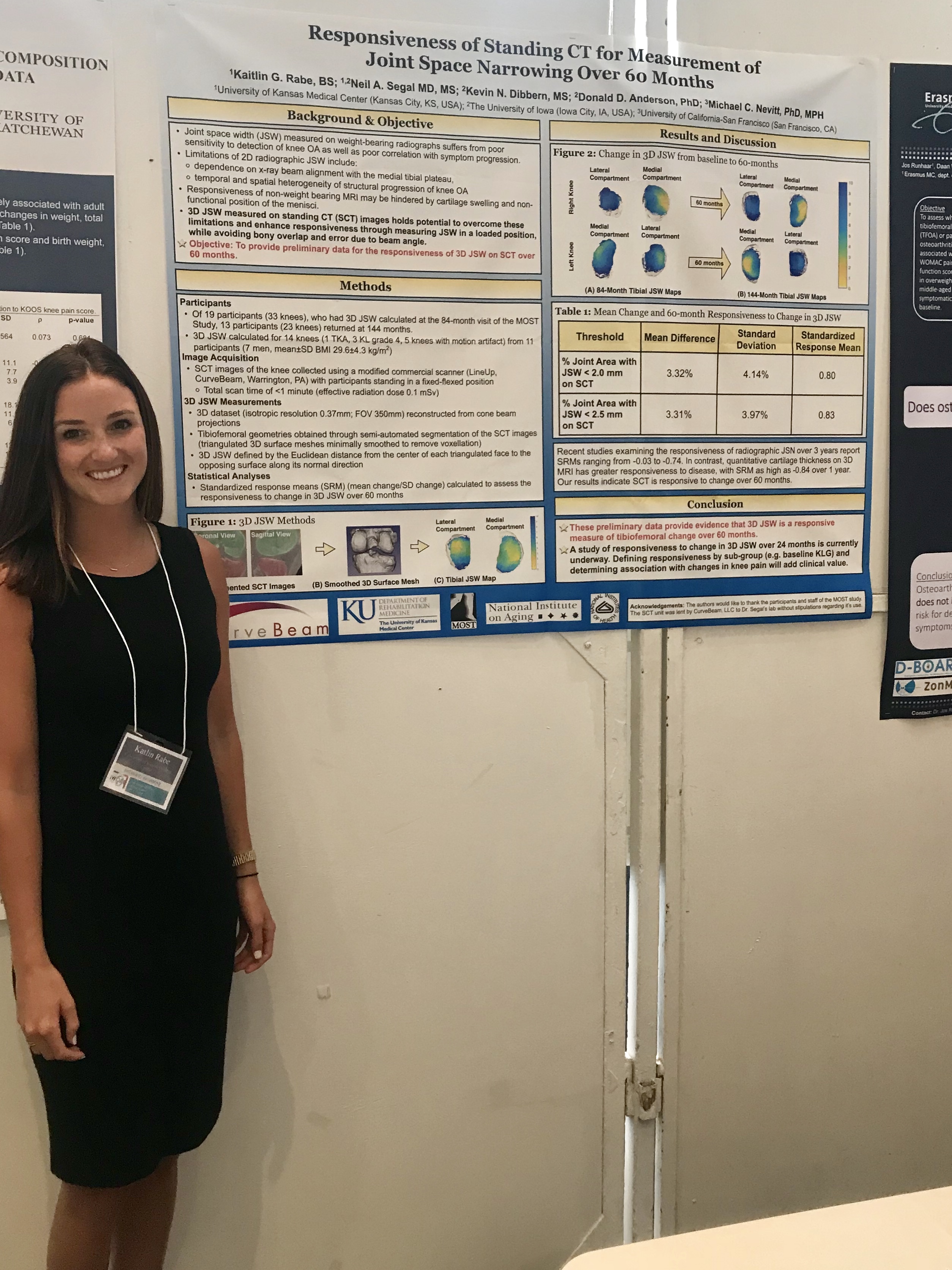
Kaitlin Rabe, Biomedical Research Assistant at the University of Kansas Medical Center, will be presenting a poster titled, “Responsiveness of Standing CT for Measurement of Knee Joint Space Narrowing Over 60 Months.” The study was sponsored by the National Institutes of Health, the University of Iowa, and the University of California, San Francisco.image2

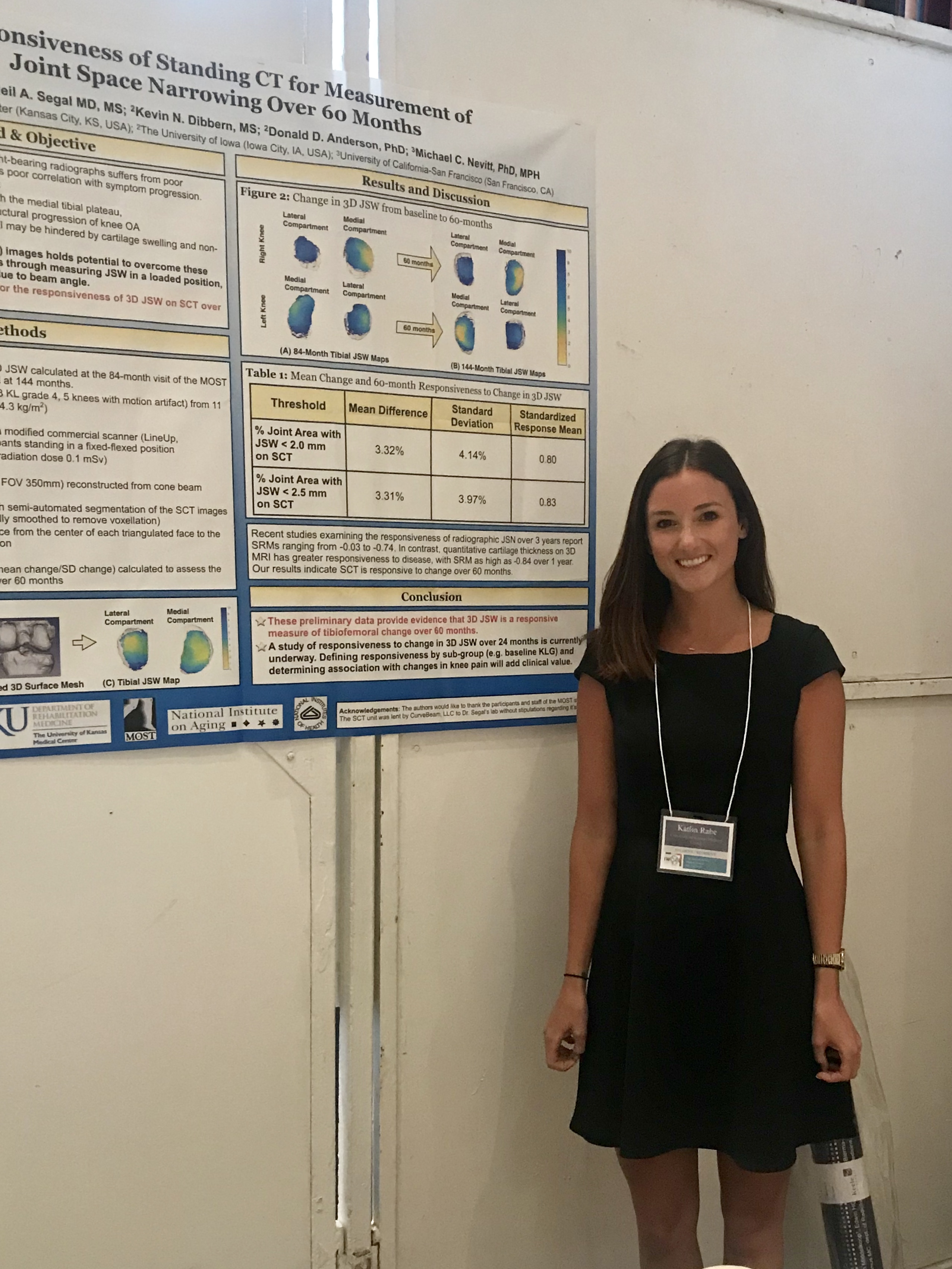
In the workshop, Ms. Rabe will discuss the limitations of two-dimensional radiography when measuring joint space width (JSW). These 2D weight-bearing radiographs traditionally suffer from poor sensitivity when used to detect knee osteoarthritis and produce poor correlation with symptom progression.

Much of these issues can be attributed to the inherent limitations in the 2D radiography procedure, such as the dependence on X-ray beam alignment with the medial tibial plateau. In addition, the temporal and spatial heterogeneity of structural progression of knee OA limits the responsiveness of radiographic JSW to diagnose disease progression. MRI offers important advantages over 2D radiography, but is typically non-weight-bearing.

Standing CT (SCT) imaging holds the potential to overcome these limitations and enhance responsiveness in three-dimensional JSW measurements. This is because JSW is measured in a loaded position while avoiding bony overlap and error due to beam angle variations.

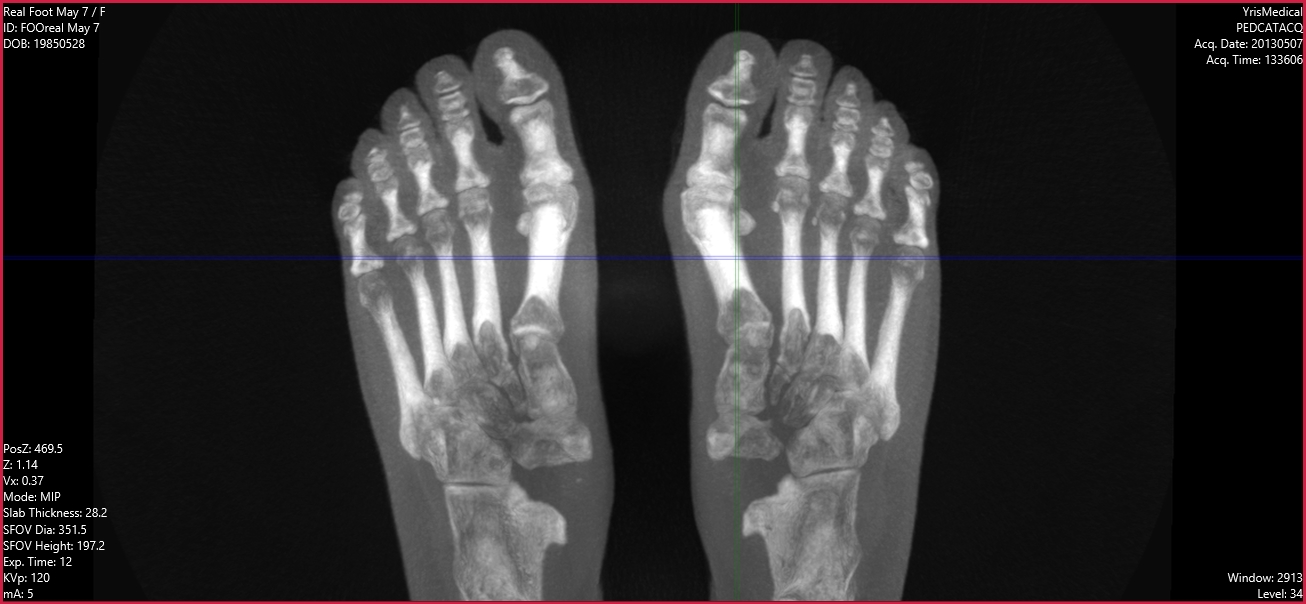
In her workshop, Ms. Rabe will provide preliminary data for the responsiveness of 3D JSW on SCT over 60 months using an early prototype of the CurveBeam LineUP system. The LineUP is a compact, ultra-low dose Cone Beam CT imaging system specifically designed for the orthopedic and podiatric specialties. Preliminary data calculated for 33 knees from 19 participants over 60 months support the hypothesis that 3D JSW exhibits a greater responsiveness than has been reported for radiography, similar to that for quantitative cartilage thickness measured on MRI over 12-months.

To get all the details on this important study, be sure to attend Ms. Rabe’s workshop at the IOWAI in Menton, France, July 5-8. For more details on accommodations and the all the workshops at this important conference, go to https://www.iwoai.org/.

WBCT ISG Special Session takes place next week in Boston

nettoGuest post by Dr. Cesar de Cesar Netto, MD, PhD

Dr. Netto is currently a foot and ankle fellow at Hospital for Special Surgery (HSS). He is part of the WBCT ISG board. His main research interests are weight bearing CT imaging and adult acquired flatfoot deformity. 

Weight-Bearing Cone Beam CT (WB CBCT) is a reality. This relatively new imaging technology has quickly transformed the ability of orthopedic surgeons to assess simple and complex deformities of the foot and ankle.  It is following the path to soon become the standard imaging study for foot and ankle pathologies.

The Weight-Bearing CT International Study Group (WBCT ISG) was formally founded in 2017. Our mission is to promote dialogue and collaboration on WB CBCT research initiatives, and to develop standardized protocols for WB CBCT measurements and imaging analysis and is jointly sponsored by Carestream, CurveBeam and Planmed.

The Group is comprised of active and passive members from relevant international surgical and radiology foot and ankle societies, and research projects are conducted and published in collaboration.  The main tasks of the WBCT ISG are:

  • To promote and improve research using three-dimensional imaging with WB CBCT
  • To standardize the methodology regarding measurement methods on WB CBCT images
  • To serve as a platform of communication for foot and ankle surgeons, radiologists and other healthcare providers with scientific interest in this field
  • To offer guidelines and reviews regarding the indications and use of WB CBCT at an international level

The WBCT ISG has been promoting Scientific Sessions at least once or twice a year, usually during the most important orthopedic foot and ankle societies meetings in the US and Europe. The sessions have been a great opportunity for people to get to know the technology and learn from the experience of the most important authors that have been publishing on the field. Our next WBCT ISG Scientific Session will take place in Boston (Massachusetts, US) during the American Orthopedic Foot and Ankle Society (AOFAS) Annual Meeting. We are meeting on July 12th, 2018, from 5:00 p.m. to 6:30 p.m., at the Hynes Convention Center, Room 310.

We are having a great line-up with very interesting talks of experienced speakers on the field, with the following agenda:

  • Moderation: Martinus Richter MD, Ph. D
    • (05:00-05:10 PM) – Opening and Message of the President

Professor Richter is the current president International WBCT Study Group and the most experienced researcher on the field. He is Associate Professor at the Hannover Medical School and Head of the Department for Foot and Ankle Surgery Nuremberg and Rummelsberg at the Hospital Rummelsberg and Sana-Hospital Nuremberg.

  • (05:10-05:22 PM) Francois Lintz, MD: Surface Interaction Maps in the Foot and Ankle Joints using Distance Mapping and WBCT

Dr. Lintz a Foot and Ankle Surgeon at the Clinique de L’Union in Toulouse, France. He is part of the WBCT ISG board and a leader on the field.

  • (05:22-05:34 PM) Daniel Guss MD, MBA: Weight Bearing CT and the Syndesmosis: A Nice-To-Have or Must-Have?

Dr. Guss is Foot and Ankle Surgeon at Massachusetts General Hospital (MGH), In Boston-MA, US. He is Assistant Professor of Orthopedic Surgery at Harvard Medical School. He has been actively looking for answers in the diagnosis and treatment of the challenging ankle syndesmotic injuries.

  • (05:34-05:46 PM) Alberto Leardini, Ph. D: Preliminary experience in the use of WB CBCT at the Istituto Ortopedico Rizzoli in Bologna.

Professor Leardini is a world famous and recognized author in foot and ankle biomechanics. He is the Director of the Laboratory of Movement Analysis at the Instituto Ortopedico Rizzoli, Bologna, Italy. He has been studying the role of the WB CBCT in the research and clinical environment.

  • (05:46-05:58 PM) Alastair Younger, MB, ChB, MSc, ChM, FRCSC: Relationship between percentage of bone healing on WBCT and clinical outcomes for fractures and fusions. 

Dr. Younger is world recognized surgeon and researcher from St. Paul’s Hospital in Vancouver, British Columbia, Canada.  He has enormous experience with complex deformities of the foot and ankle and special interest in bone healing and non-union.

  • (05:58-06:10 PM) Cesar de Cesar Netto: How weightbearing cone beam CT is going to change the treatment of flatfoot deformity?

Dr. de Cesar Netto is currently a Foot and Ankle Fellow at Hospital for Special Surgery (HSS). He is part of the WBCT ISG board and his main research interest are WB CBCT and adult acquired flatfoot deformity.

  • (06:10-06:25 PM) Questions and Answers
  • (06:25-06:30 PM) Adjourn

We would be glad to have you attend! Please RSVP today; see you in Boston!

CurveBeam Announces Exhibit Presentation Schedule for AOFAS

Blog-94---AOFAS-In-Booth-Presentation-Preview (1)

The American Orthopaedic Foot & Ankle Society (AOFAS) will hold its annual meeting from July 11 to July 14 in Boston, Massachusetts. The event will feature exhibits and targeted educational opportunities along with the chance to network with professionals in the orthopaedic specialties.

CurveBeam is honored to be exhibiting at this year’s event and will host the following presentations in booth #512:

  • Thursday, July 12, 9:30 AM to 9:45 AM: Francois Lintz presents “Automatically measuring hindfoot in Three-Dimensions.”
  • Thursday, July 12, 3:45 PM to 4:00 PM: Cesar de Cesar Netto presents “Weight Bearing Cone Beam CT: The Road to Somewhere!”
  • Friday, July 13, 10:00 AM to 10:15 AM: Arne Burssens presents “Understand what you diagnose, know what you treat using weightbearing CT.”
  • Friday, July 13, 1:15 PM to 1:30 PM: Martinus Richter presents “What we have learned from more than 8,000 scans at a foot and ankle center.”

​The American Orthopaedic Foot & Ankle Society (AOFAS) is an organization of more than 2,200 orthopaedic surgeons from the US and abroad who specialize in the medical and surgical treatment of injuries, diseases, and​ other conditions of the foot and ankle. Their annual meeting is a premier event in the orthopaedic community, and CurveBeam is pleased to be participating alongside some of the industry’s most distinguished leaders. CurveBeam’s highly innovative work in CT imaging has been well received by foot and ankle specialists, and we are proud of our collaboration in efforts toward improved patient outcomes.

Learn more here about CurveBeam’s work with the orthopaedic community.

CurveBeam Plans Weight Bearing CT Presentation with a Special Focus on Sports Med at AOSSM Annual Meeting 2018

Folded_Card_Front_LineUP1Revision

The American Orthopaedic Society for Sports Medicine (AOSSM) Annual Meeting will soon open its doors to welcome 1,300 sports medicine specialists, practitioners, and society members. Meeting attendees receive four days of leading research and education programming from elite orthopaedic sports medicine experts and medical innovators.

While you attend AOSSM, be sure to check out a presentation by Dr. Kent Feldmen titled ‘Weight Bearing CT: Shining New Light on Common Lower Extremity Sports Injuries.” This presentation will occur on Thursday, July 5 from 10:55 a.m. to 11:05 a.m. at AOSSM Booth #401.

AOSSM 2018 takes place this year on July 5 through July 8 in San Diego, California at the Manchester Grand Hyatt. The Annual Meeting plays a crucial role in providing insights on frontline research to the orthopaedic sports medicine community, which is why CurveBeam is excited to exhibit.

Learn More About pedCAT and LineUP

Visit booth 401 to learn about how pedCAT and LineUP could improve your practice. Bilateral, weight bearing scans of the feet, ankles and knees give physicians the information they need to assess the biomechanical spatial relationships and alignment of the lower extremities.

Also learn about the InReach, an upper extremity imaging solution for immediate point-of-care diagnosis of scaphoid fractures, distal radius fractures, and other easily misdiagnosed injuries.

Weight Bearing CT and Lower Extremity Sports Injuries Presentation

The CurveBeam team is looking forward to the opportunity to meet, educate and connect with top physicians in the field of sports medicine. To learn more about our weight bearing CT technology and find out where the team will be this year, please visit curvebeam.com/events-and-webinars/events-and-trade-shows/.

Curvebeam Achieves ISO 13485:2016 & MDSAP Certification

QualityCurvebeam announced the company’s ISO 13485 certification has been updated to the latest revision of the standard. The company’s quality system is now certified to the ISO 13485:2016 standard, which represents the most recent revision of the widely recognized manufacturing quality standard issued by the international Organization for Standardization (ISO).

In addition to 13485:2016 requirements, this certification covers the requirements of the Therapeutic Goods Administration (TGA) of Australia; Brazil’s Agencia de Vigilancia Sanitaria (ANVISA); Health Canada; the U.S. Food and Drug Administration (FDA) under the Medical Device Single Audit Program (MDSAP).

“These latest accomplishments are the result of a significant effort by our entire quality, technical and operations teams,” said Ryan Conlon, Director of Quality and Regulatory Affairs for Curvebeam. “CurveBeam’s quality system lays a strong operational foundation for continuous innovation in orthopedic imaging.”

The ISO 13485 standard is specifically designed to certify medical device manufacturers and their key suppliers. The International Standard outlines requirements for a quality management system where an organization needs to demonstrate its ability to provide medical devices and related services that consistently meet customer and applicable regulatory requirements.

ESSR in Amsterdam: Muscles & Nerves

Are you heading to Amsterdam to attend ESSR this week? The focus of this year’s meeting is “Muscles & Nerves”, and there are some great opportunities to expand your knowledge on some critical issues at this event.

ESSR Overview

The European Society of Musculoskeletal Radiology (ESSR) is an organization whose mission is to advance the knowledge, diagnosis and treatment through imaging of both the normal and abnormal musculoskeletal system. The ESSR is the primary organization in Europe in this field, and they provide a European forum for education and research of musculoskeletal radiology and cooperation among medical and non-medical professionals in these endeavors. The annual meeting is the major European forum for sharing advances in musculoskeletal radiology and offers excellence in education.

Recommended Sessions

The team at CurveBeam would like to recommend some interesting sessions as you plan your schedule at ESSR:

  • Tibial Tuberosity—Trochlear Groove Distance: Basic Positioning Errors of Current Measurement and Methods of Correction

Presented: Saturday, June 16, at 2:12 p.m. in Room 2.

  • Low Dose CT Technique and Radiation Dose: Does the ALARA Principle Still Apply?

Presented: Saturday, June 16, at 12:50 p.m. in Room 1.

  • Standardised Reporting: Guidelines for the Radiologist, Benefit for the Clinician

Presented: Saturday June 16, at 12:20 p.m. in Room 2.

CurveBeam Commitment

CurveBeam makes it a priority to stay abreast of the latest research and development in the field of imaging in order to best serve your practice and your patients.

Learn more here about the innovative imaging and diagnostic technologies available at CurveBeam.

CurveBeam Unveils New Logo

Logo
On the heels of a recent FDA approval for its LineUP system, CurveBeam unveiled a fresh, new look as it poises itself to enjoy a remarkable leadership position in the healthcare industry.

CurveBeam unveiled a new logo design that it will transition to using in all branded content and materials.

“CurveBeam’s primary focus is innovation,” explains Marketing Manager Vinti Singh. “Our new logo incorporates subtle design updates that reflect our culture of constant adaptation and change. However, the color scheme and swoop motif are preserved as a symbol of a commitment to our core values.”

With a line of cone beam scanning devices, CurveBeam allows clinics to offer weight bearing CT scanning to their patients to improve their quality of care. Here is the latest overview of the CurveBeam product line:

  • The InReach is an extremity cone beam CT scanner that images the hand, wrist, forearm, elbow and the lower extremities. The InReach is the most compact orthopedic extremity CT scanner available.
  • The LineUP provides bilateral, weight-bearing CT imaging of the knee and lower extremities to redefine orthopedic care.
  • The pedCAT  is a compact, ultra-low dose CT imaging system that is ideal for orthopedic and podiatric clinics. Patients benefit from the convenience of point-of-care advanced diagnostic imaging. Scan time is one minute, and the pedCAT automatically generates all standard X-Ray views in addition to the full CT volume.
  • CubeVue software provides powerful visualization tools to enhance diagnosis. The pedCAT system comes complete with CubeVue installed. CubeVue automatically generates all standard X-Ray views, possibly eliminating the need for your plain X-Ray system altogether. Insta-X views are free from magnification, distortion or patient positioning inaccuracies.

CurveBeam  has refreshed its brand and is committed to staying at the forefront of the orthopedic field as the healthcare industry evolves. As its team continues to push forward with new technology, CurveBeam is continuously evolving, always innovating, and keeping its approach to product development fresh.

CurveBeam was founded in 2009 by a group of individuals with a proven track record in the advanced and compact 3D imaging device domain. Learn more about the exciting things happening at CurveBeam today by our blog.

The Weight-Bearing CT International Study Group, Brought to You by CurveBeam

This year CurveBeam will be co-sponsoring the Weight-Bearing CT International Study Group Thursday, July 12, 5:00-6:30 PM at the Hynes Convention Center, Room 310, in Boston, MA.

Dr. Martinus Richter from Krankenhaus Rummelsberg will be moderating a panel of experts from around the world. Professor Alberto Leardini from the Rizzoli Institute, Dr. Francois Lintz from Clinique de L’Union, Dr. Cesar de Cesar Netton from the Hospital for Special Surgery, and Dr. Alastair Younger from the Footbridge Centre for Integrated Orthopedic Care will join and discuss the latest thinking in weight-bearing CT.

The goal of the study group is to promote dialogue and collaboration on weight-bearing CT research, as well as create standardizations for weight-bearing CT measurements and analysis.

Weight-bearing CT scans use low doses of radiation while creating an image of the foot and ankle while the patient is standing in a natural standing position. Having patients in a natural standing position is beneficial for doctors because they can see the bones and tendons of the foot and ankle under normal conditions. Patients with pain and deformities seem to benefit most from using weight-bearing CT scans.

While many years of research have shown the benefits of these types of scans, we know there are many out there who still have questions. This International Study Group will provide you with answers. We would also love to have orthopedics and podiatrists with experience with weight-bearing CT to join us and share your own experiences with the technology.

Anyone interested in learning more about weight-bearing CT, including the Cone Beam CT imaging equipment for orthopedics and podiatrists designed by CurveBeam, is invited to join us for presentations from these experts.

Space is limited. RSVP today at wbctstudygroup.com!

Co-Sponsored by Curvebeam

Weight-Bearing CT May Improve Diagnoses in Hallux Valgus Patients

Hallux Valgus

A recent analysis of studies titled Imaging of Hallux Valgus by James Welck and Naji Al-Khudairi has shown that newer, three-dimensional imaging techniques including weight-bearing computer tomography (WBCT) may permit a more thorough understanding of the hallux valgus (HV) deformity and provide greater information to foot and ankle surgeons prior to corrective surgery.

Traditional two-dimensional AP, lateral, and oblique radiographs are the current standard for imaging HV deformity. Two-dimensional radiographic images can be used to measure several radiographic angles which are commonly used to quantify the extent of HV deformity. However, recent research shows that some of these angle measures show low levels of reliability and accuracy. Coughlin et. al. found that measures of the hallux valgus angle and the intermetatarsal angle assessed using 2-D imaging had high intraobserver and interobserver reliability. In contrast, the distal metatarsal articular angle was measured to 5° or less in only 58.9% of cases, and measurement of metatarsophalangeal joint congruency led to a wide variation in cases identified. The primary reason that two-dimensional imaging techniques may fail to be reliable is that HV is a triplanar deformity with rotational aspects that two-dimensional images cannot account for.

Fortunately, research shows three-dimensional HV imaging techniques have promise. Conventional CT imaging is not weight-bearing, but weight-bearing cone beam CT provides better insight into HV. This means that many patients imaged through traditional CT may not have been diagnosed correctly.

  • Welck and colleagues have found WBCT to better display the sesamoids, which are difficult to represent accurately using traditional AP radiographs.
  • Collan and colleagues demonstrated the importance that CT is weight-bearing by finding that there were significant differences in HVAs and IMAs between HV and control groups only when patients were in a weight-bearing stance.

Two other research groups found that WBCT provided insight into the effects of joint hypermobility in HV patients which would not typically be measurable using two-dimensional imaging techniques.

To learn more about how three-dimensional imaging techniques like WBCT can benefit your foot and ankle practice, visit http://www.curvebeam.com/products/pedcat/. CurveBeam’s PedCAT systems are at the cutting edge of WBCT innovation and can help provide your patients with the best HV care that current science has to offer.