Latest WBCT ISG Scientific Meeting Topics Include Distance Mapping and Syndesmosis Modeling

landscape-3719918_1280


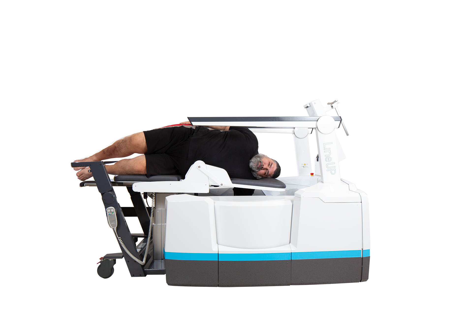
The WBCT International Study Group’s most recent scientific meeting was held during the 3-day European Foot and Ankle Society (EFAS) International Congress. Expert speakers came together from across the globe to impart their knowledge and clinical findings as it pertains to a wide variety of studies surrounding weight bearing CT imaging. Speakers and topics included:

  • Shadpour Demehi, MD (Maryland, USA): “Advanced imaging of syndesmotic injuries: where are we now and what can we do with weight bearing CT”
  • Sorin Siegler, PhD (Pennsylvania, USA): “The use of distance mapping in combination with WBCT”
  • Andrew Goldberg, MD (London, UK): “Center of rotation of the subtalar joint: novel research in the use of WBCT to assess dynamic function”
  • Kris Buedts, MD (Antwerp, Belgium): “Deformity correction and 3-D planning based on the WBCT”
  • Oliver Michelsson, MD (Helsinki, Finland): “Use of low dose WBCT in cartilage and bony lesions of the ankle”
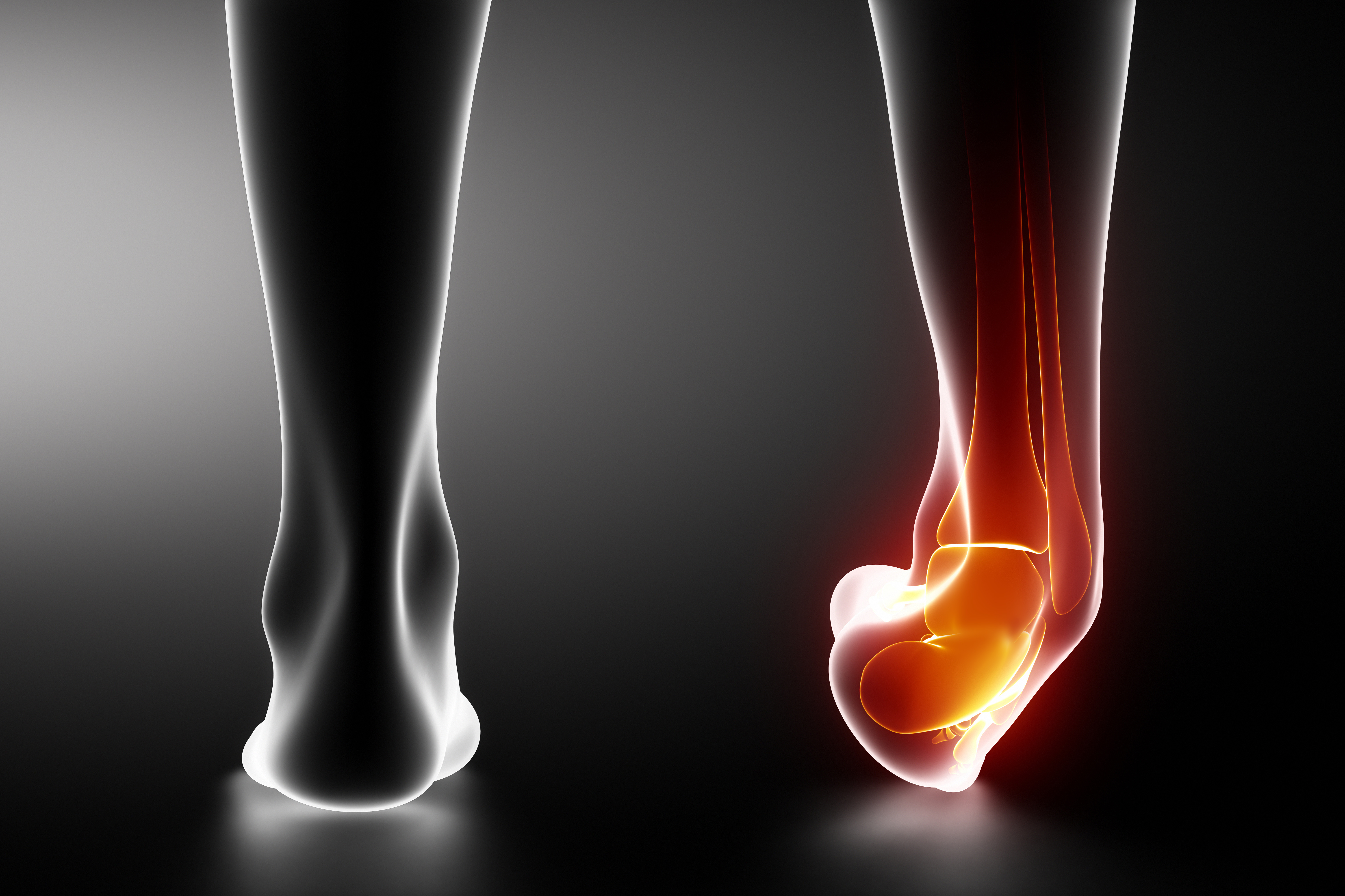
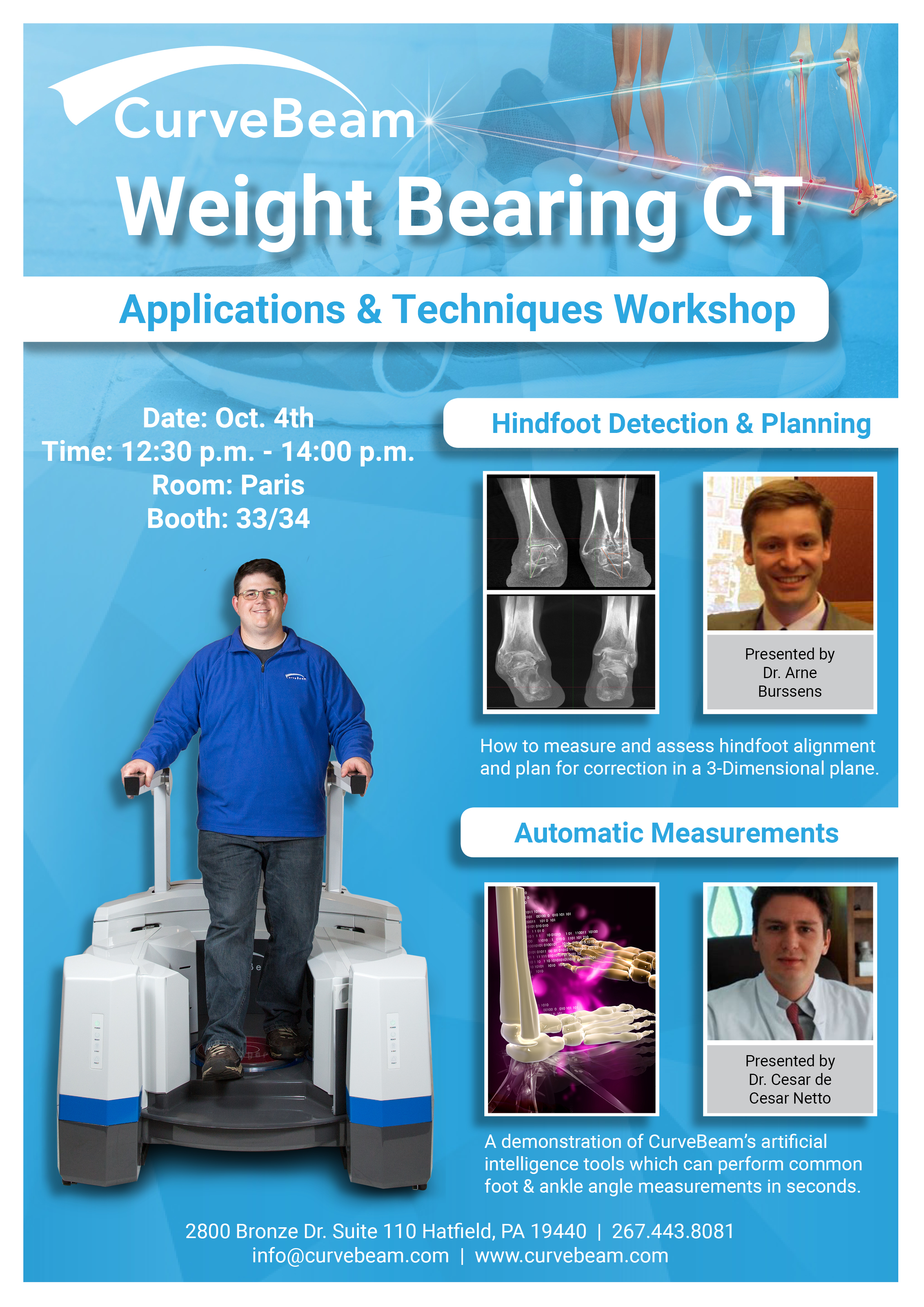
  • Arne Burrsens, MD (Utah, USA): “Three-dimensional modeling of the weight bearing ankle syndemosis”
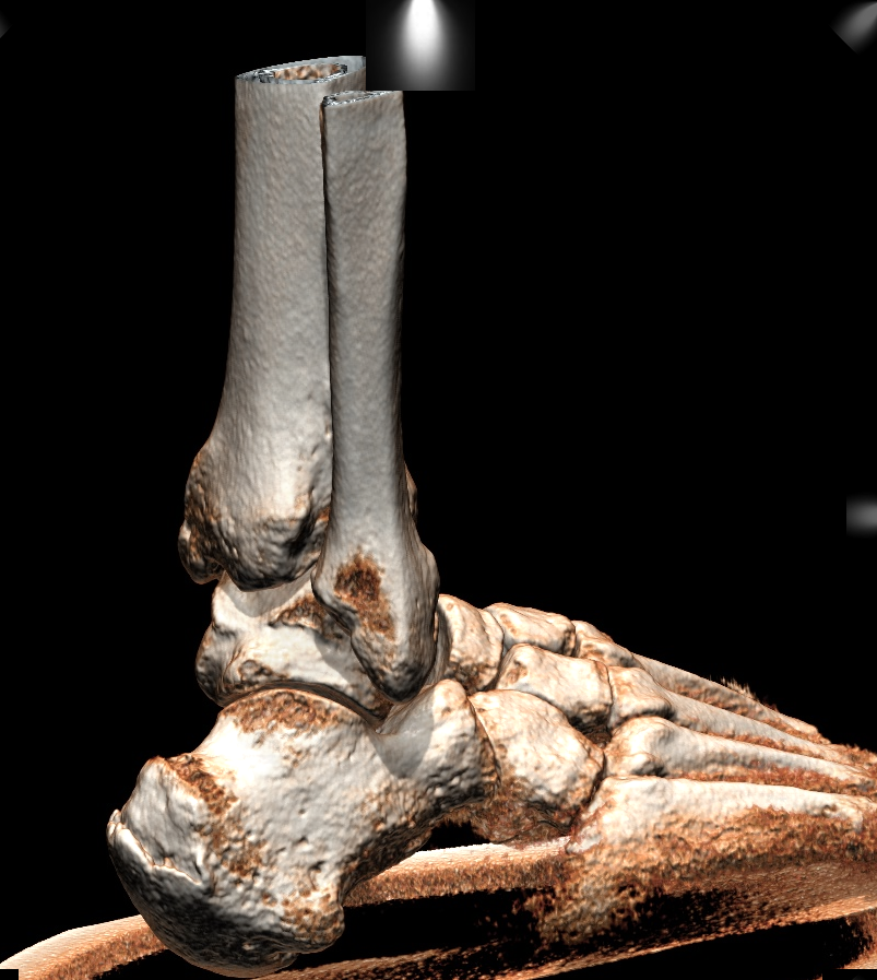
  • Martinus Richter, MD, PhD, & International WBCT Study Group President (Rummelsberg, Germany): “Results of a 5-year/10,000 scans experience with WBCT: impact on costs, radiation exposure, and time spent”

Completely independent from the industry but cooperative with different manufacturers of different WBCT devices, the Weight Bearing CT International Study Group is comprised of lower extremity orthopedic surgeons, radiologists, and biomedical engineers. The group’s mission is to promote dialogue and collaboration on weight bearing CT research initiatives and is working to create standardized protocols for weight bearing CT measurements and analysis.

Curvebeam is proud of their collaborative role as an industry sponsor in bringing together this unique group of educated, experienced professionals to discuss both the potential and proven successes of WBCT modality. We want to express our thanks to the group’s speakers and members for sharing their unparalleled expertise, and to our co-partners—Planmed and Carestream—for ensuring a clear-cut future for the most promising innovation in foot and ankle radiographic imaging. Watch the presentation below.

Join CurveBeam at the 2018 Orthopaedic Summit in Las Vegas

las vegas

Orthopaedic Summit Evolving Techniques (OSET) is the premier gathering of spine and trauma surgeons, physician assistants, nurse practitioners, athletic trainers, and physical and occupational therapists from across the globe. This year’s event will be held at the Bellagio in Las Vegas from December 5-8 and includes cutting-edge, industry-sponsored, surgical demonstrations, symposia, debates, and small group discussions on the latest techniques in orthopaedics.

CurveBeam, an industry leader in the design and manufacturing of CT imaging equipment for the orthopedic and podiatric specialties, is proud to be a part of this year’s summit. Attendees can stop by Booth 323 on the exhibition floor to check out the company’s innovative products, such as the LineUP, firsthand. The LineUP allows bilateral, weight-bearing CT imaging of the knee and lower extremities and is helping to redefine orthopedic care.

WapnerIn addition, Dr. Keith Wapner, MD, will be presenting December 6th during the “Innovation in Foot & Ankle Surgery” section from 2:20 pm to 2:27 pm. Dr. Wapner, Director of the Division of Foot and Ankle Orthopaedic Surgery at the University of Pennsylvania, will be sharing valuable information on weight bearing CT scans for foot and ankle surgeons.

The 2018 Orthopaedic Summit promises to be an exciting event. Schedule your LineUP demo at the Curvebeam booth at the summit today by clicking here.

3 Reasons to Visit CurveBeam at RSNA 2018

chicago-1791002_1280

The Radiological Society of North America 104th Annual Meeting commences November 25 at McCormick Place in Chicago. Attendees will get hands-on experience with cutting-edge tech, including artificial intelligence, 3D printing and virtual reality in the radiology specialty.

  1. See the complete LineUP system on display

CurveBeam15SMALL

CurveBeam will feature its largest exhibit to date at the 2018 RSNA meeting. CurveBeam’s LineUP system, which was cleared by the FDA for sale earlier this year, will be on display. CurveBeam will showcase the Multi-Extremity chair, which permits imaging of the hand and elbow, as well as non-weight bearing foot and ankle imaging. Radiology administrators will be able to get a first-hand look at the multi-extremity chair’s ergonomic design that is comfortable for both patients and operators.


2. Improved Metal Artefact Reduction

Before MAR Lateral After MAR Lateral

CurveBeam unveiled its proprietary metal artefact reduction (MAR) tool earlier this year with the launch of its LineUP system. CurveBeam’s MAR employs a high- density detection filter, which enables more authentic reconstructions in the vicinity of metal hardware. Users have the option to activate MAR prior to the start of each scan. Attendees can get a demo of before and after MAR datasets at CurveBeam’s exhibit.

3. Cinematic Renderings

4-ProgressiveFancyLighting


CurveBeam will display a cinematic rendering feature, currently under development, that will be offered as a part of its visualization software, in the future. Cinematic renderings simulate lifelike lighting techniques to provide a more photo-realistic 3D rendering of the bones and joints. In a recent study by Swiss researchers, radiologists and surgeons preferred cinematic renderings to volume renderings for a majority of ankle injuries.

If you are interested in setting up an appointment for a personal demo at RSNA, click here.

Dr. Alexej Barg WBCT ISG FOOTinnovate Webinar Nov. 12, 9 P.M. EST

WBCTFOOTinnovateLogo

Webinar: Monday, November 12, 2018

9:00 PM EST

CLICK HERE TO REGISTER TODAY!

BargEmailBlastGraphicLONG

Dr. Alexej Barg, MD is a foot and ankle surgeon and Assistant Professor for orthopaedic surgery at the University of Utah. He specializes in the care of the foot and ankle and traumatic injuries to the lower extremity. Dr. Barg’s expertise is to perform reconstructive surgery of the foot and ankle. Special interests include total ankle replacement and joint preserving procedures in patients with ankle osteoarthritis, sports injuries of the foot and ankle and patients with sequalae of trauma of the lower extremity. Prior to joining the foot and ankle team of the University of Utah, Dr. Barg was Head of Foot & Ankle Surgery at the University of Basel in Switzerland. He is current vice-president of the Weight-Bearing CT International Study Group (WBCT ISG). He is working on several studies addressing clinical use of WBCT in hindfoot disorders In the planned webcast, he will share his approach how to possibly use the WBCT in patients with hindfoot instability.

CLICK HERE TO REGISTER TODAY!


A FOOTInnovate account is required to register. FOOTInonovate membership is free for foot and ankle specialists.

CurveBeam Heads to Germany for Medica

Im Rahmen der weltgrößten Medizinmesse MEDICA informieren sich 130.000 Fachleute, davon 60 Prozent internationale Besucher, über Innovationen und Weiterentwicklungen aus allen Bereichen der ambulanten und stationären Versorgung. 5.000 Aussteller aus 66 Nationen präsentieren sich zur MEDICA 2017. http://www.medica.de | 5,000 exhibitors from 66 countries will be showcasing their products and services at MEDICA 2017. Clearly structured, with each market segment assigned its own exhibition hall, MEDICA covers the entire range of medical equipment needs in practices and hospitals. http://www.medica-tradefair.com

Medica is the leading international trade fair for the medical industry. Attracting more than 5,000 exhibitors from over 70 countries, Medica gathers leading business professionals, researchers, and thousands of decision makers from around the globe to network, discuss and learn about the latest healthcare trends.

Scheduled for November 12-15 in Düsseldorf, Germany, Medica boasts a schedule filled with forums, conferences, and special presentations covering a wide array of medical technology topics. Medica is a can’t miss opportunity for companies to present their latest innovations, reenergize existing partnerships, and forge new ones. And this year mark’s CurveBeam’s inaugural role as an independent exhibitor at Medica with a booth location in Hall 9 E 43.

With a core team that pioneered Cone Beam CT imaging technology, CurveBeam has been setting new standards in orthopedic 3D imaging worldwide since 2012. At Booth E 43 in Hall 9, both the LineUp and InReach will be on display. The InReach is CurveBeam’s revolutionary low dose point-of-care extremity CT imaging system. Featuring a small footprint with minimum siting requirements and less than 25 second scan time, its cone beam technology allows for imaging of hands, wrists, elbows, forearms, and lower extremities.

Cleared by the FDA in May 2018, the cutting-edge LineUp is the complete multi-extremity CT imaging system, with easy-operation and less than 30 second scan times. The LineUp’s capability for bilateral, weight-bearing CT imaging of the knee and lower extremities makes it a unique solution that is redefining orthopedic care.

We look forward to seeing you at Medica. To learn more about CurveBeam’s groundbreaking imaging technology before the show, visit https://www.curvebeam.com/products/pedcat/ today.


Barry University School of Podiatric Medicine Acquires the pedCAT for Research

DrArmstrongpedCATWeight-bearing CT imaging systems are quickly approaching the standard of podiatry care. The pedCAT bilateral weight bearing CT scanner from CurveBeam is a both a valuable research tool and clinical asset. The Barry University School of Podiatric Medicine in Miami, Florida, recently became the first medical school in the southeastern U.S. and the second accredited podiatry school in the country to acquire the pedCAT. Under the leadership of Dean Dr. Albert Armstrong, the faculty and students have planned several research initiatives to improve understanding of foot and ankle injuries and conditions.

The Prestige of Barry University

 Barry University’s School of Podiatric Medicine is a highly respected medical school. Founded in 1985 with aims to graduate highly skilled podiatric physicians, the school has educated more than 1,000 podiatric physicians and earned a reputation for excellence. The school offers students the opportunity to earn a Doctor of Podiatric Medicine (DPM) degree in four years and become certified as a practitioner of podiatric medicine, making them eligible to enter postgraduate residency training in podiatric medicine. It is one of only nine such schools in the United States.

Barry University Acquires the pedCAT System

 The pedCAT system was installed at Mercy Hospital inside one of the University’s clinics. This innovative technology provides 3D, weight-bearing CT scans of the foot and ankle, which gives podiatric physicians the information they need to assess the biomechanical spatial relationships and alignment of the lower extremities.

“We are so very fortunate to have acquired the pedCAT machine for one of our clinics at Barry University School of Podiatric Medicine,” said Dr. Albert V. Armstrong Jr, DPM, MSC, BSRS, CWS, Dean of the School of Podiatric Medicine, Associate Professor of Radiology, and a podiatric physician expert in lower extremities. He added, “The images are absolutely amazing!” He considers the pedCAT a “great investment for our school, our clinics, our students, and our patients.”

Benefits over Traditional CT Scans

 Traditional CT scanning, which requires patients lie down for the procedure, has been used in medicine for decades, making weight bearing position impossible. Traditional CT scans of the lower extremities expose patients to higher dose than pedCAT scans.

Kent State Faculty and Students Research the Use of Weight Bearing CT to Effectively Assess Magnitude of Hallux Valgus Deformity

Hallux valgus, bunion in foot on white background

Hallux valgus is a triplane deformity. In patients with this deformity, the sesamoids displace from their normal alignment. Recent evidence suggests that, according to researchers, “the magnitude of this displacement can be determined by the coronal plane sesamoid rotation angle.” Podiatric doctors often use weight-bearing radiographs to determine the magnitude of the hallux valgus deformity. This is a crucial step in planning surgical correction. However, conventional foot radiographs have long been problematic due to:

  • geometric distortion
  • unreliable measurements made between different observers
  • limited imaging in the coronal plane
  • measurable differences between weight-bearing and non-weight-bearing images

CT scanning addresses these problems. It allows “cross-sectional imaging of the anatomical parts in all three planes of the foot without typical radiographic distortion.” In other words, doctors are able to look at slices of the foot for better data. CT scanners also allow for three-dimensional reconstruction of the foot.

The Kent State University College of Podiatic Medicine recently acquired a CurveBeam pedCAT bilateral weight-bearing CT scanner via a grant funded by the Ohio College of Podiatric Medicine (OCPM) Foundation. In the wake of this acquisition, the College of Podiatric Medicine students and faculty proposed three research projects. Each one has the potential to break new ground in the specialty. A team of KSUCPM researchers has completed the first project, which focuses on the evaluation of hallux valgus deformity in the coronal plane of the foot.

Differences in Rotation Angle Between Two Extreme Weight-Bearing Positions

Using the weight-bearing CT scanner, this study was designed to determine, according to the researchers, “the effect of different weight-bearing foot positions on the coronal plane sesamoid rotation angle as compared with standard sesamoid axial studies.” Study data demonstrated “significant differences in the rotation angle between the two extreme weight-bearing positions.” Sesamoid rotation angles, the researchers noted, were “significantly higher in the pronated foot position.” Sesamoid rotation angles from the weight-bearing CT supinated position correlated with those values.

Weight-Bearing CT Scan Determination Should Replace Forefoot Axial Studies

These results strongly suggest that “weight-bearing CT scan determination with the foot in a non-affected weight-bearing position should replace forefoot axial studies as the accepted imaging standard.”

Learn more about how Weight-Bearing CT in diagnosing Hallux Valgus patients here.

Researchers propose reproducible method to quantify syndesmosis displacement based on spatial WBCT data

Sprained ankle black x-ray

The syndesmosis is located just above the ankle where the tibia and fibula meet, providing stability to the ankle joint while allowing for its motion. A sprain, twist, rotational injury, or break to the ankle can stretch and tear the ligaments that support the syndesmosis. Syndesmotic injuries occur in up to 18 percent of all ankle sprains and 23 percent of all ankle fractures. However, the limitations of 2D imaging make a diagnosis and operative treatment of syndesmotic ankle injuries challenging.

Despite high accuracy and sensitivity, CT scans may underestimate the extent of syndesmotic lesions because of non-weight bearing conditions.

Weight bearing cone beam CT (WBCT) is an alternative imaging technique with numerous advantages, including relatively low radiation dose.

Researchers in the United States and Belgium aimed to develop a reproducible method using WBCT to quantify displacement, translation and rotation of the fibula caused by subtle syndesmostic injuries. Current methods use a single slice of a CT volume. The researchers proposed segmenting a volume out of bilateral ankle CTs superimposing the healthy ankle on the contralateral ankle to compare the deviation of the fibula to quantify the extend of the lesion.

The researchers conducted a study on eighteen patients with a unilateral syndesmotic lesion. The results were described in a study titled,  “Templating of Syndesmotic Ankle Lesions by Use of 3D Analysis in Weight-bearing and Nonweightbearing CT”.

For those patients with a high ankle sprain (n = 12), bilateral imaging was performed with weight-bearing cone-beam computed tomography (CT), while non-weight-bearing CT was used for those with fracture-associated syndesmotic lesions (n = 6). To quantify the syndesmotic lesions, changes between the most lateral aspect of the lateral malleolus and the anterior and posterior tubercle in the healthy, stationary fibula were compared to those of the affected patients, using a control group of seven studies.

Deviations were calculated using defined anatomical landmarks on computer assisted design (CAD) software, rather than via manual methods.

The study found there were significant differences in the tibiofibular configuration between injured and healthy ankles.

The study concluded that

  • The method was accurate in assessing subtle syndesmotic injuries.
  • In the case of fracture associated with syndesmotic injury, it offered a precise description of the displacement related to the integrity of the distal tibiofibular syndesmosis.
  • In a case with pronounced fibular comminution, the amount of shortening could be preoperatively calculated, facilitating reconstruction of the fibula.

Click here to read about a previous study in which cadavers were scanned via WBCT imaging in an effort to shed light on the rotational dynamics in syndesmosis.

EFAS Workshop: Weight Bearing CT Applications and Techniques

Curvebeam will host an Applications and Techniques Workshop on  October 4th at the 12th EFAS International Congress in Geneva, Switzerland.

Dr. Arne Burssens, who recently completed a fellowship at the University of Utah in Salt Lake City,  will be presenting on Hindfoot Detection and Planning. He will discuss how to measure and assess hindfoot alignment and plan for correction in a 3-Dimensional plane.

Dr. Cesar de Cesar Netto, a fellow at Johns Hopkins University in Maryland, will demonstrate CurveBeam’s Automatic Measurements toolset, which can perform common foot and ankle measurements in seconds

. The event will be hosted in the Paris Room from 12:30 p.m to 2 p.m.

CurveBeamEFASInsertWBCTISGFront

Visit the CurveBeam exhibit at EFAS to learn more about our weight bearing CT imaging systems.

Also, plan to attend the WBCT ISG Scientific Session on Oct. 5.

Weight Bearing CT A Key Topic at EFAS 2018

2-EFAS-PromoToolkit_SocialMediaBanner_1024X512October 4-6 marks the 12th EFAS International Congress in Geneva, Switzerland. This year’s theme, State of the Art Foot and Ankle Surgery, focuses on the latest clinical and scientific findings in the foot and ankle community.

Weight bearing CT imaging has emerged as an essential tool for the proper evaluation of deformities of the ankle, midfoot, forefoot, and hindfoot. EFAS attendees will have multiple opportunities to learn about the modality in the main program, including the following podium lectures:

  • 3D biometrics: A prospective comparative evaluation of the Foot Ankle Offset using weight bearing CT semi automatic software”, Presented by Dr. Francois Lintz
  • How are coronal alignments of the knee and hindfoot correlated ? A clinical study of 124 lower limbs using 3D weight bearing imaging”, Presented by Dr. Francois Lintz
  • Weightbearing CT and MRI findings of Stage II Flatfoot Deformity: Can We Predict Patients at High-Risk for Foot Collapse?”, Presented by Dr. Cesar de Cesar Netto

In addition, several posters on display will review recent research efforts involving weight bearing CT, including:
– “Distance mapping analysis of the Foot and Ankle complex joints using Weight Bearing CT in varus and valgus deformities”, Dr. Francois Lintz

Comparative study of 3D versus 2D first intermetatarsal M1M2 angle measurement using Weight Bearing CT”, Dr. Francois Lintz
Templating of Acute and Chronic Syndesmotic Ankle Lesions in Weightbearing and Nonweightbearing CT”, Dr. Arne Burssens
Clinical and Radiological Outcome after a Medial Calcaneal Osteotomy Assessed by Weightbearing CT”, Dr. Arne Burssens

Intrinsic Anatomical Risk Factors in High Ankle Sprains Determined by Computed 3D CT Analysis”, Dr. Arne Burssens

Foot Alignment in Basketball and American Football Elite Athletes. Are they different?”, Dr. Cesar de Cesar Netto

Instability of the First Ray and Hallux Valgus in Patients with Adult Acquired Flatfoot Deformity (AAFD): A Weightbearing CT Study”, Dr. Cesar de Cesar Netto

The Weight-Bearing CT International Study Group will be hosting a special session on Friday, October 5th featuring presentations by Dr. Arne Burssens, Dr. Cesar de Cesar Netto, and Dr. Francois Lintz. To RSVP, click here.